Objective To identify the type of iron deposition and describe its amount,distribution and associated lesions,in order to support an etiologic diagnosis for hemochromatosis.Methods Hematoxylineosin (HE) stain,reticular fiber stain,Masson's stain and Perl's iron stain were used to assess liver biopsies from 31 patients with hemochromatosis.The Ishak scoring system and Deugnier scoring system were used to assess the histological change in liver and to semi-quantify the excess of hepatic iron.Genetic testing results were received from a portion of the patients and used in analysis.Results One patient had hereditary (-HFE) hemochromatosis complicated with Gilbert's syndrome,for which the pattern of iron deposition was similar to that of the four patients with Gilbert's syndrome.Iron accumulation appeared as fine granules predominating at the biliary pole of cells and was distributed throughout the lobule with a decreasing gradient spanning from the periportal to centrolobular areas.Mild chronic inflammation was found to be commonly associated with low stage fibrosis.One patient had HFE hemochromatosis complicated with hepatitis B virus infection,and the pattern of iron deposition resembled that in the eight patients with viral hepatitis,wherein the deposition was mainly in the sinusoidal cells and/or portal macrophages.Histological grading and fibrosis staging differed among patients.The five patients with blood disordered showed iron accumulation mainly in the periportal hepatocytes,but mesenchymal iron deposits were also present.The grade of inflammation,as well as of fibrosis,was mild.The five patients with alcoholic disease and the five patients with drug-induced hepatitis showed hepatic iron deposition in swollen or ballooned hepatocytes.The two patients with excessive iron supply showed iron deposition localized within the parenchymal and mesenchymal cells.Conclusion Etiologic diagnosis of hemochromatosis relies on both the type of iron deposition and the nature of associated lesions.Liver biopsy is necessary for both diagnosis and prognosis.
>病历摘要患儿,女,8岁,辽宁省瓦房店人,学生。因"反复肝功能异常3年"到北京佑安医院就诊。2008年9月,患儿因"支气管肺炎"当地医院给予阿奇霉素静脉滴注、后口服罗红霉素,1周后出现乏力、食欲减退,尿黄如浓茶色,于当地医院就诊,查谷丙转氨酶(alanine transatninase,ALT)993 IU/L,天冬氨酸转氨酶(aspartate aminotransferase,AST)819 IU/L,总胆红素(total bilirubin,TBiL)103μmoL/L。甲型肝炎病毒(HAV IgM、IgG)、丙型肝炎病毒(HCV IgM、IgG)、戊型肝炎病毒(抗-HEV IgM、IgG)抗体均阴性;乙型肝炎表面抗体(Hepatitis B surface antibody,HBsAb)阳性,乙型肝炎其他指标均阴性。由于患儿肝功能异常原因不明,发病前用阿奇霉素、罗红霉素治疗,当地诊断"药物性肝炎"可能性大,予保肝药物治疗,1个月后肝功能正常。2008年12月患儿再次因"上呼吸道感染"用"抗生素"输液治疗3 d(药名不详)
患者女,43岁.因"纳差、尿黄2个月"于2012年4月10日入院.既往患有失眠症10年,长期服用安定5~10片/次,服用螺旋藻等保健品1年.无手术输血史,近2年曾多次于当地私人诊所洗牙.在化工厂工作7年,经常接触到粉尘等,未采取防护措施.无饮酒史,无慢性肝病家族史,无输血史,否认食物药物过敏史.
<正>自身免疫性肝病是指由于机体免疫系统攻击自体肝组织引起的肝组织损伤和肝功能异常的一组免疫性疾病,包括有自身免疫性肝炎(AIH)、原发性胆汁性肝硬化(PBC)和原发性硬化性胆管炎(PSC)。通常情况下这三种肝脏疾病独立存在,约有6%~9%的患者可以在同一时段或病程中出现两种疾病的临床表现、血清学和组织学特征,称为重叠综合征。近年来随
Objective To evaluate the application of Glypican 3(GPC3),heat shock protein 70(HSP70) and glutamine synthetase(GS) in hepatocellular carcinoma.Methods Morphology of 85 cases of HCC were evaluated;three immunohistochemical markers-GPC3,GS,HSP70 were detected in these cases.Results In the 85 HCC cases,GPC3 was positive in 88.24%(75/85) cases and its expression was higher in poorly differentiation HCC than that in well/moderately differentiated HCC,the expression difference was statistically significant,individual cells in regenerative nodules adjacent HCC were GPC3 positive in 7 HCC;HSP70 was positive in 94.12%(80/85) cases,their expression had no significant correlation with HCC differentiation,and individual cells in regenerative nodules adjacent HCC were positive in 3 HCC;GS expression had different distribution modes between HCC and surrounding liver tissues,GS was positive in 67.06%(57/85) HCC and its expression had no significant correlation with HCC differentiation.Conclusion HSP70,GS,GPC3 for the diagnosis of HCC have their own advantages and disadvantages respectively.In daily work,diagnosis should base on HE morphology,and application of a panel of immunohistochemistry markers which can further improve the reliability of HCC diagnosis.
Objective To study the expression of adiponectin and α-smooth muscle actin (α-SMA) in liver from the patients of chronic hepatitis B(CHB) overlapped non-alcoholic fatty liver disease( NAFLD), and to explore the mechanism of CHB patients with NAFLD. Methods 94 patients of CHB overlapped NAFLD (case group)underwent liver biopsy, 119 cases of patients with CHB alone (control group) as control, the fasting venous blood sample was taken for liver biochemical and virological indicators. Liver biopsy specimens were immunohistochemically stained for histology, adiponectin and α-SMA, and integral absorbance. Results The serum ALT, total cholesterol, triglyceride, low density lipoprotein cholesterol and fasting glucose levels in case group were significantly higher than that in control group (Z = 3.425,4.488,4.858、2.265, P < 0.05) .There was no significant difference in HBV DNA viral load and HBeAg between the two groups( Z = 0.825, χ2 = 0.323, P > 0.05). In case group, adiponectin expression was no significant correlation with steatosis, inflammation and fibrosis(r = 0.032, -0.107, -0.133, P>0.05); however, in control group adiponectin was negatively related to inflammation and fibrosis(r= -0.223, -0.259,P<0.05). In case group,α-SMA expression was significant correlation with inflammation and fibrosis( r = 0.323,0.355, P < 0.05). In control group, its expression was no correlation with inflammation and fibrosis( r = 0.172,0.155, P > 0.05). Conclusions The occurrence of NAFLD is mainly related to metabolic factors and no relation with viral factors. Adiponectin is not directly related to liver injury of patients of CHB overlapped NAFLD. α-SMA may be important indicator of liver injury,and NAFLD may promote the progress of liver injury in patients with CHB.
OBJECTIVE To detect and compare the PD-1/PD-L1 (programmed death 1/programmed death 1 ligand) expressions in the liver tissues of chronic HBV infection patients in immune tolerant phase and those in immune clearance phase. METHODS Liver biopsy samples were divided into two groups: 25 samples from patients in immune clearance phase and 19 samples from patients in immune tolerant phase. PD-1/PD-L1 expressions on T lymphocytes in these liver biopsy specimens were detected by immunohistochemistry method. Percentage of PD-1/PD-L1 positive cells among CD3 positive cells was calculated by semi-quantitative evaluation. Differences between the two groups were statistically analyzed. RESULTS PD-1/PD-L1 expressions were significantly higher in the patients in immune tolerant phase as compared to that in immune active phase (P < 0.05). No statistical difference found between the two groups for PD-L1 expression in Kupffer cells (P > 0.05). CONCLUSION PD-1/PD-L1 expression level can reflect the immune functions of chronic hepatitis B patients.
患者女,33岁,因皮肤瘙痒7年,伴乏力、尿黄3年,加重10d于2008年4月21日入院.患者于2001年7月开始间断出现皮肤瘙痒,未予重视.5年前因皮肤瘙痒加重在当地医院检查发现肝功能轻度异常(具体不详),肝脏病毒学指标均阴性,未予明确诊断,间断服用保肝药物及中药治疗,肝功能仍轻度异常.
患者,女性,43岁,因“反复双下肢水肿2月,乏力、纳差1周”于2008年8月28日入院.患者于2008年6月底无明显诱因出现双下肢水肿,就诊当地诊所,静脉滴注人血白蛋白治疗好转1周后再次出现双下肢水肿,未行进一步检查及治疗,并于8月21日感乏力、纳差,进食量减至平时的1/3.查体:神志清楚、面色晦暗、全身皮肤无黄染、未见皮疹、无肝掌、蜘蛛痣.浅表淋巴结无肿大;双手静止性颤抖、巩膜轻度黄染;颈软、甲状腺无肿大、心肺检查未见异常;腹部平坦、腹壁静脉无曲张、腹部软,无压痛、反跳痛,腹部移动性浊音阳性,肝、脾肋下无触及,墨菲氏征阴性,肝区、脾区、双肾区无叩击痛,腹部移动性浊音阴性,双下肢凹陷性水肿.实验室检查:血常规WBC 2.75×10⁹/L、NEU 1.43×10⁹/L、NEU% 52.1%、RBC 3.7×10¹²/L、Hb 115 g/L、PLT 58×109/L;PT 19.8 s;血沉15 mm/h;甲状腺指标T3 1.23 mg/mL、T4 8.3 μg/dL、TSH 0.55 IU/mL、FT3 1.91 pg/mL、FT4 1.20 ng/dL;血生化Alb 26.5 g/L、Glb32.8 g/L、TC 4.34 mmol/L、GLU 4.47 mmol/L、UREA 4.92 μmol/L、 CRE 64.4 μmol/L、UA 146.2 μmol/L、TBil 25.98 μmol/L、DBil 10.76 μmol/L、ALT 23 U/L、AST 63.4 U/L、ALP 110.9 U/L、GGT 185 U/L;HBV DNA阴性;乙型肝炎病毒标志物:抗-HBs、抗-HBe、抗-HBc阳性;AFP16.23 μg/L;抗-HCV阴性;抗-SSB(-)、抗-SSA(-)、ANA(+)、AMA-M2弱阳性、2周后复检AMA-M2阴性.
Objective To study the clinic-pathological characteristics and related factors in the patients of chronic hepatitis B with hepatic steatosis.Methods A total of 306 cases of chronic hepatitis B were chosen for this study,and divided into two groups according to the percentage of hepatic steatosis less than 5% or more than 5%.The two groups were compared regarding the presence of risk factors for steatosis and clinical laboratory,virological and histological characteristics.Results Among 306 chronic hepatitis B patients,there were 141 cases with steatosis(46.1%) and 165 cases without steatosis(53.9%),respectively.High body mass index(BMI) and sex were independent factors and significantly higher in steatosis group than that in the group without steatosis(P0.0001 and P=0.008,respectively).No significant difference were found in the other parameters between two groups,including age,blood sugar,cholesterol,viral load,histological activity(HAI) and stage(P0.05).Conclusion The risk factors mainly related to steatosis were high body mass index and sex in chronic hepatitis B with steatosis.
Studies have suggested that glutamine synthetase (GS) is a potential marker of hepatocellular carcinoma (HCC). We aimed to evaluate the expression of GS in non-malignant liver tissue and serum GS levels in HCC, liver cirrhosis (LC), chronic hepatitis B (CHB), five kinds of extrahepatic diseases patients and healthy subjects. Immunohistochemistry (IHC) was used to assess GS expression in 260 liver tissue samples (from 120 HCC, 90 CHB stage 4, and 50 CHB stage 1–3 patients). Enzyme-linked immunosorbent assays of 325 samples (from 100 healthy donors, 33 CHB stage 1–3, 43 CHB stage 4, 111 HCC, and 45 extrahepatic diseases patients) were used to further analyze GS levels in serum. IHC studies showed the expression of GS in 70% of HCC patients, 46.7% of CHB stage 4 patients and 38% of CHB stage 1–3 patients. The χ2 tests showed significant difference between HCC samples and non-tumor tissues (P = 0.001 for HCC vs. CHB stage 4, P = 0.000 for HCC vs. CHB). Consistent with this, serum GS levels are increased in HCC and CHB stage 1–4 patients. There are significant differences among all samples (P = 0.000 for all), except CHB stage 1–3 versus CHB stage 4 (P = 0.552). Based on multiple linear regressions, HCC, CHB stage 1–4 and AFP were significantly associated with serum GS levels. In addition, in HCC group, TNM and Child-Pugh were significantly associated with GS levels. Expression of GS is increased in HCC, LC, and CHB. It may be a new serum marker for liver disease.
OBJECTIVE:To evaluate the efficacy and safety profiles of patients with hepatitis B e antigen (HBeAg)-positive chronic hepatitis B (CHB) treated with adefovir dipivoxil (ADV) or ADV plus bicyclol, and to optimize the treatment strategy for CHB patients. PATIENTS AND METHODS:A total of 250 patients with HBeAg-positive CHB were randomized to ADV plus bicyclol combination group and ADV monotherapy group. The patients in the ADV plus bicyclol combination therapy group (n = 125) received ADV 10 mg orally q.d. and bicyclol 25 mg orally t.i.d. for 48 weeks, and those in the ADV monotherapy group (n = 125) were administered ADV 10 mg orally q.d. alone for 48 weeks. The serum aminotransferases (ALT/AST), HBV DNA, HBeAg/HBeAb, and liver biopsy were conducted before and after therapy. RESULTS:The serum aminotransferase levels were decreased significantly in both groups. The serum aminotransferase level in ADV plus bicyclol combination therapy group decreased greater than that in ADV monotherapy group (P < 0.01). The virological response rate in ADV plus bicyclol combination therapy group was not significantly different from that in ADV monotherapy group (P > 0.05). After treatment for 48 weeks, the Knodell necroinflammatory score of the two groups were all alleviated significantly, and the Knodell score in the combination group was significantly lower than that in the ADV monotherapy group (P < 0.05). There were no remarkable adverse events probably related to the drug in this study. CONCLUSION:Adefovir dipivoxil plus bicyclol combination therapy is a safe and superior treatment regimen for patients with HBeAg-positive CHB when compared with ADV monotherapy.
Objective To investigate the changes of CD4+ CD25+ regulatory T lymphocyte (Treg) and expressions of folkhead helix transcription factor 3 (FoxP3) in intestinal mucosa in human immunodeficiency virus (HIV) infected patients. Methods Twenty-one HIV infected patients and 17 control subjects without HIV infection were included in this study. The expression of FoxP3, which was considered as a specific marker of CD4+ CD25 + Treg, was detected in intestinal mucosa specimens from HIV infected patients by immunohistochemistry. Meanwhile, the in situ expression of CD4+ T lymphocyte was also determined by immunohistochemistry. The data were analyzed by t test. Results The positive labeling index of CD4+ T lymphocyte in intestinal mucosa was significantly lower in HIV infected patients compared to the controls (11. 56%±4. 44% vs 43. 49% ±8. 90% ,t=-11. 86,P<0. 01). The positive labeling index of FoxP3 in intestinal mucosa was also significantly lower in HIV infected patients compared to the controls (0.46% ± 0.20% vs 1. 18% ± 0. 44% ,t= - 5. 98,P<0.01). Conclusion The depletion of CD4+ CD25+ Treg is accompanied with the depletion of CD4 + T lymphocyte and the reduction of FoxP3 expression in intestinal mucosa of HIV infected patients.
Purpose To observe the tissue regeneration of reconstruction with artificial esophagus made of medical polyurethane,and to study the feasibility of the new artificial esophagus in reconstruction of the cervical esophageal defect in a dog model.Methods Electric flexible esophagography was used to observe the artificial esophagus and granulation tissue,Gross and HE staining were used to observe regeneration of the esophageal tissues.Results The esophageal epithelium can regenerate,and it can cover the surface of defect in 3 months,The regeneration of gland,smooth muscle and striated muscle can not be found from 1 to 6 months after operation.After the articial esophagus ablated,it should be taken out to protect the epithelium.Conclusion The artificial esophagus made of medical polyurethane can be used to reconstruct the esophageal defect,The esophageal epithelium can regenerate.
卡氏肺孢子菌肺炎(pneumocystis Carinii pneumocystis)是获得性免疫缺陷综合征(AIDS)患者最常见的机会性感染[1].卡氏肺孢子菌(以往称为卡式肺囊虫,缩写为PCP)虽然广泛分布在土壤等周围环境中,但是很少感染免疫系统功能正常的人.
Objective To study the expression of IgM and IgG in plasma cells of liver biopsy tissues from primary biliary cirrhosis(PBC) and autoimmune hepatitis(AIH) and to investigate its role in differentiating diagnosis between two diseases.Methods Seventeen and twenty biopsies cases from untreated patients diagnosed as PBC and AIH were examined by immunostaining with anti-polyclonal antibody for IgM and IgG.Positive expression in same portal tracts of IgM and IgG in plasma cells were detected with semiquantitative scoring.Results There were 15 cases(15 /17,88.2%) of PBC with predominantly expressing IgM in plasma cells of liver biopsy tissues and 18 cases(18 /20,90.0%) of AIH with predominantly expressing IgG(P 0.0001).The expression of IgM was more prominent in the moderate and marked inflammatory biopsies than mild inflammatory biopsies in PBC(P 0.05) and also of IgG in AIH(P 0.05).Conclusion Immunostaining detection for IgM and IgG in plasma cells of liver tissue can be helpful in differentiating PBC from AIH.
<正>1病例摘要患者,女,20岁,主因"肝功能异常4年,间断腹胀、双下肢水肿9个月,加重10天"于2008年3月24日入院。
1 病史 张某,男,40岁,因"发现艾滋病病毒(Human immunodeficiency virus,HIV)抗体阳性10年余,大、小便失禁7小时",门诊以"艾滋病"收入院.患者10年前体检时发现HIV抗体阳性,CD4230个/μl.一年前查CD499个/μl,CD8477个/μl,CD4/CD8:0.27.出现食欲不振,疲劳,体重下降5公斤.
 Ronghua Jin合作论文数Capital Med Univ, Beijing Youan Hosp, Beijing, Peoples R China25Bing Shen (沈冰)合作论文数25Xin Meng (孟忻)合作论文数22
 Huiping Yan (闫惠平)合作论文数Beijing YouAn Hospital, Capital Medical University21DeZhuang Huang (黄德庄)合作论文数19MengDong Lan (兰孟东)合作论文数19
 Jun Cheng (成军)合作论文数Beijing Ditan Hospital, Capital Medical University17XiaoHong Shi (石晓虹)合作论文数16DeJun Xu (许德军)合作论文数16XiaoYan Dang (党晓燕)合作论文数16ChunHua Wang (王春花)合作论文数16
 Yusen Zhou (周育森)合作论文数Academy of Military Medical Sciences16JunQiang Li (李俊强)合作论文数15Zhaoxia Luo (罗朝霞)合作论文数14PeiQing Ma (马佩卿)合作论文数14ChenChao Song (宋晨朝)合作论文数14ShiJie Zhang (张世杰)合作论文数14LiJie Zhang (张立洁)合作论文数13Liang Zhang (张亮)合作论文数11Xingang Zhou合作论文数10XiaoYing Teng (滕晓英)合作论文数10
 Lin Sun (孙琳)合作论文数Beijing Children's Hospital, Capital Medical University10WenBin Dao (刀文彬)合作论文数9JianJun Wang (王建军)合作论文数9
 Yan Liu (刘妍)合作论文数Chinese PLA General Hospital9
 Dong Ji (季东)合作论文数Chinese PLA General Hospital9
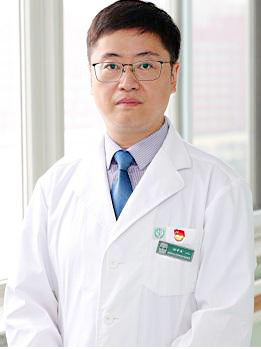 Zhongjie Hu (胡中杰)合作论文数Beijing Youan Hospital, Capital Medical University9HuiYu Liao (廖慧钰)合作论文数8Qian Yang (杨倩)合作论文数8LiXiang He (贺立香)合作论文数7LiYing Zhang (张黎颖)合作论文数7HaiTao Wang (王海涛)合作论文数7
 Hongxin Zhao (赵红心)合作论文数Beijing Ditan Hospital, Capital Medical University7
 Xinhui Guo (郭新会)合作论文数Beijing YouAn Hospital, Capital Medical University7Hongbo Ma (马洪波)合作论文数6RongHua Jin (金荣华)合作论文数6Rui Jin (金瑞)合作论文数6Song Chenzhao (宋晨朝)合作论文数6
 Zhongping Duan (段钟平)合作论文数Beijing Youan Hospital, Capital Medical University6Peizhi Wang (王培之)合作论文数6Zhichun Ma (马志春)合作论文数6Cheng Jun (成军)合作论文数5ChengLin Li (李承霖)合作论文数5ChunHui Zhao (赵春惠)合作论文数5RuiPing Xu (徐瑞平)合作论文数5
 Chun Huang (黄春)合作论文数Beijing Center for Disease Prevention and Control5RenPing Zhu (朱仁平)合作论文数4YanBin Guo (郭雁宾)合作论文数4




