肱骨头坏死是临床上常见的骨坏死之一。肱骨头的血供主要来源于旋肱前动脉和旋肱后动脉。由多因素导致的肱骨头血液供应受损,进而发生局部缺血时,肱骨头就会出现骨坏死。肱骨头坏死的病因可分为创伤性和非创伤性两大类,常见的创伤性因素有:骨折、脱位、肩部手术等;非创伤性因素有:糖皮质激素、血红蛋白病、沉箱病、戈谢病、吸烟、酗酒、系统性免疫病等。肱骨头坏死的主要症状是肩关节疼痛、活动受限。由于疾病早期通常无症状或症状较轻,不能引起患者重视,从而延误诊治。因此在临床工作中碰到高风险患者时,医务工作者要提高警惕,避免漏诊、误诊。肱骨头坏死可根据患者病史、临床表现、影像学检查来诊断。影像学检查是疾病分期的依据,疾病分期对治疗具有指导作用。早期肱骨头坏死可通过保守治疗取得较好效果,而晚期多需要手术治疗。无论采用何种治疗方法,其目的均是缓解疼痛、改善患肢功能。
Objective To report the clinical efficacy of absorbable suture assisted fixation in the treatment of elbow joint cartilage fractures.Methods The clinical data of 21 patients with cartilage fractures of the elbow joint admitted from June 2016 to March 2021 were retrospectively analyzed,including 1 case of dislocation of the elbow joint with fracture of the medial condyle of humerus and radial capitulum,6 cases of fracture of the radial capitulum with partial cartilage detachment,6 cases of fracture of the radial capitulum with fracture of the capitulum of humerus,3 cases of intra-articular fracture of the distal humerus,and 4 cases of dislocation of the elbow joint with fracture of the ulna coronoid process and the radial capitulum,1 case of fracture of ulna olecranon,radial capitulum and ulna coronoid process.All the patients were conducted by absorbable sutures to assist in the fixation of articular cartilage fracture blocks during surgery.After surgery,the elbow function was evaluated according to the Broberg-Morrey elbow function scoring standard,and the elbow flexion and extension activity and forearm rotation activity were recorded.Results All the patients were follow-up from 13 to 64 months with an average of 23 months.One patient underwent secondary arthrolysis due to ectopic ossification around the joint.One case had calcification of the medial collateral ligament and stiffness of the elbow joint.At the last follow-up,the average flexion of the affected elbow joint was 119.8°(90° to 140°),and the extension was-3.8°(-45° to 0°).The forearm pronation averaged 78.3°(46° to 84°)and supination averaged 79.6°(62° to 87°).The Broberg-Morrey score of the elbow joint was 59 to 95 points,with an average of 91.2 points.There were 12 excellent cases,8 good cases,and 1 poor case.Conclusion The repair of elbow joint cartilage fractures with suture fixation technology can maximize the preservation of articular cartilage integrity,and early joint functional exercise can achieve satisfactory results.
目的 探讨微型锚钉治疗尺骨茎突骨折的临床疗效.方法 自2020年6月至2021年11月,对18例尺骨茎突骨折的患者,采用微型锚钉技术进行手术治疗.结果 术后所有患者均获得随访,时间为6~22个月,平均12个月,伤口均Ⅰ期愈合,未出现锚钉松动、断裂、脱出、固定失效等并发症.无神经损伤及骨折再移位发生,X线片显示骨折均骨性愈合.根据改良Mayo腕关节功能评定方法评定疗效:优15例,良2例,可1例.结论 应用微型锚钉技术治疗尺骨茎突骨折,手术操作简便,固定可靠,利于术后早期功能锻炼.
锤状指损伤是指由于终末伸肌装置连续性中断而形成以远指间关节(DIP)屈曲为特征的畸形,关节丧失主动背伸功能.一般是由创伤造成的手指伸肌腱I区断裂或远节指骨基底部背侧撕脱骨折所致.目前锤状指的治疗选择存在争议,治疗方式选择不当不仅不能纠正指体的畸形,改善患指功能,甚至会引起一些难以弥补的并发症.本文结合国内外的文献讨论锤状指的非手术治疗及手术治疗的指征、固定方式等诊疗要点,以期提高及改善患者的预后.
患者 女,34岁,1个月前无明显诱因出现右肩部活动时疼痛伴活动受限.患者曾于2007年在外院因"右肱骨干骨纤维结构不良"行病灶切除+植骨术.临床检查:右肩部轻度肿胀,压痛(+),皮肤无红肿破溃,皮温不高,肩关节上举130°、外展156°、后伸30°,末梢血液循环及感觉未见明显异常.X线示:右肩胛骨、肱骨全长及桡骨近端多处呈膨胀性改变伴畸形,皮质菲薄,内见磨砂玻璃样改变,边界尚清晰(图1).
Objective:To investigate the clinical efficacy of ultrasound guided elastic intramedullary nail in the treatment of double fractures of ulna and radius in children.Methods:From January 2015 to April 2019, 25 cases of double fractures of ulna and radius in children treated by ultrasound guided elastic intramedullary nail were retrospectively analyzed. The clinical efficacy was analyzed from the indexes of incision length, operation time, intraoperative bleeding, intraoperative C-arm fluoroscopy times, fracture healing time, wrist and elbow mobility and so on.Results:All the cases were follow-up for 12 to 18 months, with an average of 15.6 months. The incision length was (2.8±0.5) cm. The operation time was (42.5±3.8) minutes. The intraoperative bleeding volume was (5.2±1.3) ml. The times of C-arm fluoroscopy were 3.5±0.7. The fracture healing time was (5.1±1.2) weeks. At the last follow-up, Cooney wrist and Mayo Elbow function scores were (97.2±2.1) points and (97.8±1.1) points respectively. No fracture nonunion and malunion, neurovascular injury, early epiphyseal closure, needle tail irritation symptoms occurred.Conclusion:The ultrasound guided elastic intramedullary nail in the treatment of double fractures of ulna and radius in children has the characteristics of minimally invasive, less bleeding, less times of intraoperative C-arm fluoroscopy, accurate curative effect, and it is convenient for secondary removal, so as to reduce the pain of children.
Objective:To compare the clinical efficacy of antibacterial suture fixation through the distal phalanx tunnel and Kirschner wire alone in the treatment of old bony mallet fingers.Methods:From February 2016 to December 2020, 45 patients with old bony mallet fingers were treated with antibacterial suture fixation through the distal phalanx tunnel (23 cases in group A) and simple Kirschner wire (22 cases in group B), respectively. The perioperative conditions, postoperative finger mobility and complications of the patients were recorded, and the clinical efficacy was evaluated according to Crawford function score.Results:The postoperative follow-up ranged from 12 to 20 months, with an average of 14.7 months. The range of motion of the affected finger joint and the excellent and good rate of postoperative function in group A were better than those in group B ( P<0.05). There was no significant difference in the incidence of total complications between the two groups ( P>0.05), but the incidence of recurrent mallet fingers in group B was higher than that in group A. Conclusion:The treatment of old bony mallet fingers with antibacterial suture fixation through distal bone tunnel is simple and reliable. The function of the affected fingers is good after operation, and the clinical effect is satisfactory. It is worth popularizing.
Objective:To compare the clinical effects of bridge combined internal fixation system and locking plate system in treating closed humeral shaft fracture.Methods:From August 2017 to August 2020, there were 45 patients with closed humeral shaft fracture treated with surgery. Bridge combined internal fixation system were performed in 19 patients (bridge combined group, aged 38.3±11.9, range 21-72 years), including 13 males and 6 females. Further, there were 26 patients underwent locking plate system (locking plate group, aged 41.2±14.7, range 20-79 years), including 20 males and 6 females. The operation was performed by minimal invasive percutaneous osteosynthesis with fixators. The patients were followed up every 4 weeks. The fracture healing condition was recorded. Shoulder function was assessed according to University of California at Los Angeles (UCLA) shoulder rating scale and elbow function was assessed according to Mayo elbow performance score (MEPS).Results:The follow-up duration of all patients were 14.2±4.7 (range 10-32) months. All wound were healed completely. The operation duration in bridge combined group (68.1±12.5 min) was longer than that in locking plate group (56.3±11.6 min) ( t=3.32, P=0.002). The blood loss of bridge combined group and locking plate group were 112.6±20.2 ml and 104.2±14.1 ml, respectively ( t=1.65, P=0.107). The volume of drainage of bridge combined group was 68.4±16.2 ml and that of locking plate group was 73.1±14.9 ml ( t=1.00, P=0.323). The hospitalization time of bridge combined group was 9.7±2.3 d and the locking plate group was 9.4±1.9 d ( t=0.57, P=0.573). All above parameters had no statistical significance between the two groups. Fracture nonunion occurred in 1 of 19 patients in bridge combined group. The fracture healing time was 15.3±4.3 weeks in another 18 cases, while that of locking plate group was 15.9±3.9 weeks ( t=0.47, P=0.638). At 4-weeks postoperative follow-up, the UCLA shoulder score (18.1±3.9) and the MEPS (55.4±6.8) of bridge combined group were lower than those of locking plate group ( P<0.05). At 8-weeks postoperative follow-up, the shoulder score (26.2±4.3) and the MEPS (70.9±6.5) of bridge combined group were lower than those of locking plate group ( P<0.05). At 12-weeks postoperative follow-up, the UCLA shoulder score (33.6±1.0) and the MEPS (91.0±3.7) of bridge combined group had no statistical difference with those of locking plate group ( P>0.05). Conclusion:The bridge combined internal fixation system could provide personalized and multidimensional fixation according to the fracture type. The clinical effects of bridge combined internal fixation system on humeral shaft fracture are similar to the locking plate system at 12 weeks postoperatively. However, the operation duration of bridge combined system is longer and with increased the risk of nonunion.
Following the publication of this paper, it was drawn to the Editors' attention by a concerned reader that certain of the cell invasion and migration assay data shown in Fig. 2E and 4E, and the immunohistochemical data shown in Fig. 5C were strikingly similar to data appearing in different form in other articles by different authors. Owing to the fact that the contentious data in the above article had already been published elsewhere, or were under consideration for publication, prior to its submission to Molecular Medicine Reports, the Editor has decided that this paper should be retracted from the Journal. After having been in contact with the authors, they agreed with the decision to retract the paper. The Editor apologizes to the readership for any inconvenience caused. [Molecular Medicine Reports 16: 5157‑5164, 2017; DOI: 10.3892/mmr.2017.7273].
Objective:To establish an animal model of rat sciatic nerve injury (SNI) by traction and observe the sciatic nerve function index, electrophysiological and pathological changes after sciatic nerve traction.Methods:Totally, 24 SPF rats were randomly divided into 4 groups: control group (A), light (B), moderate (C), and severe (D) traction groups, with 6 rats in each group. The left sciatic nerve was exposed. The wound was closed after 10 min of exposure in group A, and the wound was closed after 10 min of pulling the sciatic nerve with different tensions in groups B, C and D. The sciatic nerve function index, Rotarod test and Hargreaves test, nerve target muscle compound muscle action potential, and sciatic nerve traction segment histopathology were observed.Results:SFI measurement showed that 2 weeks after nerve injury, the severe group (-100.042±4.667) completely lost nerve function ( t= 200.900, P<0.01), the moderate group (-65.250±23.705) had partial nerve function ( t= 4.830, P<0.05), and the mild group (-26.792±18.762) had complete recovery of nerve function ( t=2.246, P>0.05). CMAP measurement found that the nerve conduction in the severe group (10.250±7.146) was completely blocked ( t=198.900, P<0.01), and the action potential of the moderate injury group (39.875±20.259) decreased in the first 20 days after the injury, and began to recover 20 days after the injury ( t=8.944, P<0.05). No significant difference was found between mild group (86.125±9.094) and control group ( t=1.980, P>0.05). The results of the Rotarod test gradually returned to normal after 10 days in the mild traction group (2.692±0.240), while the recovery in the moderate (1.891±0.328) and severe (0.804±0.280) groups was not obvious. There was significant difference between control group and experimental groups ( t=11.330, 12.220, 30.490, P<0.05). Hargreaves test found that the thermal response was significantly reduced after injury and there was no recovery in the severe group. There was significant difference between control group and experimental groups ( t=3.623, 8.994, 215.749, P<0.05). Conclusion:Using tension as a parameter can stably establish different degrees of traction injury models of the sciatic nerve.
The present study was designed to detect possible biomarkers associated with diabetic foot ulcer (DFU) incidence in an effort to develop novel treatments for this condition. The GSE7014 and GSE29221 gene expression datasets were downloaded from the Gene Expression Omnibus (GEO) database, after which differentially expressed genes (DEGs) were identified between DFU and healthy samples. These DEGs were then arranged into a protein-protein interaction (PPI) network, and Kyoto Encyclopedia of Genes and Genomes (KEGG) pathway and Gene Ontology (GO) term enrichment analyses were performed to explore the functional roles of these genes. In total, 1192 DEGs were identified in the GSE7014 dataset (900 upregulated, 292 downregulated), while 1177 were identified in the GSE29221 dataset (257 upregulated, 919 downregulated). GO analyses revealed these DEGs to be significantly enriched in biological processes including sarcomere organization, muscle filament sliding, and the regulation of cardiac conduction, molecular functions including structural constituent of muscle, protein binding, and calcium ion binding, and cellular components including Z disc, myosin filament, and M band. These DEGs were also enriched in the adrenergic signaling in cardiomyoctes, dilated cardiomyopathy, and tight junction KEGG pathways. Together, the findings of these bioinformatics analyses thus identified key hub genes associated with DFU development.
目的 比较分析两种不同锁骨钩置钩方法在治疗Rock-WoodⅢ、Ⅳ、Ⅴ型肩锁关节脱位治疗中的临床效果.方法 将本研究纳入的48例患者按治疗方式不同分为两组:采用常规置钩方式治疗24例(常规组),其中男17例、女7例,平均年龄42.4岁;采用改进置钩法治疗24例(改进组),其中男16例、女8例,平均年龄44.7岁.通过对比常规组及改进组不同置钩方法对Rock-WoodⅢ、Ⅳ、Ⅴ型肩锁关节脱位的治疗,比较两组在肩锁间隙、喙锁间隙的差别,并进行视觉模拟评分(visual analogue scale,VAS)及肩部功能的Constant评分.结果 所有患者获得平均13.4个月随访,改进组术后喙锁间隙与常规组术后无明显差别,在肩锁间隙上两组有明显差别,改进组在VAS及Constant评分优于常规组.结论 改进后的置钩在治疗肩锁关节脱位上与常规方法相比固定更为稳定,在减小术后肩锁关节增宽、控制肩锁关节前后移位方面有其优势,为应用锁骨钩钢板治疗Rock-WoodⅢ、Ⅳ、Ⅴ型肩锁关节脱位提供了新的思路.
朗格汉斯组织细胞增生症是一组原因未明的组织细胞增生性疾患,目前研究表明其可能与感染、免疫调节、遗传、肿瘤及骨髓基质环境局部失衡等因素相关[1].多发于婴儿和儿童,罕见于成人,成人的发病率为0.0001%~ 0.0002%[2].朗格汉斯组织细胞增生症一般分为三类:(1)嗜酸性肉芽肿,好发于白种人和男性;(2)韩-薛-柯病,多灶性骨损伤;(3)勒-雪病,爆发性侵犯网状内皮系统.现将本院收治的1例成人肩胛骨朗格汉斯细胞增生症患者诊治经过报道如下.
尺骨冠状突对维持肘关节稳定性十分重要,对于骨折块较小且粉碎性的冠状突骨折,首先要选择合适手术入路,肘关节前方经血管神经间入路安全、创伤小、显露范围广。尺骨近端都是松质骨,螺钉固定不紧容易拔脱,单纯钢丝固定去套易形成切割,常规固定方式难以有效固定。针对这些不足,笔者设计经尺骨隧道"T"形钛板与钢丝相结合固定O'Driscoll Ⅱ、Ⅲ型尺骨冠状突骨折。笔者对收治的48例O'Driscoll Ⅱ、Ⅲ型尺骨冠状突骨折患者,均采用经尺骨隧道"T"形钛板钢丝内固定治疗,探讨该固定方式治疗O'DriscollⅡ、Ⅲ型尺骨冠状突骨折的临床疗效。
Objective:To explore the effects of lateral closed wedge osteotomy plus locking plating via a small incision and posterior lateral approach on adult cubitus varus.Methods:A retrospective analysis was conducted of the 19 adults with cubitus varus who had been admitted to Department of Hand Surgery, Wuhan Fourth Hospital from July 2013 to September 2017.They were 3 males and 16 females, aged from 17 to 27 years (average, 20.3 years). The left side was affected in 13 cases and the right side in 6 ones. The angle of cubitus varus was 20.7°±2.8°. The lifting angle on the healthy side and the cubitus angle on the affected side were accurately measured on the Medical Image Archiving and Transmission System (PACS) to calculate the osteotomy angle. Lateral closed wedge osteotomy plus locking plating was conducted to correct the deformity via a small incision and posterior lateral approach. The lifting angle on the affected side was measured at the last follow-up. The Mayo elbow performance scores(MEPS) was used to evaluate the elbow function and the Bellemore scoring system the effects of elbow correction surgery.Results:The 19 patients were followed up for 9 to 32 months (mean, 14 months). A good healing was observed at the osteotomy site in all patients. At the last follow-up, the elbow lifting angle was 11.4°±1.6° and the surgical scars averaged 4 cm (from 3 to 5 cm). The cubitus varus was significantly improved after operation, leading to fine elbow function. According to the MEPS, 10 cases were excellent, 7 good and 2 moderate. According to the Bellemore scoring system, 13 cases were excellent and 6 good. After operation, injury to partial ulnar nerve was observed in 2 cases and one patient developed mild elbow stiffness. Follow-ups revealed no implant failure like loosening or breakage and none cases of delayed fracture union or non-union.Conclusions:The osteotomy angle should be accurately measured on the PACS before operation. Lateral closed wedge osteotomy plus locking plating should be performed via a small incision and posterior lateral approach. These procedures can ensure the orthopedic angle and postoperative recovery of the elbow joint, resulting in a limited surgical scar, beautiful appearance and fine efficacy.
Background: Diabetic wound (DW) treatment is a serious challenge for clinicians, and the underlying mechanisms of DWs remain elusive. We sought to identify the critical genes in the development of DWs and provide potential targets for DW therapies. Material/Methods: Datasets of GSE38396 from the Gene Expression Omnibus (GEO) database were reviewed. Pathway analysis was performed using the Kyoto Encyclopedia of Genes and Genomes (KEGG), Gene Ontology term analyses were carried out, and Cytoscape software (Cytoscape 3.7.2) was used to construct the protein interaction network. Serum samples from patients with diabetes and control participants were collected, and the expression of estrogen receptor 1 (ESR1) was measured by quantitative reverse-transcription polymerase chain reaction. In addition, the function of ESR1 in human skin fibroblasts was investigated in vitro. Results: Eight samples were analyzed using the Morpheus online tool, which identified 637 upregulated and 448 down- regulated differentially expressed genes. The top 5 KEGG pathways of upregulated differentially expressed genes were associated with sphingolipid metabolism, estrogen signaling, ECM-receptor interaction, MAPK signaling, and PI3K-Akt signaling. The hub genes for DWs were JUN, ESR1, CD44, SMARCA4, MMP2, BMP4, GSK3B, WDR5, PTK2, and PTGS2. JUN, MMP2, and ESR1 were the upregulated hub genes, and ESR1 was found to be consistently enriched in DW patients. Inhibition of ESR1 had a stimulative role in human skin fibroblasts. Conclusions: ESR1 was identified as a crucial gene in the development of DWs, which suggests potential therapeutic targets for DW healing.
Objective:To explore the clinical efficacy of ultrasound-guided injection combined with extracorporeal shock wave in the treatment of calcified tendinitis of supraspinatus muscle.Methods:From September 2018 to March 2019, 40 patients with calcified tendinitis of supraspinatus muscle were randomly divided into two groups: experimental group (20 cases) and control group (20 cases). The visual analogue scale (VAS), Constant-Murley shoulder function score and the reduction rate of calcification in 2 weeks, 4 weeks, 3 months and 6 months after treatment were compared between the two groups.Results:The follow-up time of the two groups ranged from 6 to 13 months with an average of 8.4 months. After 2 weeks, 4 weeks, 3 months and 6 months of the treatment, the score of shoulder joint function in the two groups was improved and the calcification was reduced compared with that before treatment ( P<0.05). There was no significant difference in VAS, Constant-Murley shoulder function score between the two groups at 2 and 4 weeks after treatment ( P>0.05). There was no significant difference in the reduction rate of calcification under ultrasound between the two groups at 4 weeks after treatment ( P>0.05). All the indexes of the experimental group were better than those of the control group 3 and 6 months after treatment ( P<0.05). Conclusion:Ultrasound-guided injection combined with extracorporeal shockwave has a good clinical effect in the treatment of calcified tendonitis of supraspinatus muscle. Compared with single steroid injection, there is no significant difference in the short-term period (<4 weeks) between the two groups. In the middle and long-term period (>4 weeks), experimental group can better alleviate pain, promote the absorption of calcified focus, and recover shoulder joint function.
 Dong Ren合作论文数52ZhengRen Peng (彭正人)合作论文数51ZhiMing Zhao (赵志明)合作论文数28Wei Feng (冯伟)合作论文数24Zhihong Xiao合作论文数20WuSheng Kan (勘武生)合作论文数19BiGuang Zhou (周必光)合作论文数15SuPing Liao (廖苏平)合作论文数13Lei Wei (危蕾)合作论文数11qiang lin (林强)合作论文数6Hao Pan (潘昊)合作论文数6ZhongHua Yang (杨中华)合作论文数6Ming Zhang (张明)合作论文数5JunWen Wang (王俊文)合作论文数5Fang Xia (夏芳)合作论文数5Xiang Yu (余湘)合作论文数4Huan Wang合作论文数4RuoKun Huang (黄若昆)合作论文数4Jin Chen (陈金)合作论文数4Ting Rao合作论文数4Sha Qi合作论文数3Feng Wei合作论文数3feng wei (冯玮)合作论文数3Wei FENG (冯炜)合作论文数3Fei WU (吴飞)合作论文数3QiPeng Wu (吴其鹏)合作论文数3GuangYong Wang (王光勇)合作论文数3Qiong Zheng (郑琼)合作论文数3Fan Li (李凡)合作论文数3Wenjun Cheng (程文俊)合作论文数3
 Yan Chen (陈燕)合作论文数Union Hospital, Tongji Medical College, Huazhong University of Science and Technology3
 Fei Wu (吴非)合作论文数Wuhan National Laboratory for Optoelectronics, Huazhong University of Science and Technology3
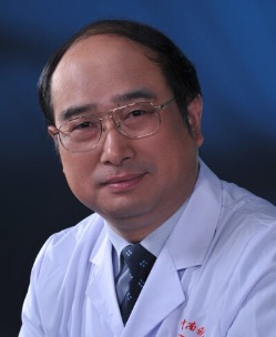 Aixi Yu (喻爱喜)合作论文数Zhongnan Hospital of Wuhan University3Long Qian (钱隆)合作论文数2Peng LI (李鹏)合作论文数2Qiong Han合作论文数2Zhi-Gang Zhao合作论文数2Lin Xie合作论文数2shiwei yu (俞诗威)合作论文数2Yunbao Zhu (朱云宝)合作论文数2wenwu li (李文武)合作论文数2




