In the study of electroencephalography (EEG)-based motor imagery (MI) brain-computer interfaces (BCIs), neurorehabilitation technologies hold significant potential for recovering from intracerebral hemorrhage (ICH). However, the rehabilitation process is hindered as the clinical practicality of such systems is reduced considerably due to their lengthy setup procedures caused by excessive number of channels. Accordingly, this study proposes a channel selection method based on an adaptive recursive learning framework, which establishes a comprehensive evaluation metric by combining time-frequency domain features. Experimental results demonstrate that, upon using 37.50% fewer channels, the average accuracy of MI classification increased from 65.44% to 69.28% in healthy subjects and from 65.00% to 67.64% in patients with ICH. This study presents the pioneering EEG-based MI BCI channel selection process specifically designed for ICH patients, paving the way for personalized rehabilitation protocols and facilitating the translation of neurotechnology into clinical practice.
Electrical stimulation of tiny peripheral nerve trunks for near-organ neuromodulation enables precise electroceutical therapy for refractory diseases, but long-term stable neuromodulation of these delicate, fragile nerve trunks remains an engineering challenge. Here, we report the development of NeuroWrap Ultrasonic Stimulator (NWUS), an untethered, self-adhesive thin-film neurostimulator capable of conformally wrapping around tiny nerve trunks. The untethered architecture can help avoid the damage or rupture of tiny nerves due to uncontrollable micromotion of lead wires during bodily movements. The NWUS is an ultrasound-responsive acoustoelectric converter with optimized impedance matching, which enables highly effective wireless electrical stimulation. The electroceutical application of NWUS was further illustrated in chronic vagus nerve stimulation for immunomodulation therapy in a rat model. The wireless neuromodulation therapy of rat experimental autoimmune myocarditis was proven to restore left ventricular function, suppress proinflammatory cytokine expression as well as macrophage infiltration within cardiac tissue, and promote regulatory T cell recruitment along with increased anti-inflammatory cytokine levels.
Motor imagery (MI)-based neurorehabilitation shows promise for intracerebral hemorrhage (ICH) recovery, yet conventional unimodal brain-computer interfaces (BCIs) face critical limitations in cross-subject generalization. This study presents a multimodal electroencephalography (EEG)-functional near-infrared spectroscopy (fNIRS) fusion framework incorporating a Wasserstein metric-driven source domain selection method that quantifies inter-subject neural distribution divergence. Through comparative neuroactivation analysis of 17 normal controls and 13 ICH patients during MI tasks, the transfer learning model achieved 74.87% mean classification accuracy on patient data when trained with optimally selected normal templates. Cross-validation on two public hybrid EEG-fNIRS datasets demonstrated generalizability, increasing baseline accuracy to 82.30% and 87.24%, respectively. The proposed system synergistically combines the millisecond temporal resolution of EEG with the hemodynamic spatial specificity of fNIRS, establishing the first clinically viable multimodal analytical protocol for ICH rehabilitation. This paradigm advances neurotechnology translation by paving the way for personalized rehabilitation regimens through robust cross-subject neural pattern transfer while addressing the critical barrier of neurophysiological heterogeneity in post-ICH populations.
INTRODUCTION:Bilateral subthalamic nucleus (STN)-deep brain stimulation (DBS) has been proven to be effective for treating probable rapid eye movement (REM) sleep behavior disorder (pRBD) symptoms in patients with Parkinson's disease (PD). However, the underlying cortical mechanism has yet to be elucidated. OBJECTIVES:To investigate the relation of structural connectivity of the STN with fiber tracts and cortical regions to postoperative outcomes of RBD symptoms. METHODS:Forty PD patients with preoperative pRBD who underwent bilateral STN-DBS were enrolled and divided into a pRBD-group and a pRBD + group according to postoperative scores on the RBD Screening Questionnaire (RBDSQ). The volume of tissue activated (VTA) was calculated for each electrode to locate the sweet spot correlated with the decrease in the RBDSQ score. Tractography was applied to confirm differences in cortico-STN connectivity and to further search for critical fiber tracts and cortical regions associated with treatment effects. RESULTS:Patients in the pRBD-group showed better improvement on RBDSQ (adjusted P = 0.042), better sleep quality (adjusted P = 0.081) and less restlessness (adjusted P = 0.093) after STN-DBS. VTA analyses revealed a positive correlation between the RBDSQ score decrease and the overlap volume of the left associative subnucleus (adjusted P = 0.090). pRBD-patients were found to have more stimulation scope on VTA-anterior thalamic radiation-9p (adjusted P = 0.096), VTA-dentatorubrothalamic tract (DRTT)-SFL (adjusted P = 0.092), and VTA-superior thalamic radiation-8BL (adjusted P = 0.088) fibers. CONCLUSION:The effectiveness of STN-DBS for PD-RBD is significantly related to the VTA of electrodes. An appreciable effect may be achieved from VTA circumscribing the STN associative subnucleus, anterior thalamic radiation-9p, DRTT-SFL, and superior thalamic radiation-8BL fibers.
In recent years, robot-assisted stereotactic system has been widely used in stereo-electroencephalogram (SEEG) surgery. However, current registration mainly based on bone-fiducial registration (CBR) is limited by the inconvenient of repeated contact during the process. Here, a novel contactless optical-tracking registration (OTR) were applied for SEEG using robot-assisted stereotactic system. To demonstrate the accuracy and convenience of the novel OTR method compared with contact CBR method during the robot-assisted SEEG in phantom and animal model study. A head phantom and 12 Bama pigs (Sus scrofa domestica) with six under CBR and six under OTR were selected for the SEEG. Procedures were performed using the robot-assisted stereotactic system in different registration methods. The positioning error and time consumption during the registration process were assessed to compare the accuracy and convenience of OTR and CBR. Besides, two new users for the robot-assisted stereotactic system were selected for the learning curve analysis. The mean positioning errors in OTR group of the target and entry points were 1.68 ± 0.80 mm and 0.76 ± 0.39 mm. And in CBR group, mean positioning errors of the target and entry points were 1.49 ± 0.79 mm and 0.70 ± 0.33 mm. The registration time of OTR method (99.71 ± 1.08 s) was significantly shorter than that using CBR method (241.29 ± 28.95 s) (p value < 0.001). During the learning curve analysis, it is earlier for the users to go under OTR than CBR to reach a preferable entry error of 0.5 mm. The contactless OTR method can effectively reduce the time consumption during the registration process while maintaining the accuracy with CBR method. The novel method not only simplify the procedure by optical-tracking but also shorten the new user’s learning curve compared with current method.
BackgroundThe mental health of patients with chronic liver disease (CLD) warrants greater attention and understanding, especially concerning its risk factors.MethodPatients from our hospital’s hepatology clinic were consecutively enrolled and completed a questionnaire assessing anxiety, depression, and sleep quality using the GAD-7, PHQ-9, and PSQI scales, respectively. Reliability and validity were evaluated with Cronbach’s α and Kaiser-Meyer-Olkin (KMO). Continuous and categorical variables were analyzed using the Mann-Whitney U and Chi-square tests. Univariate and multivariate logistic regression were used to identify risk factors, while restricted cubic spline (RCS) were used to explored non-linear associations. Subgroup analyses were stratified by sex, age, and education.ResultA total of 1030 questionnaires were collected, and after quality control, 1003 were included. 56.2% (564/1003), 53.2% (534/1003), and 67.4% (676/1003) individuals had anxiety, depression, and sleep disorders. Differences in age, gender, and education level were observed (P<0.05). Subgroup analysis showed similar demographic trends. Univariate and multivariate regression analysis found age negatively correlated with anxiety (OR=0.98, 95%CI=0.97-0.99, P=0.02) and depression (OR=0.98, 95%CI=0.96-0.99, P<0.001), but positively correlated with sleep disorders (OR=1.03, 95%CI= 1.01-1.05, P< 0.001); males are less prone to anxiety (OR=0.68, 95%CI=0.52-0.88, P=0.004) and sleep disorders (OR=0.72, 95%CI: 0.55-0.94, P=0.02); university degree is more susceptible to depression (OR=1.36, 95%CI=1.04-1.77, P=0.02) and anxiety (OR=1.45, 95%CI=1.11-1.89, P=0.007). RCS analysis suggested a linear relationship between the age and affective disorders among different population.ConclusionYoung individuals, female, and those with higher education are more vulnerable to mental health, warranting increased attention.
Implantable neural stimulation devices are becoming prevalent in bioelectronic medicine for the precise treatment of various clinical diseases. Nevertheless, the limited lifespan and buckling size of the implanted devices remain significant obstacles for chronic clinical application. In this study, we developed an ultrasound-driven battery-free neurostimulator based on a high-performance mini-sized nanogenerator and demonstrated its successful application for the deep-brain-stimulation (DBS) therapy of Parkinson's disease in a rat model. This soft piezoelectric-triboelectric hybrid nanogenerators (PTNG) are made of porous thin-films of molecular piezoelectric materials, which have great advantages of facile, scalable, low-temperature, and flexible processing. Without any bucky accessory control circuits, the subcutaneously implanted soft PTNG can function as a wirelessly powered neurostimulator, allowing for the adjustment of stimulation parameters through external programmable ultrasound pulses. This DBS electroceutical application of energy-harvesting thin-film devices based on molecular piezoelectric materials provides valuable insight into the development of a soft high-performance bioelectronic device.
A robust neural interface with intimate electrical coupling between neural electrodes and neural tissues is critical for stable chronic neuromodulation. The development of bioadhesive hydrogel neural electrodes is a potential approach for tightly fixing the neural electrodes on the epineurium surface to construct a robust neural interface. Herein, we construct a photopatternable, antifouling, conductive (∼6 S cm-1), bioadhesive (interfacial toughness ∼100 J m-2), soft, and elastic (∼290% strain, Young's modulus of 7.25 kPa) hydrogel to establish a robust neural interface for bioelectronics. The UV-sensitive zwitterionic monomer can facilitate the formation of an electrostatic-assembled conductive polymer PEDOT:PSS network, and it can be further photo-cross-linked into elastic polymer network. Such a semi-interpenetrating network endows the hydrogel electrodes with good conductivity. Especially, the photopatternable feature enables the facile microfabrication processes of multifunctional hydrogel (MH) interface with a characteristic size of 50 μm. The MH neural electrodes, which show improved performance of impedance, charge storage capacity, and charge injection capability, can produce effective electrical stimulation with high current density (1 mA cm-2) at ultralow voltages (±25 mV). The MH interface could realize high-efficient electrical communication at the chronic neural interface for stable recording and stimulation of a sciatic nerve in the rat model.
Autophagy is well-known to be a lysosome-mediated catabolic process for maintaining cellular and organismal homeostasis, which has been established with many links to a variety of human diseases. Compared with the therapeutic strategy for inhibiting autophagy, activating autophagy seems to be another promising therapeutic strategy in several contexts. Hitherto, mounting efforts have been made to discover potent and selective small-molecule activators of autophagy to potentially treat human diseases. Thus, in this perspective, we focus on summarizing the complicated relationships between defective autophagy and human diseases, and further discuss the updated progress of a series of small-molecule activators targeting autophagy in human diseases. Taken together, these inspiring findings would provide a clue on discovering more small-molecule activators of autophagy as targeted candidate drugs for potential therapeutic purposes.
Background: Pseudolymphoma is a rare, benign, nonspecific condition that forms a mass-like lesion characterized by the proliferation of non-neoplastic lymphocytes. Lacking of specific clinical symptoms, serological markers, and imaging features, the diagnosis is difficult. We reporte five cases of hepatic pseudolymphoma and provide a systematic review of existing literatures to improve our understanding of this rare liver disease. Methods: We followed-up five cases of hepatic pseudolymphoma in West China Hospital from January 2002 to January 2022. We also summarized the cases of hepatic pseudolymphoma from January 1981 to December 2021 through the PubMed database and comprehensively analyzed the characteristics of the cases. Results: The pathologic features of the five cases were characterized by benign lymphoid tissue hyperplasia, lymphoid follicle formation, and a polarized germinal center. Immunohistochemistry, in situ hybridization, and gene rearrangement revealed non-malignant lymphoma. Besides, a total of 116 cases have been reported in the PubMed database from 1981 to 2021. The incidence of hepatic pseudolymphoma is higher in middle-aged and elderly women and has been reported more frequently in Asia. All cases were pathologically diagnosed, among which 85.95% of the patients were treated by surgery. Conclusions: Hepatic pseudolymphoma is an extremely rare benign disease, mainly in middle-aged and elderly women. Without distinctive clinical and imaging characteristics, pathological diagnosis is the highly reliable method at present. Thus, in the absence of risk factors for a primary liver tumor or metastatic tumor in middle-aged and elderly women, the possibility of pseudolymphoma should be considered to avoid extensive treatments.
目的 探讨血清S100钙结合蛋白B(S100B)联合神经元特异性烯醇化酶(NSE)检测在高血压脑出血(HICH)中的应用效果.方法 选取武汉科技大学附属天佑医院收治的HICH患者50例作为观察组,根据格拉斯哥昏迷量表(GCS)评分将其分为轻度组(GCS评分 13~15分,15例)、中度组(GCS评分 9~12分,19例)和重度组(GCS评分 3~8分,16例).另选取同期15例健康者作为对照组.随访3个月,根据患者格拉斯哥预后(GOS)评分将患者分为预后良好组27例(GOS评分 4~5分)和预后不良组23例(GOS评分 1~3分).采用酶联免疫吸附法分别测定S100B和NSE水平,Spearman法分析S100B、NSE与预后的相关性.结果 各时间段轻度组、中度组、重度组S100B水平与对照组比较,差异有极显著性统计学意义(P<0.01);各时间段中度组、重度组S100B水平与轻度组比较,差异有统计学意义(P<0.05);入院时、入院24 h及入院48 h,中度组S100B水平与重度组比较,差异有统计学意义(P<0.05).各时间段轻度组、中度组、重度组NSE水平与对照组比较,差异有极显著性统计学意义(P<0.01);入院48 h及72 h重度组NSE水平与轻度组比较,差异有统计学意义(P<0.05).Spearman相关性分析显示,S100B、NSE水平与HICH患者入院时GCS评分呈负相关(r=-0.876、-0.721,P<0.01),与随访3个月时GOS评分呈负相关(r=-0.421、-0.309,P<0.05).预后良好组与预后不良组患者S100B、NSE水平比较,差异有统计学意义(P<0.05).结论 S100B联合NSE水平用于评估HICH患者疾病严重程度及判定患者预后的效果较好.
Background: Brainstem tumors are rare and extremely heterogeneous and present significant challenges in surgical treatment. Thus, biopsies often set the foundation for the diagnosis of brainstem tumors. Multimodal, image-guided, robot-assisted frameless stereotactic biopsies are increasingly popular in neurosurgery centers. This study aimed to compare the safety, efficacy, and duration of the Remebot robot-assisted (Remebot) frameless brainstem tumor biopsy versus those of frame-based stereotactic biopsy. Method: A retrospective analysis of 33 patients with brainstem tumors who underwent stereotactic brainstem biopsies in the department of neurosurgery from January 2016 to January 2021 was conducted. The patients were divided into two groups: the Remebot group (n = 22) and the frame-based group (n = 11). The clinical characteristics, trajectory strategy, duration of procedure, diagnostic yielding, histopathological diagnosis, and postoperative complications were retrospectively analyzed and compared between the groups. Results: More pediatric patients performed Remebot frameless brainstem tumor biopsy than frame-based biopsy, with a mean age of 17.3 ± 18.7 vs. 32.8 ± 17.1 (p = 0.027). The diagnostic yield had no significant difference in the two groups, with the diagnostic yield of frame-based biopsy and Remebot frameless brain biopsy being 90.9% and 95.5%, respectively. The time of the total process was 124.5 min for the frame-based biopsy and 84.7 min for the Remebot frameless brain biopsy (p < 0.001). There were no significant differences with respect to the occurrence of complication or the duration of the operation between the two groups. Conclusion: Remebot frameless stereotactic brainstem biopsy is as safe and efficacious as frame-based stereotactic biopsy. However, Remebot frameless biopsy can reduce the total duration of the procedure and has better application in young pediatric patients. Remebot frameless stereotactic biopsies can be a better option towards the safe and efficient treatment of brainstem tumors.
Objective:To investigate the efficacy of deep brain stimulation of the subthalamic nucleus (STN-DBS) in the treatment of advanced Parkinson′s disease (PD).Methods:Twenty-eight patients with advanced PD who received STN-DBS at the Neurosurgery Department of Tongji Hospital, Tongji Medical College, Huazhong University of Science and Technology from May 2015 to October 2019 were enrolled. Postoperative overall and classified scores of Unified PD Rating Scale Part Ⅲ (UPDRS-Ⅲ), improvement of non-motor symptoms, improvement of levodopa equivalent daily dose (LEDD) and drug complications were evaluated retrospectively.Results:One year after operation, the LEDD[ M( Q1, Q3)] was 393.8 (250.0, 500.0) mg, which was significantly different compared with 812.5 (750.0, 1 168.9) mg before operation ( P<0.001). The incidences of drug-induced wearing-off effect, dyskinesia and on-off phenomenon were all decreased (all P<0.05). The total score of UPDRS-Ⅲ at 1 month and 1 year after operation was 15.0 (9.3, 18.0) points and 26.0 (21.3, 28.0) points respectively, which were significantly different compared with 37.0 (35.0, 43.8) points before operation (both P<0.001). In terms of the UPDRS-Ⅲ classified items, the scores of resting tremor, postural tremor, limb movement, bradykinesia, and posture were significantly improved at 1 month and 1 year after operation, and the differences were statistically significant (all P<0.001). However, the scores of rigidity, speech, countenance and gait were significantly improved only at 1 month after operation (all P<0.001), and the difference was not statistically significant at 1 year after operation (all P>0.05). The dysphagia score showed a worsening trend ( P=0.006). Some non-motor symptoms improved at 1 year after operation (all P<0.05). Conclusions:STN-DBS significantly improves motor symptoms in patients with advanced Parkinson′s disease in the short term, and has limited effect on some motor symptoms and non-motor symptoms at 1-year follow-up after surgery. It can also reduce drug-related complications.
Parkinson's disease (PD) presents clinical heterogeneity in late-stage patients who show a varied responsivity to subthalamic nucleus deep brain stimulation (STN-DBS) on motor symptoms. To explore the heterogeneity of STN-DBS effect on motor symptoms in late-stage PD patients and to categorize the patients, basic characteristics, preoperative motor symptoms, and motor symptoms one month and one year after the operation of 28 late-stage PD patients were collected and analyzed with agglomerative hierarchicalclustering. Preoperative motor complications, non-motor symptoms, and late-stage related manifestations were also collected and compared among the clusters. The analysis revealed three patient clusters with different motor features: 1) high response; 2) low response, and 3) moderate response with fast progression. Clinical characteristics showed significant differences in disease duration, postoperative UPDRS-III, decrease in UPDRS-III, and index of progression. Patterns of the six major motor symptoms can clearly differentiate the three clusters. Preoperative dyskinesia, memory decline, and visual hallucination were also found to differ in the three clusters. The current study identified three clusters of late-stage PD patients showing distinct responses to STN-DBS on motor symptoms. These clustering patterns may relate with diverse dopaminergic/non-dopaminergic impairments and hopefully help to categorize late-stage PD patients and to select adequate STN-DBS candidates.
Background: Medulloblastoma (MB) is the most common intracranial malignant tumour in children, but genes and pathways involved in its pathogenesis are still under investigation. This study was designed to screen and identify biomarkers of MB to provide markers for clinical diagnosis and prognosis assessment. Methods: The data sets of GSE109401 and GSE42656 were acquired from Gene expression omnibus (GEO). Limma package in R was adopted to identify the differentially expressed genes (DEGs), and the GSE30074 data set was adopted to analyse their prognostic role. Children with MB (n=55) diagnosed in Affiliated Ezhou Central Hospital were enrolled and assigned to the patient group, and healthy children (n=30) who received physical examination in our hospital during the same time period were assigned to the control group. The two groups were compared in serum NLGN2 and PTGDS levels, and all patients were followed up for three years to understand the associations of Neuroligin 2 (NLGN2) and Prostaglandin D2 synthase (PTGDS) with the survival of patients. Results: With Limma, 247 DEGs were screened out. The LASSO Cox regression analysis revealed that 6 genes were associated with MB prognosis, and the established model revealed a lower survival rate in the high risk group. According to Cox regression analysis, NLGN2 and PTGDS may be independent prognostic factors of MB. Similar to the data sets, the Real time-quantitative polymerase chain reaction (RT-qPCR) assay revealed lowly-expressed NLGN2 and PTGDS levels in MB patients, and patients with lower expression of them showed a lower 3-year survival rate. Conclusion: With low expression in MB cases, NLGN2 and PTGDS have high prognostic value for MB.
In vivo electrical stimulation (ES) has shown great promise in promoting tissue repair for various tissue engineering applications. However, a significant limitation of current long-term ES technique is that the existing postoperative protocols with transcutaneous leads have great risk of infection and need second operation to remove the tethered electrical-interface. Herein, we explored an ultrasound-driven in vivo ES technique based on the biodegradable piezoelectric nanogenerator (PENG) without any transcutaneous leads for the repair of peripheral nerve injuries. The piezoelectric nanogenerator contains biodegradable piezoelectric materials, including potassium sodium niobate (KNN) nanowires, poly (L-lactic acid) (PLLA), and poly(3-hydroxybutyrate-co-3-hydroxyvalerate) (PHBV), biodegradable encapsulation layers, such as Poly (lactic acid) (PLA) or poly-ɛ-caprolactone (PCL) films, as well as biodegradable magnesium (Mg) electrodes and molybdenum (Mo) wires. Owing to the merits of ultrasound (US) in biomedical engineering, such as deep tissue penetration and predominant clinical security, US was selected as an exterior wireless energy source to drive the implantable nanogenerators which were fabricated with dissolvable piezoelectric films. With mechanical excitation remotely activated by programmable US pulses, the implanted piezoelectric nanogenerator can deliver adjustable ES to the biodegradable conductive conduits of peripheral nerves beyond the intraoperative period. Moreover, upon in-situ ES of the recovered nerves by the implanted nanogenerator, the nerve repairing process can be monitored in real-time with recorded muscle electrophysiology response. With a sciatic nerve injury model, our comprehensive investigation on neurologic function recovery analysis, histological assessment and microstructure analysis confirmed the great enhancement in nerve regeneration by the ultrasound-driven in vivo ES. This work provides a novel strategy with ultrasound-responsive biodegradable piezoelectric nanogenerator to deliver in vivo ES for tissue engineering applications.
Abstract Introduction Anaplastic meningiomas constitute about 2% of all intracranial meningiomas and are known to be aggressive with a mean overall survival and mean relapse-free survival at 3.3 years and 2.7 years respectively. We treated a patient with a World Health Organization (WHO) grade III Anaplastic Meningioma, a rare histological variant of convexity meningiomas in which intraoperatively we found the tumor mass was uniquely of mixed characteristics particularly peritumoral subdural effusion Research question We show in this case report that convexity lesions presenting with subdural lesions should be highly suspected for WHO III anaplastic meningiomas backed with the immunohistopathological findings. Material and methods Routine computed tomography for patients with neurologic signs and symptoms which reveal a convexity peri-tumoral subdural collection should be suspected for WHO III anaplastic meningiomas. Surgery and adjuvant radiotherapy offered a good clinical outcome. Results Postoperative the patient described a gradual improvement of the of expressive aphasia, deficits in speech, detectable personality changes or motor/sensory deficits. MRI at the three-month, six-month, one-year and two-year postoperative follow-up, revealed no evidence of residual tumor nor any observed recurrence nor recurrence of pre-operative symptoms. Discussion and conclusion In this paper, we present the rare radiological presentation and immunohistopathological characteristics of this WHO III Grade III anaplastic meningioma. WHO III anaplastic meningiomas should be considered in the differential diagnosis of intracranial tumors which present with peritumoral subdural collection on routine contrast tomography imaging. Our findings may contribute to the literature of the histopathological nature and behavior of this tumor subtype.
目的 探讨睿米机器人及术中B超在颅内微小病变手术中的临床应用效果.方法 回顾性分析2018年10月至2020年9月在睿米机器人及术中B超辅助下显微手术切除的21例颅内微小病变的临床资料.结果 21例病变最大径1~3 cm,平均1.8 cm;位于额叶8例、颞叶4例、顶叶5例、枕叶4例.21例病灶定位准确率为100%,机器人系统显示定位误差在2 mm以内.术后复查MRI显示病灶均全切除.术后症状改善19例,无明显变化2例.术后均没有严重的并发症,无新发神经功能障碍,无手术死亡病例.术后病理检查结果显示低级别星形细胞瘤6例,间变性星形细胞瘤3例(WHO Ⅲ级),胶质母细胞瘤3例,海绵状血管瘤5例,脑膜瘤4例.21例术后随访3~12个月,复查MRI未见复发.结论 睿米机器人及术中B超有助于精确定位颅内微小病灶,减少手术创伤,提高全切除率.
 Kai Shu (舒凯)合作论文数Tongji Hospital, Tongji Medical College, Huazhong University of Science and Technology45
 Ting Lei (雷霆)合作论文数Tongji Hospital, Tongji Medical College, Huazhong University of Science & Technology34
 GuoHu Kuai (郐国虎)合作论文数11Shiqiang Wu合作论文数8Ju Zhu (朱聚)合作论文数8
 Xu Chen (陈旭)合作论文数Tongji Hospital, Tongji Medical College, Huazhong University of Science & Technology8Feng Hu合作论文数7
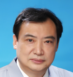 Jincao Chen (陈劲草)合作论文数Zhongnan Hospital of Wuhan University7Sun Shoujia合作论文数7Weihua Liu合作论文数6Jie Liu (刘杰)合作论文数6
 Jian Chen (陈坚)合作论文数Tongji Hospital, Tongji Medical College of Huazhong University of Science and Technology6
 Mingxin Zhu合作论文数5Kai Zhao (赵恺)合作论文数5Dongbo Wu合作论文数5
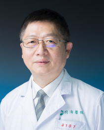 Zhengming Yang (杨正明)合作论文数Tongji Hospital, Tongji Medical College of Huazhong University of Science and Technology5ZHANG Fuchi (张复驰)合作论文数4WANG Yuan-xing (王元星)合作论文数4Ying LI (李应)合作论文数4
 Guido Nikkhah合作论文数4LiDong Cheng (程立冬)合作论文数4Papazoglou Anna合作论文数4
 Fabian Buchele合作论文数4Pan Gao (高盼)合作论文数4
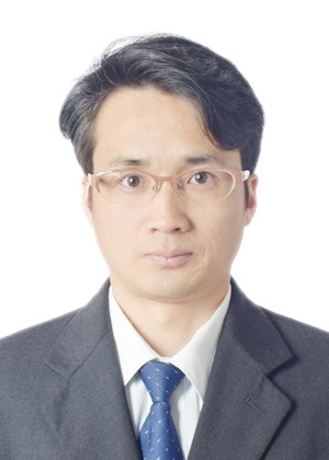 Zhiqiang Luo (罗志强)合作论文数College of Life Science & Technology, Huazhong University of Science and Technology4
 James Lubuulwa合作论文数3Mate Doebroessy合作论文数3Kun Yang合作论文数3Liwu Jiao (焦利武)合作论文数3Ping Chen合作论文数3Pan GAO (高攀)合作论文数3Xiao-jing ZHANG (张晓静)合作论文数3ZuoWei Lei (雷琢玮)合作论文数3Shan Peng (彭姗)合作论文数3ADan Fu (付阿丹)合作论文数3ShengWen Liu (刘胜文)合作论文数3Jie Ma (马杰)合作论文数3
 Huicong Kang (康慧聪)合作论文数Tongji Hospital, Tongji Medical College, Huazhong University of Science & Technology3
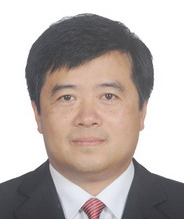 Suiqiang Zhu (朱遂强)合作论文数Tongji Hospital, Tongji Medical College, Huazhong University of Science & Technology3
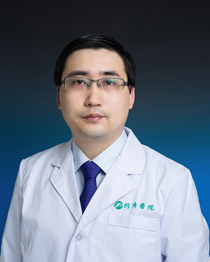 Qungen Xiao (肖群根)合作论文数Tongji Hospital, Tongji Medical College, Huazhong University of Science and Technology3Zhuang Miao合作论文数2Sheng Wang合作论文数2Zhuangzhuang Miao合作论文数2Kuan Huang合作论文数2Xiaojing Zhang合作论文数2Xiaopeng Li (李晓鹏)合作论文数2Changli Lu合作论文数2Rajluxmee Beejadhursing合作论文数Tongji Hosp, Huazhong Univ Sci & Technol2Hang Hu (胡航)合作论文数2




