Background The optimal rehabilitation strategy after acute Achilles tendon rupture (AATR) repair remains controversial. Although early weight-bearing (EWB) may promote functional recovery, the clinical implementation of such protocols is currently limited by a lack of objective criteria to guide progression. This study evaluated the safety and effectiveness of an ultrasound-guided early weight-bearing (USEWB) protocol following open repair of AATR. Methods This retrospective cohort study included 64 patients who underwent open repair for AATR between March 2025 and September 2025. Patients were divided into a USEWB group (n = 31) or a conventional rehabilitation (CR) group (n = 33) according to the postoperative rehabilitation protocol. Functional outcomes were evaluated at 2, 4, 6, 8, 12, and 16 weeks postoperatively. The calf circumference difference (CCD), Achilles tendon resting angle (ATRA), Achilles Tendon Rupture Score (ATRS), and postoperative complications were subjected to statistical analysis. Linear mixed-effects models were used for longitudinal analysis. Results In both the ITT and PP populations, USEWB significantly accelerated early functional recovery, demonstrating higher ATRS scores at 4, 8 and 12 weeks and smaller CCD at 8, 12, and 16 weeks postoperatively compared with CR. In the PP population, USEWB exhibited a significantly lower ATRA at 8 weeks postoperatively (p = 0.017). No significant differences were observed in complication rates between groups. Conclusion USEWB after open AATR repair appears to accelerate early functional recovery without increasing the risk of tendon elongation or postoperative complications. This strategy may provide a practical approach for individualized rehabilitation after Achilles tendon repair.
Background:Pathological complete response (pCR) after neoadjuvant immunochemotherapy (nICT) confers a significant survival advantage in locally advanced esophageal squamous cell carcinoma (ESCC), yet remains difficult to anticipate preoperatively. This study aimed to develop and validate a noninvasive, multimodal model integrating intratumoral and peritumoral radiomic features with clinical variables to predict pCR after nICT. Materials and Methods:We retrospectively analyzed 122 consecutive patients with ESCC who underwent contrast-enhanced computed tomography (CT) before nICT followed by surgical resection. Radiomic features were extracted from manually delineated intratumoral and peritumoral regions, and corresponding radiomic scores were constructed. Independent clinical predictors were combined into a clinical score. These three scores were integrated using multivariable logistic regression to develop a multimodal nomogram. Model performance was evaluated using receiver operating characteristic analysis, calibration curves, and decision curve analysis. Feature contributions were assessed using SHapley Additive exPlanations (SHAP). Results:Clinical T stage and tumor location constituted the clinical score. The peritumoral radiomic score showed numerically higher AUCs than the intratumoral score in the training (0.818 vs. 0.774) and validation sets (0.808 vs. 0.656). The multimodal nomogram showed good discrimination, with AUCs of 0.901 and 0.862 in the training and validation sets, respectively. SHAP analysis indicated that the peritumoral radiomic score had the largest relative contribution among model components. Conclusions:A CT-based multimodal nomogram incorporating intratumoral and peritumoral radiomic features with clinical variables showed good performance for preoperative prediction of pCR after nICT in ESCC. Peritumoral radiomic features may provide complementary imaging information, although their biological interpretation remains exploratory.
PURPOSE:This study evaluates a 3-step image guidance strategy (bone-prostate-rectal anterior wall alignment) to reduce posterior clinical target volume-planning target volume (CTV-PTV) margins in hypofractionated prostate radiation therapy while preserving target coverage and minimizing rectal toxicity. METHODS AND MATERIALS:Twenty-three patients (575 daily cone beam computed tomography scans) underwent a 3-step alignment protocol: bony alignment, prostate soft-tissue matching, and rectal anterior wall fine-tuning. A dose calculation and hybrid deformable image registration-based workflow was created to accumulate the delivered dose. A 5-mm CTV-PTV margin (0 mm posteriorly) with 70 Gy/25 fractions and strict rectal constraints was used. The deformable image registration uncertainty has been thoroughly evaluated, covering geometric indicators, deformation vector field physical properties, spatial uncertainty, and dosimetric uncertainty indicators. Normal tissue complication probability for grade ≥2 gastrointestinal (GI) and genitourinary (GU) toxicity was calculated using the Lyman-Kutcher-Burman model. Statistical comparisons employed paired t-tests or Wilcoxon rank tests (P < .05). RESULTS:The dice similarity coefficient for CTV, rectum, and bladder exceeded 0.8, with mean distance to agreement under 3 mm. Jacobian determinant analysis showed biomechanical validity, with most voxels deforming within physiological ranges (92.23% for CTV, 89.63% for rectum, and 88.96% for bladder) and no nonphysical deformations. The distance discordance metric varied from 0.01 mm to 10.25 mm, and the δ index showed a 90.04% average passing rate within the PD80%. Compared to prostate-only alignment, rectal normal tissue complication probability was significantly reduced with the 3-step strategy (7.95% vs 13.61%, P < .01), with comparable CTV coverage (D95%:69.98 Gy delivered vs 70.98 Gy planned, P = .08). Deformable dose accumulation confirmed adequate dominant intraprostatic lesion (DIL) coverage (median D100 %: 71.51 Gy), except for DILs <0.7 mm from the rectal wall, or the rectal volume overlapping with DIL2 cm (DIL extends uniformly outward by 2 cm) >1.5 cm³ (requiring 2-3 mm margin expansion). CONCLUSIONS:A 3-step image guidance protocol enhanced rectal protection while maintaining target coverage compared to prostate-only alignment. Reducing the posterior PTV margin preserved CTV coverage, but DIL coverage varied by location. If the DIL is less than 0.7 mm from the rectum or overlaps more than 1.5 cm³ within 2 mm of the rectum, the reduced margin may not be suitable, potentially compromising DIL coverage, suggesting the need for personalized margins and adaptive radiation therapy to prevent underdosage.
We established and validated an innovative two-phase pipeline for automated detection and segmentation on multi-parametric cervical cancer magnetic resonance imaging (MRI) and investigated the clinical efficacy. The retrospective multicenter study included 125 cervical cancer patients enrolled in two hospitals for 14,547 two-dimensional images. All the patients underwent pelvic MRI examinations consisting of diffusion-weighted imaging (DWI), T2-weighted imaging (T2WI), and contrast-enhanced T1-weighted imaging (CE-T1WI). The deep learning framework involved a multiparametric detection module utilizing ConvNeXt blocks and a subsequent segmentation module utilizing 3-channel DoubleU-Nets. The pipeline was trained and tested (80:20 ratio) on 3,077 DWI, 2,990 T2WI, and 8,480 CE-T1WI slices. In terms of reference standards from gynecologic radiologists, the first automated detection module achieved overall results of 93
Breast cancer is the most common malignant tumor in women; breast ultrasound is crucial for its diagnosis, but its accuracy and standardization (especially in BI-RADS application) are operator-dependent. Peking University Third Hospital faced challenges related to suboptimal ultrasound reporting compliance and excessively high positive predictive value (PPV) of BI-RADS 4 A lesions. This study aimed to evaluate the effectiveness of the Plan-Do-Check-Act (PDCA) cycle as a management tool in postgraduate medical education to bridge this skill gap. A retrospective study was conducted in a multi-campus academic institution. Throughout 2024, an educational intervention based on the PDCA cycle was implemented. Key educational outcome metrics, including BI-RADS categorization rate, reporting compliance rate, ultrasonographic-pathologic concordance rate, and PPV, were compared between the pre-PDCA and post-PDCA groups. The reporting compliance rate significantly increased from 87.18
Alignment, functionalization and detection of carbon nanotube (CNT) bundles are vital processes for utilizing this one-dimensional nanomaterial in electronics. Here, we report a polymer-assisted wet shearing method to acquire super-aligned crater-patterned CNT arrays by nanobubble (NB) self-assembly with a “migrate and aggregation” mechanism and use craters to controllably mold even-sized nanodisks periodically along CNT bundles with tunable densities. This green, low-cost method can be extended to diverse substrates and fabricate different nanodisks. As an example, the Ag-nanodisk-patterned CNT arrays are utilized as substrates of surface-enhanced Raman scattering (SERS) for rhodamine 6G (R6G) and methylene blue (MB) in which a linear correlation is found between the SERS intensity and the CNT bundle density due to the periodic distribution of hot spots, enabling a spectral detection of CNT bundles and their densities by conventional dye molecules. Distinguishing from routine morphological characterization, this spectral method possesses an enhanced accuracy and a detection range of 0.1–2 µm−1, showing its uniqueness in the detection of CNT bundle density since the intensity of traditional spectral merely relates to the quantity of CNTs, exhibiting its potential in future CNT-bundle-based electronics.
Purpose To investigate the utility of combined tumour and lymph node (LN) radiomics features in predicting disease-free survival (DFS) among patients with locally advanced esophageal squamous cell carcinoma (ESCC) after neoadjuvant chemotherapy and resection. Methods We retrospectively enrolled 176 ESCC patients from January 2013 to December 2016. Tumour and targeted LN segmentation were performed on venous phase CT images. Models were constructed using LASSO Cox regression: a clinical model, a clinical-tumour radiomics model, and a clinical-tumour-LN radiomics model. Model fitting was evaluated using Akaike information criterion and likelihood ratio (LR), while performance was assessed using Harrell’s concordance index (C-index) and time-dependent receiver operating characteristic analysis. Results The clinical model included clinical stage and neutrophil-to-lymphocyte ratio (NLR). Integration of tumour features significantly improved prognostic accuracy (clinical-tumour model vs. clinical model, LR: 17.84 vs. 11.84, P = 0.049). Subsequent integration of LN features further augmented model performance (clinical-tumour-LN model vs. clinical-tumour model, LR: 24.48 vs. 17.84, P = 0.009). The final model included clinical stage, NLR, two tumour features (Conventional_mean and GLZLM_HGZE), and one LN feature (GLCM_entropy). The C-index was 0.68 for the training set and 0.70 for the test set. The nomogram based on these features effectively stratified patients into high- and low-risk groups (P < 0.001). Conclusions The clinical-tumour-LN model, integrating clinical stage, NLR, and radiomics features, outperformed simpler models in predicting DFS among ESCC patients after neoadjuvant chemotherapy and resection. This underscores the potential of radiomics data to enhance prognostic models, offering clinicians a more robust tool for assessment.
Objective:To explore the application effect of mentorship combined with mini-clinical evaluation exercise (Mini-CEX) in standardized residency training in ultrasound.Methods:A total of 21 residents who were trained in the ultrasound department of Peking University Third Hospital from June 2018 to October 2019 were selected as the research subjects. Mentorship combined with Mini-CEX teaching method was adopted. When the training finished, the final and initial Mini-CEX scores of the residents were compared, and a questionnaire survey was conducted on the residents and 10 mentors. Paired sample t-test were used for grades analysis. Results:The final and initial evaluation scores in Mini-CEX of the residents was recorded in 7 aspects that contained consultation skills, ultrasound scanning skills, ultrasound image description and reporting, diagnostic and differential diagnostic skills, communication and humanistic care skills, organizational effectiveness, overall clinical competence. The final scores of the 7 aspects were higher than the initial scores [(7.52±0.75) vs. (6.38±1.47) , (7.67±0.73) vs. (6.47±1.29) , (7.61±0.74) vs. (6.80±1.25) , (7.38±0.59) vs. (6.29±1.35) , (8.00±0.77) vs. (7.14±1.28) , (7.33±0.58) vs.(6.52±1.12) , (7.52±0.81) vs. (6.52±1.17), all P<0.05]. All residents and 9 tutors were satisfied with the teaching method. Conclusions:Mentorship combined with Mini-CEX practices has practical value in training effect improvement of ultrasonic residents. It can provide a reference for the teaching reform in residents training.
OBJECTIVE:We developed an intelligent assistance system for shoulder ultrasound imaging, incorporating deep-learning algorithms to facilitate standard plane recognition and automatic tissue segmentation of the rotator cuff and its surrounding structures. We evaluated the system's performance using a dedicated data set of rotator cuff ultrasound images to assess its feasibility in clinical practice.METHODS:To fulfill the system's primary functions, we designed a standard plane recognition module based on the ResNet50 network and an automatic tissue segmentation module using the Mask R-CNN model. The modules were trained on carefully curated data sets. The standard plane recognition module automatically identifies a specific standard plane based on the ultrasound image characteristics. The automatic tissue segmentation module effectively delineates and segments anatomical structures within the identified standard plane.RESULTS:With the use of 59,265 shoulder joint ultrasound images, the standard plane recognition model achieved an impressive recognition accuracy of 94.9% in the test set, with an average precision rate of 96.4%, recall rate of 95.4% and F1 score of 95.9%. The automatic tissue segmentation model, tested on 1886 images, exhibited a commendable average intersection over union value of 96.2%, indicating robustness and accuracy. The model achieved mean intersection over union values exceeding 90.0% for all standard planes, indicating its effectiveness in precisely delineating the anatomical structures.CONCLUSION:Our shoulder joint musculoskeletal intelligence system swiftly and accurately identifies standard planes and performs automatic tissue segmentation.
Colorectal liver metastases (CRLM) patients combined with hepatic lymph node metastases was a negative prognostic factor associated with outcomes. Up to now, there was still lack of a reliable method to identify the status of hepatic lymph nodes. The aim of this study was to investigate the predictive ability of mono-exponential, bi-exponential, and stretched-exponential diffusion-weighted imaging (DWI) models to distinguish between benign and malignant hepatic lymph nodes in CRLM patients who underwent neoadjuvant chemotherapy prior to surgery. In total, 97 CRLM patients with pathologically proved hepatic lymph node status were included. Various quantitative parameters, including the apparent diffusion coefficient (ADC) from mono-exponential model, D, D*, and f derived from intravoxel incoherent motion (IVIM) model, as well as DDC and α from stretched-exponential model (SEM), were measured. Multivariate analysis revealed that the pre- treatment DDC value and the short diameter of the largest lymph node after treatment were independent predictors of metastatic hepatic lymph nodes. A nomogram combining these two factors demonstrated excellent performance in distinguishing between benign and malignant lymph nodes in CRLM patients, with an area under the curve (AUC) of 0.873. Therefore, the nomogram can serve as a preoperative assessment tool for determining the status of hepatic lymph nodes and aiding in the decision-making process for surgical treatment in CRLM patients.
目的 探讨超声在股直肌腱钙化性肌腱炎诊断中的应用价值.方法 回顾性分析35例因腹股沟区疼痛在我院行超声检查并诊断为股直肌钙化性肌腱炎的患者,观察股直肌腱的病变部位、厚度,内部回声、钙化灶形态、局部血流情况及有无其他并发症.结果 股直肌钙化性肌腱炎主要累及直头(91.4%),患侧股直肌腱较健侧多肿胀增厚(74.3%),腱体内结构紊乱(71.4%),回声减低(91.4%),局部血流信号增多(42.8%),腱体内钙化灶以Ⅱ型和Ⅲ型为主(65.7%),边缘不清(68.6%),在超声扫查过程中35例患者局部均有明显压痛.结论 超声能显示病变肌腱的形态和回声变化,动态观察钙化部位及其与周围组织的关系,可作为该病的理想影像检查方法.
MRI因具有多平面、多序列、高分辨率等特点,对肿瘤疾病评估具有重要价值.随着肿瘤诊疗逐渐精准化,肿瘤影像的定量评价逐渐成为研究热点.T2定量成像(T2 mapping)技术是一种可以在体素水平上对组织T2值进行定量分析的方法 ,并可通过后处理生成空间对应的伪彩图,以不同颜色反映T2值的高低,提高观察的客观性及直观性.良恶性肿瘤组织细胞密度的不同及肿瘤治疗过程中发生水肿、坏死等因素均可导致T2值改变.故T2 mapping具有极高的临床应用价值.本文就目前T2 mapping技术在肿瘤中的研究进展进行较全面的阐述.
目的 研究CBL结合SP教学法在超声科住院医师医患沟通能力培养中的作用,探索有效提高医患沟通能力的教学方式.方法 将2019年9月1日-2020年8月31日参加规范化培训的超声专业住院医师28人作为研究组,2018年9月1日-2019年8月31日接受传统方式医患沟通教学的住院医师26人作为对照组.根据超声科特有的医患沟通场景及案例,对研究组先进行以分组案例讨论为主的CBL教学,再进行以情景对话为主的SP教学.教学后应用评估量表对研究组及对照组进行考核,并对研究组进行问卷调查.结果 医患沟通能力评估量表显示,研究组的总分及沟通技巧和综合评价部分的得分均显著高于对照组(87.4±5.2 vs 82.7±3.2,44.2±2.9 vs 41.9±2.3,26.1±2.1 vs 24.4±1.6),差异均具有统计学意义(P<0.05).调查问卷显示,住院医师认为CBL结合SP教学法能有效提高医患沟通能力并具有趣味性.结论 研究组医患沟通能力评分高于对照组,研究组住院医师调查问卷提示CBL结合SP的教学模式得到住院医师的普遍认可,在超声科具有较强的可行性,有助于提高住院医师医患沟通能力培训的效果,值得继续推广.
目的 调研分析超声专业住院医师规范化培训(住培)所需的疾病系统及知识能力系统,为住培结业考核设计提供依据.方法 选择国家卫生健康委员会"住院医师规范化培训结业考核效度研究(超声医学专业)"专家组进行调研,组建超声住培疾病系统,共计5部位分类及34类具体疾病,评估者独立评估各疾病建议例数及重要性,得到各疾病总权重;制订知识能力系统,包含19项超声住培所需掌握的知识、技能与能力,评估各项指标权重.上述两系统各指标权重相乘,构成疾病系统-知识能力系统联表.结果 超声住培疾病系统共计34类疾病,总权重得分最高的前10位疾病为肝局灶性病变、甲状腺疾病、乳腺疾病、肝弥漫性病变、胆囊疾病、卵巢疾病、心脏瓣膜病及冠心病、颈部血管、房/室间隔缺损、心肌疾病.重要性得分最高的前5位疾病为卵巢疾病、心脏瓣膜病及冠心病、房/室间隔缺损、乳腺疾病、甲状腺疾病.知识能力系统权重得分最高的前四项为诊断及鉴别诊断技能,扫查与图像识别技能,解剖、病理及病理生理知识,报告书写技能.疾病系统-知识能力系统联表总权重得分最高的前3位疾病-知识能力项目为肝局灶性病变、甲状腺疾病、乳腺疾病的诊断及鉴别诊断技能.结论 本项目评估超声住培疾病系统及知识能力系统各指标权重,通过疾病系统-知识能力系统框架建立考题的结构模型,可为全国超声住培结业考核设计提供依据.
患者女,45岁,2020年10月体检发现双肺多发结节,无发热、咳嗽、咳痰、体质量减轻等;于2011年接受子宫肌瘤剥离术、2014年因新发子宫肌瘤接受子宫切除术,无家族性遗传疾病史.入院查体及实验室检查均未见异常.胸部正位片见双肺多发结节(图1A).胸部增强CT:双肺多发结节,呈轻度强化(图1B).针对左肺上叶舌段胸膜下结节行CT引导下穿刺活检术,所获组织呈灰白色,光镜下见分化良好的平滑肌细胞呈束状排列,细胞呈梭形(图1C);免疫组织化学:CD10(+),Ki-67 (2%+),CK(+),雌激素受体(estrogen receptor,ER)(>90%+,图1D),孕激素受体(progesterone receptor,PR)(>90%+,图1E),SMA(+),TTF-1(上皮+),Caldesmon(+),Des(+).病理诊断:肺转移性子宫平滑肌瘤.综合诊断为肺良性转移性平滑肌瘤(benign metastasizing leiomyoma,BML).
OBJECTIVE:To investigate the performance of quantitative CT analysis in predicting the prognosis of patients with locally advanced oesophageal squamous cell carcinoma (ESCC) after two cycles of induction chemotherapy before definitive chemoradiotherapy/radiotherapy.METHODS:A total of 110 patients with locally advanced ESCC were retrospectively analysed. Baseline chest CT and CT after two cycles of induction chemotherapy were analysed. A multivariate Cox proportional-hazard regression model was used to identify independent prognostic markers for survival analysis. Then, a CT scoring system was established. Time-dependent receiver operating characteristic (ROC) curve analysis and the Kaplan-Meier method were employed for analysing the prognostic value of the CT scoring system.RESULTS:Body mass index, treatment strategy, change ratios of thickness (ΔTHmax), CT value of the primary tumour (ΔCTVaxial) and the short diameter (ΔSD-LN), and the presence of an enlarged small lymph node (ESLN) after two cycles of chemotherapy were noted as independent factors for predicting overall survival (OS). The specificity of the presence of ESLN for death after 12 months was up to 100%. Areas under the curve value of the CT scoring system for predicting OS and progression-free survival (PFS) were higher than that of the RECIST (p < 0.05). Responders had significantly longer OS and PFS than non-responders.CONCLUSION:Quantitative CT analysis after two cycles of induction chemotherapy could predict the outcome of locally advanced ESCC patients treated with definitive chemoradiotherapy/radiotherapy. The CT scoring system could contribute to the development of an appropriate strategy for patients with locally advanced ESCC.KEY POINTS:• Quantitative CT evaluation after two cycles of induction chemotherapy can predict the long-term outcome of locally advanced oesophageal cancer treated with definitive chemoradiotherapy/radiotherapy. • A CT scoring system provides valuable imaging support for indicating the prognosis at the early stage of therapy. • Quantitative CT evaluation can assist clinicians in personalising treatment plans.
Objective:Based on the research results of the national continuing medical education project "musculoskeletal ultrasound and new technology quality class" held by the Department of Ultrasound Diagnosis of Peking University Third Hospital, to explore and analyze the effect of basic and applied teaching method based on musculoskeletal ultrasound.Methods:Totally 109 participants attending the classes held on April 14-18, 2019 and on September 9-13, 2019 were selected as study objects, and the training effects of quality private classes were evaluated by questionnaires, operational tests and theoretical tests. SPSS 21.0 was used for t test. Results:The results showed that more than 80% of students were satisfied with the teaching content, teaching plan and teaching materials. Through the class training, the greatest gains of the students were that the operation ability [67 cases (30.7%)] and the theoretical level [53 cases (24.3%)] had been significantly improved. All 109 students passed the operation test. There were no statistical differences in the average scores of theoretical tests among the students with different professional titles, academic qualifications and whether they were from primary hospitals (75-80 points, P>0.05) . Conclusion:The musculoskeletal ultrasound quality private class can improve the students' operating ability and theoretical level in the musculoskeletal system. The class model can guarantee the teaching quality and provide a new direction for continuing medical education.
目的 探讨超声在肠道淋巴瘤诊断中的应用价值和扫查技巧.方法 回顾性分析2013年6月至2020年4月北京大学第三医院收治的18例经手术或内镜病理证实的肠道淋巴瘤的病理类型、病变部位和超声表现分型信息,并比较用超声和CT分别测量的病变处的肠壁厚度的差异.结果 病理类型:18例均为非霍奇金淋巴瘤,其中弥漫性大B细胞淋巴瘤14例,套细胞淋巴瘤2例,单形性嗜上皮性肠道T细胞淋巴瘤1例,Burkitt淋巴瘤1例;原发15例,继发3例.病变部位:5例位于小肠,1例位于回盲部,11例位于结肠,1例位于直肠.超声表现分型:肠壁增厚型8例,肠道肿块型7例,囊性病变型1例,非特异型2例.可见肠套叠、穿孔、扩张等并发征象,多数合并腹腔或腹膜后淋巴结肿大.肠壁增厚型淋巴瘤超声测得的病变最厚处肠壁厚度均值(1.13±0.69)cm,小于CT测量均值(2.21±1.06)cm(P<0.05).结论 肠道淋巴瘤的超声表现具有一定特征,超声可以较准确地显示病变部位、累及范围及并发症情况,对于肠道淋巴瘤的诊断有重要价值.应用超声评估肠道淋巴瘤的病变时,需注意全面扫查并应用逐级加压手法.
Objective:To make a survey of the job tasks of China's ultrasound residents, in order to provide a basis for improving the standardized training program for ultrasound residents and the design of final examination.Methods:We first established a job task list for ultrasound residents to reflect their clinical work, scientific research training, and ability training needed in their actual work. A total of 1369 residents from 11 provinces who have completed the standardized residential ultrasound training program were enrolled in the study. A stratified sampling investigation was conducted according to the proportion of residents by province. The frequency and importance of each job task in the current work and during the residence training period were investigated, and the comprehensive score of each task was calculated. Kappa consistency test was used to evaluate the consistency between the task frequency in the current work and during the training period.Results:The task list of ultrasound residents included 18 job tasks, of which the top five with the highest comprehensive score were: abdominal and pleural effusion ultrasound, standardized writing of ultrasound report, superficial organ ultrasound, peripheral blood vessel ultrasound, and obstetrics and gynecology ultrasound. The frequency of each job task in the current work and during the residence training period had a moderate consistency (Kappa=0.477~0.580, P<0.001). The top three tasks with the largest difference were emergency ultrasound (3.88 vs 3.34), obstetrics and gynecology ultrasound (4.00 vs 3.69), and peripheral vascular ultrasound (4.08 vs 3.87), which were performed more frequently in the current work than in the training period. The frequency consistency of job task assisting interventional ultrasound between actual work and residential training was poor (Kappa=0.200, P<0.001), and the current frequency was significantly lower than that during residential training (1.67 vs 2.87).Conclusion:There is a moderate consistency for the frequency of most job tasks between current work and the training period. The assessment of job tasks could provide a basis for formulating the final examination of residency training program. It is suggested that the training for emergency ultrasound, obstetrics and gynecology ultrasound, and peripheral vascular ultrasound should be enhanced during residency training period, and the proportion of interventional ultrasound be reduced in the final examination.
Objective To evaluate PDCA circulation method in the management of teaching rounds of residents in department of diagnostic ultrasound. Methods PDCA circulation method was applied to the teaching rounds in department of ultrasound in Peking University Third Hospital from 2013—2019, and each circulation lasted for one year. PDCA circulation consists of four stages: plan, performance(do), check, and action. During the practice, the problems of the teaching round forms and contents were reviewed and modified to pick up content which were more suitable to department of diagnostic ultrasound. The improvement in teachers'assessment and training was also made according to the limitations in teacher training. Results PDCA circulation method improved the training quality of residents. All the residents in the third training year were competent to clinical performance and the residents all passed the phase Ⅰ skill examination of the standardized training in the past three years. Conclusions PDCA circulation method improves quanlity of teaching rounds and the training outcomes of residents in the department of diagnostic ultrasound.
 Ligang Cui (崔立刚)合作论文数Peking University Third Hospital;Institute of Medical Technology, Health Science Center, Peking University21
 Jinrui Wang (王金锐)合作论文数Peking University Third Hospital17
 Yingshi Sun (孙应实)合作论文数Beijing Cancer Hospital16
 Xiaoting Li (李晓婷)合作论文数Beijing Cancer Hospital12LiYing Miao (苗立英)合作论文数11Wen Chen (陈文)合作论文数11
 Ling Jiang (江凌)合作论文数Department of Ultrasound, Peking University Third Hospital11
 Xian-Shu Gao (高献书)合作论文数Department of Radiation Oncology, Peking University First Hospital;Institute of Medical Technology, Peking University Health Science Center11
 Huiyu Ge (葛辉玉)合作论文数Beijing Chao-Yang Hospital, Capital Medical University;Molecular Imaging and Minimally Invasive Therapy Laboratory, Peking University10Kun Cao (曹崑)合作论文数9
 Zhiqiang Li (李志强)合作论文数Department of Ultrasound Medicine, Peking University Third Hospital9Xiuli Yue (岳秀丽)合作论文数School of Environment, Harbin Institute of Technology9HaiTao Zhu (朱海涛)合作论文数8
 Zhanwen Xing (邢占文)合作论文数School of Mechanical and Electrical Engineering, Soochow University8
 Hengte Ke (柯亨特)合作论文数College of Pharmaceutical Sciences, Soochow University8
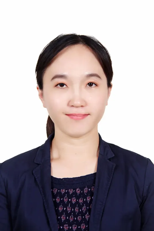 Jie Jiang (蒋洁)合作论文数Department of Ultrasound, Peking University Third Hospital8
 Heng Xue (薛恒)合作论文数Department of Ultrasound, Peking University Third Hospital7
 Mingwei Ma合作论文数7
 Zhifei Dai (戴志飞)合作论文数College of Future Technology, Peking University6Yan-Jie Shi合作论文数5JianWen Jia (贾建文)合作论文数5
 Ji-Bin Liu合作论文数Department of Radiology, Thomas Jefferson University5X. Gao合作论文数3X. Qi合作论文数3Shangbin Qin合作论文数3Mu Xie合作论文数3Jia-Yan Chen合作论文数3Siwei Liu合作论文数3
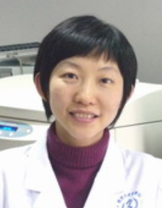 Yun Bai (白云)合作论文数School of Basic Medical Sciences, Peking University3
 Shumin Wang (王淑敏)合作论文数Department of Ultrasound Medicine, Peking University Third Hospital3
 Shaoqin Liu (刘绍琴)合作论文数School of Medicine and Health, Harbin Institute of Technology3
 Ping He (何萍)合作论文数Department of Ultrasound, Peking University Third Hospital3M.W. Ma合作论文数2M. Zhang合作论文数2Yu-Hong Qu合作论文数2Ranran Zhao合作论文数2Yi-Yuan Wei合作论文数2Zhang Fan合作论文数2Li ZHANG (张莉)合作论文数2Min Zhang合作论文数2Xue-Ying Ren合作论文数2Qi Xin合作论文数2Enze Qu合作论文数2
 Yaqi Wang合作论文数2Xiaoying Li合作论文数2zhilei zhao (赵智磊)合作论文数2Sheng Cai (蔡胜)合作论文数2Xiao Yang (杨筱)合作论文数2




