Exosomes, a type of extracellular vesicle derived from the endosomal system, have received considerable attention for their potential therapeutic applications. This review focuses on the utilization of both native and engineered exosomes in the treatment of complex tissue defects in the oral and maxillofacial regions and highlights both basic research and clinical applications. Firstly, the regenerative mechanisms and engineering strategies associated with exosome-based regenerative medicine are systematically reviewed. Given their substantial potential for clinical translation, this review also summarizes recent advances in research on their application in various aspects of tissue regeneration within the oral and maxillofacial regions. Finally, it addresses the current state, challenges, and future outlook of relevant clinical trials. The aim of this review is to provide a thorough overview of the application of natural and engineered exosomes in regenerative medicine in the oral and maxillofacial fields while inspiring innovative strategies for effective cell-free regenerative treatments for tissue defects in this domain.
Abstract Bone remodeling requires precise coordination between osteoblast-mediated bone formation and osteoclast-driven resorption. However, directly targetable and therapeutically actionable mediators that can coordinately modulate both processes during regeneration remain relatively limited. Here, we identify immunoglobulin superfamily member 10 (IGSF10) as a dual-function modulator of bone remodeling. Igsf10- deficient mice exhibit reduced bone mass, elevated osteoclast activity, and impaired osteogenesis. Recombinant IGSF10 protein restores osteogenic capacity and suppresses osteoclastogenesis in knockout cells, while exerting pro-osteogenic and anti-resorptive effects in wild-type mesenchymal and monocyte-derived cultures. Mechanistically, IGSF10 activates a noncanonical EGFR–STAT1 signaling pathway, distinct from BMP2–Smad signaling. Co-immunoprecipitation and molecular docking confirm IGSF10–EGFR interaction, and blockade of EGFR abrogates IGSF10-induced osteogenesis and its inhibitory effects on osteoclastogenesis. In vivo, IGSF10 promotes bone regeneration in both calvarial and periodontal defect models and exhibits synergy with subtherapeutic BMP2. These findings position IGSF10 as a previously unrecognized dual-acting regulator that coordinates bone formation and resorption in a context-dependent manner, with potential therapeutic value for craniofacial and skeletal regeneration.
Prolonged periodontal inflammation and progressive alveolar bone loss are typical manifestations of periodontitis. Antioxidative therapies targeting the central role of reactive oxygen species (ROS) have been explored, but lack of subcellular specificity limits efficacy. Mitochondria function as an upstream redox hub that drives oxidative stress, inflammatory responses, and alveolar bone resorption, making mitochondrial redox modulation a promising yet underexplored strategy for periodontitis therapy. Herein, we developed a mitochondria-targeted, redox-responsive nanocomposite (TC/pSeSe) that enables programmable redox modulation of the pathological periodontal microenvironment. The antioxidative core consists of a ROS-responsive diselenide-containing copolymer (pSeSe) capable of selenium release, while the triphenylphosphine/chitosan (TC) coating confers mitochondrial-targeted, controlled redox activity, mucosal retention and cationic antibacterial properties. With preferential mitochondrial localization, TC/pSeSe undergoes diselenide bond cleavage and selenium release under oxidative stress, thereby restoring mitochondrial redox homeostasis and attenuating downstream mitochondrial DNA (mtDNA)-cGAS-STING-mediated inflammatory signaling. Through combined ROS scavenging and selenium-mediated support, TC/pSeSe mitigates ferroptosis in a partially glutathione peroxidase 4 (GPX4)dependent manner and restores osteogenic potential in bone marrow-derived stem cells. In parallel, TC/pSeSe exhibits antibacterial activity against periodontal pathogens through combined selenium and the cationic TC coating functionalities. In vivo, TC/pSeSe restored alveolar bone regeneration and attenuated periodontal inflammation. Collectively, this study proposes a mitochondria-centered redox modulation strategy, providing a comprehensive and promising therapeutic approach for periodontitis treatment.
The intractability of diabetic bone defects mainly results from derailed inflammation. While peripheral neuropathy is a common comorbidity, whether sensory dysfunction contributes to uncontrolled inflammation in diabetes is poorly understood. Here, within diabetic bone defects, we show that diminished sensory innervation is coupled with disrupted immune dynamics, characterized by both delayed neutrophil chemotaxis and abnormal neutrophil retention that resulted from impaired macrophage efferocytosis. Therefore, we design a chocolate chip cookie-like scaffold, in which the surface-embedded microspheres function as “chips” enabling burst interleukin-8 (IL-8) release, while the surrounding matrix provides sustained nerve growth factor release from silk fibroin matrix. Timely neutrophil chemotaxis induced by IL-8 triggers bone healing via stem cell recruitment, which is reinforced by sensory innervation by inducing neutrophil N2 polarization. Notably, macrophages preferentially established intimate physical proximity to outgrowing neurites to form a synapse-like structure, where they restore efferocytosis driven by neuronal Galectin-3. Moreover, spatiotemporally regulating neuroimmune circuit enhances mandibular bone regeneration in diabetic rats, highlighting the therapeutic potential of neuroimmune interaction in programming diabetic inflammation resolution.
Prosthodontics is rapidly entering the digital era, with computer-aided design and manufacturing (CAD/CAM) soon becoming the mainstream method for fixed restoration design. Recent advances in image recognition, data analytics, and decision systems are accelerating the use of artificial intelligence (AI), shifting workflows from simple automation to adaptive, learning-based design. Nevertheless, clear clinical guidance remains limited. This expert consensus sets out core principles, technology categories, and priority use cases for AI in fixed restorations, and proposes Standardized Operating Procedures that span from preoperative planning, digital impression processing, tooth preparation evaluation, to functional design and personalized esthetic design. It also defines quality control checkpoints and ethical safeguards that highlight the central role of the clinician in reviewing and validating AI outputs. Recommendations are provided for data governance, including security, privacy, and auditability. Finally, the document outlines near-term development needs such as interoperable data standards, transparent model reporting, and clinically oriented validation metrics. The goal is to support standardized, safe, and effective clinical adoption of AI in fixed dental restorations.
Bone morphogenetic protein-2 (BMP-2) has been widely applied in oral and maxillofacial bone augmentation procedures. This study aimed to evaluate the efficacy and safety associated with the use of BMP-2 in oral and maxillofacial bone augmentation procedures across available randomized controlled trials (RCTs). RCTs involving BMP-2 used for alveolar ridge preservation (ARP), alveolar bone augmentation, and maxillary sinus floor elevation were included. The primary outcomes included changes in bone width and height, proportion of newly formed bone and residual graft material, implant survival, and marginal bone level (MBL). Seven databases were searched up to May 1, 2026. The Cochrane risk-of-bias 2 assessment tool was used to assess the risk of bias in the included studies. Meta-analysis, sensitivity analysis, and subgroup analyses were performed using RevMan 5.4.1 and STATA 12.0. (PROSPERO registration number: CRD420250586726). Twenty-three RCTs involving 990 participants with 1104 sites were included. Meta-analysis revealed that the use of BMP-2 was associated with significantly greater alveolar ridge width at the apical region. However, BMP-2 did not significantly enhance the proportion of new bone nor reduce residual graft material. Subgroup analyses suggested that its performance varies according to the surgical procedures and carriers. No significant differences were found between groups in implant survival, MBL or the incidence of adverse events. The majority of included studies were assessed as having some concerns or high risk of bias. Because of substantial heterogeneity across the included RCTs, the overall pooled estimates should be considered exploratory and hypothesis-generating. The use of BMP-2, particularly in ARP, appears to be associated with modest improvements in alveolar bone width (at the apical region), whereas no significant differences were observed in the proportion of newly formed bone, the implant survival rate, or the incidence of adverse events. BMP-2 may be considered selectively in appropriate clinical scenarios after careful evaluation of cost, defect characteristics, and expected clinical benefit.
Abstract Bone defect repair remains a significant challenge in clinical orthopedics, primarily due to the limitations of traditional techniques, such as insufficient osteogenic efficiency and non‐functional bone regeneration. Recently, the regulation of energy metabolism has emerged as a promising avenue for bone regeneration. This review systematically examines the critical yet underexplored role of energy metabolism in bone regeneration, focusing on the dynamic regulatory mechanisms in osteoblasts and bone marrow mesenchymal stem cells across various metabolic pathways, and clarifies the influence of glucose metabolism and mitochondrial function on cellular proliferation and differentiation. Besides, three complementary strategies are proposed: leveraging cell derivatives (e.g., mitochondria, exosomes) to provide metabolic support; employing active factors (e.g., ions, enzymes) to target key metabolic pathways; and designing biomimetic scaffolds to reconstruct a favorable metabolic microenvironment. Furthermore, deficiencies in current research—such as the lack of systematic analysis of metabolic networks, clinical validation, and multidisciplinary collaborative innovation—are noted. Future directions should explore the interplay between metabolism and epigenetics, the development of responsive metabolic regulatory materials, and advancements in clinical translation, ultimately advocating for a new paradigm in precise bone defect repair to significantly enhance patient outcomes.
Nociceptive pain is a cardinal feature of traumatic and inflammatory bone diseases. However, whether and how nociceptors actively regulate the immune response during bone regeneration remains unclear. Here, we found that neutrophil-triggered nociceptive ingrowth functioned as negative feedback regulation to inflammation during bone healing. A unique Il4ra+Ccl2high neutrophil subset drove intense postinjury TRPV1+ nociceptive ingrowth, which in return dissipated inflammation by activating the production of pro-resolving mediator lipoxin A4 (LXA4) in osteoblasts. Mechanistically, osteoblastic autophagy activated by nociceptor-derived calcitonin gene-related peptide (CGRP) suppressed the nuclear translocation of arachidonate 5-lipoxygenase (5-LOX) to favor the LXA4 biosynthesis. Moreover, in alveolar bone from patients with Type II diabetes, we found diminished nociceptive innervation correlated with reduced autophagy, increased inflammation, and impaired bone formation. Activating nociceptive nerves by spicy diet or topical administration of a clinical-approved TRPV1 agonist showed therapeutic benefits on alveolar bone healing in diabetic mice. These results reveal a critical neuroimmune interaction underlying the inflammation-regeneration balance during bone repairing and may lead to novel therapeutic strategies for inflammatory bone diseases.
Osteoporosis is classified as a metabolic syndrome, and the consumption of fructose has been linked to various metabolic diseases. However, the specific effects and underlying mechanisms of fructose on bone health remain inadequately understood. In this study, we demonstrate that fructose intake can exacerbate bone loss in murine models by facilitating the accumulation of cholesterol within the bones. We identify Thyroid Hormone Receptor Beta (Thrb) and Protein Kinase C Zeta (Prkcz) as potential therapeutic targets for the treatment of osteoporosis. Mice subjected to a high-fructose diet exhibited a reduction in bone density and a decrease in the osteogenic differentiation of bone marrow mesenchymal stem cells (BMSCs) compared to those on a standard diet. Fructose treatment was found to decrease Thrb expression while increasing Prkcz expression, leading to cholesterol accumulation and hindering the osteogenic differentiation of BMSCs. Furthermore, our findings indicate that the activation of Thrb and the inhibition of Prkcz significantly ameliorate bone loss in mice. This study elucidates the molecular mechanisms by which fructose influences osteogenesis through the Thrb/Prkcz/cholesterol accumulation pathway in the context of osteoporosis, thereby highlighting the therapeutic potential of Thrb and Prkcz as targets for osteoporosis treatment.
ABSTRACT Mitochondrial dysfunction in chondrocytes and the loss of joint lubrication are increasingly recognized as key pathological drivers of functional deterioration in osteoarthritis (OA). However, the rational design of multifunctional biomaterials capable of concurrently regulating bioenergetic metabolism and the joint mechanical microenvironment remains highly challenging. Inspired by the natural process of mitochondrial‐derived vesicles (MDVs) formation, we rationally employed a gradient extrusion‐based strategy to fabricate biomimetic mitochondrial membrane vesicles (BMMVs), which enable efficient cartilage penetration and intrinsic mitochondrial targeting. Upon loading with the Sirtuin 3 (Sirt3) activator dihydromyricetin (DMY), the resulting DMY‐BMMVs effectively restore inflammation‑induced mitochondrial dysfunction and reprogram chondrocyte metabolism homeostasis. Furthermore, drawing inspiration from the hydration brush architecture of lubricin, PLL‑g‑PEG (polylysine‐g‐polyethylene glycol) is electrostatically assembled onto methacrylated hyaluronic acid (HAMA) microspheres to construct a bioinspired PEG brush interface. This lubricating microsphere system simultaneously serves as an intra‐articular delivery depot for DMY‐BMMVs, enabling sustained vesicle release while reconstructing the joint lubrication microenvironment. In vivo studies demonstrated that this integrated system markedly enhances cartilage regeneration, alleviates OA‐associated pain, and restores joint mobility. Overall, this work establishes a multifunctional materials‐driven platform that synergistically integrates mitochondrial‐targeted metabolic regulation with bioinspired interfacial lubrication, offering a new paradigm for OA treatment from a biomaterials engineering perspective.
Cleft palate, a prevalent congenital disorder, arises from dysregulated embryonic palatal fusion, but the posttranslational modifications (PTMs) driving this process remain poorly understood. Here, we report that lysine acetylation is a critical MSX1 proteostasis switch that governs embryonic palatal mesenchymal (EPM) cell survival. We demonstrate in vitro and in vivo that MSX1 protein stability regulation by deacetylase SIRT1-catalyzed acetylation underlies EPM apoptosis and palatal fusion. In atRA-induced cleft palate models, SIRT1 suppression drives MSX1 hyperacetylation, accelerating proteasomal degradation and culminating in EPM apoptosis. Strikingly, transcriptomic profiling revealed the exclusive proteostatic role of acetylation, indicating that MSX1’s structural stability differs from its transcriptional activity—a paradigm distinct from that of classic PTM mechanisms during development. Lentivirus-mediated delivery of the deacetylase SIRT1 or the deacetylation mimic MSX1 K139R significantly reduced cleft severity, indicating its preventive and therapeutic potential in humans. Our work establishes the MSX1 acetylation as both a pathogenic driver and a druggable target in cleft palate, redefining PTM regulation as a central etiological factor in genetic disorders.
Transcription factor (TF) networks are pivotal regulators of stem/progenitor cell fate. However, the regulatory mechanisms mediated by key TFs in adult periosteal stem/progenitor cells (PSPCs) remain poorly understood, impeding targeted therapies development for craniofacial bone regeneration. By integrating an analysis of regeneration-related, tissue-specific TF networks with insights from embryonic development, we demonstrate that the imprinted TF PLAGL1 is critical for the osteoblast differentiation of PSPCs and that the loss of Plagl1 compromises mandibular bone regeneration. Mechanistically, PLAGL1 transcriptionally activates TF Irx5 synergistically with TF KLF4, thereby inducing the expression of downstream osteogenic genes. Using the CRISPR-dCas9-Tet1-CD/sgRNA system, we develop a differentially methylated region-targeted therapeutic strategy to reactivate the maternal allele of Plagl1, leveraging its imprinted function to promote mandibular bone regeneration. This study delineates the PLAGL1-KLF4-IRX5 regulatory axis controlling osteoblast differentiation of PSPCs and further proposes an application strategy integrating TF modulation with epigenetic regulation for craniofacial bone regeneration.
Bone morphogenetic protein-2 (BMP-2) remains a cornerstone osteoinductive agent, yet its clinical translation is critically hampered by a material-driven delivery crisis necessitating supraphysiological doses that trigger severe complications. Reframing these challenges as a multifaceted material-design problem, this review proposes a transformative roadmap through synergistic strategies. Molecular engineering enhances intrinsic efficacy via BMP family alternatives, engineered BMP-2 variants, and biomimetic peptides. Additionally, it explores the enhancement of BMP-2 endogenous antagonist blockade, synergistic integration with bioactive agents, immunomodulation, and microenvironment simulation. Programmed spatiotemporal control enables material-engineered BMP-2 delivery through embedded/particulate carriers, porous scaffolds, surface immobilization, and engineered gene platforms. Smart-responsive systems enable adaptive dosing via stimuli-sensitive platforms. Collaborative microenvironment engineering orchestrates electrically amplified BMP-2 therapy, metabolic cues, and antibody-mediated osseous regeneration (AMOR), thereby amplifying endogenous signaling pathways to enhance bone regeneration. Collectively, we emphasize how rational biomaterial design—from nano-scale molecular tuning to macro-scale intelligent ecosystems—can fundamentally reprogram BMP-2's therapeutic window. This paradigm shift, from passive carriers to instructive, multifunctional platforms, charts a definitive course toward safer, more efficacious, and personalized next-generation bone regeneration therapies, underscoring the pivotal role of materials science in solving long-standing translational roadblocks.
Nanozymes with photothermal regulation capabilities facilitate spatiotemporally controlled tumor therapy. However, the generated heat stress inevitably upregulates heat shock protein 90 (HSP90), which induces thermotolerance and compromises therapeutic efficacy. To resolve this dilemma, we developed ultrathin metallic RhMo (RM) nanosheets integrating tripartite cascading functions with flexoelectric catalytic, photothermal, and multi-enzyme properties. Accordingly, a "metabolic regulation-synergistic killing" strategy is proposed. Initially, ultrasound-driven flexoelectric catalysis was used as a metabolic pretreatment. By oxidizing intracellular nicotinamide adenine dinucleotide hydride (NADH) to nicotinamide adenine dinucleotide (NAD+), it severs the substrate supply for the tricarboxylic acid cycle. The consequent depletion of intracellular adenosine triphosphate (ATP) fundamentally inhibits ATP-dependent HSP90 activity, thereby abrogating the thermal defense of tumors. Subsequently, under near-infrared irradiation, RM generates localized hyperthermia, which amplifies its intrinsic peroxidase-, oxidase-, and catalase-like activities. These activities trigger a massive burst of reactive oxygen species (ROS) from endogenous substrates. The convergence of ATP deprivation and ROS-related oxidative stress induces severe mitochondrial damage and cytochrome C release, leading to irreversible apoptosis. This study established a controllable chemotherapy-free paradigm that leverages flexoelectric metabolic interventions to overcome thermotolerance for efficient tumor catalytic therapy.
Guided bone regeneration (GBR) is widely applied in implant dentistry, employing barrier membranes to create an osteogenic space by preventing gingival tissue ingrowth. However, this method does not enhance the osteogenic capacity of osteoblasts, limiting sufficient bone volume in larger defects. Inspired by axolotl limb regeneration, abundant soft tissue-derived stem cells mobilized to the defect may facilitate comprehensive osteogenesis within a BMP-2-enriched environment. We developed a biomimetic channel system (BCS) to promote alveolar bone regeneration, using channel structures to activate gingival-derived stem cells under a BMP-2-enriched biological barrier. In a cell-tracing mouse model, Prrx1+ stem cells demonstrated a critical role in BMP-2-induced subcutaneous osteogenesis. Sequencing and histological analyses revealed that channel structures significantly enhance soft tissue cell proliferation and migration. Attributable to the biological barrier, BCS applications markedly improved bone formation in beagle mandibular defects. These results suggest a novel osteoinductive strategy for alveolar bone regeneration that functions without a traditional barrier membrane.
A vicious cycle between microbiota dysbiosis and hyperactivated inflammation, hardly disrupted by conventional therapies, remains a significant clinical challenge for periodontitis treatment. Herein, by cloaking a cascade catalysis system in an engineered macrophage membrane, a nanodecoy-based strategy, with targeted bacteria-killing and immunomodulatory abilities, is proposed for reshaping the hostile periodontitis microenvironment. Specifically, recombinant human antimicrobial peptide, LL-37, is anchored to a Toll-like receptor-enriched macrophage membrane via genetic engineering, which facilitates the specific bacteria elimination and efficient tissue retention of the nanodecoys. Moreover, the cascade catalysis system integrates L-amino acid oxidase (LAAO) with hollowed manganese dioxide (hMnO2) by reciprocal elevation of the catalytic efficiency of hMnO2 and LAAO, leading to accelerated O2 generation under a hypoxic microenvironment and disrupted metabolism of periodontopathogenic bacteria. Notably, the nanodecoys trigger the nuclear translocation of NF-E2-related factor-2 (NRF2) to reduce oxidative stress response and rewire the polarization of macrophages, thereby boosting the osteogenic differentiation of osteoblasts. Furthermore, the alveolar bone regeneration therapeutically benefits from the nanodecoys in vivo. Altogether, these results highlight the attractive functions of engineered macrophage membrane-cloaked nanodecoys for effective periodontitis treatment.
Due to the increased risk of tooth loss associated with diabetes mellitus, patients exhibit a markedly higher demand for dental implant restoration. However, hyperglycemia and the accumulation of advanced glycation end-products (AGEs) induce oxidative stress, increase the risk of peri-implantitis, and impair osseointegration, posing significant challenges to the long-term success of implants. Moreover, the incorporation of antibacterial agents, while enhancing antibacterial efficacy, often elevates oxidative stress and compromises osteogenic capacity and cytocompatibility. Herein, a single-step fabricated bioactive protean TiO2/ZnO bio-heterojunction (BJ) system was developed on carbon-fiber-reinforced polyetheretherketone (CFRPEEK) implants to dynamically modulate diabetic microenvironment through multi-functional synergy (antibacterial/osteogenic/antioxidant). The CFRPEEK was firstly titanium plasma immersion ion implanted (PIIID-Ti) to obtain a micro-nanoscaled titanium dioxide (TiO2) interface. Sequentially, enhanced functionalities were attained by incorporating zinc oxide nanoparticles (ZnO NPs) to form TiO2/ZnO BJ through polydopamine (PDA) assisted π—π covalent immobilization. The results reveal that the TiO2/ZnO BJ effectively modulates reactive oxygen species (ROS) levels. The BJ PIIID-Ti surface exhibits excellent biocompatibility, osteo-inductive potential, and antibacterial efficacy during the early stage, while featuring a ROS scavenging function facilitated by PDA@ZnO NPs during the later stage. In vivo assessments further confirmed that the modified implants possess excellent biosafety, antibacterial ability and osseointegration capacity in the diabetic rat femoral defect model over six weeks. Moreover, the modified implants alleviated oxidative stress and improved the local regenerative microenvironment through activation of the Keap1/Nrf2 pathway. The BJ PIIID-Ti modified CFRPEEK implants demonstrate potential for combating diabetic infection and self-regulating oxidative stress for tissue protection, offering a theoretical basis for future clinical application.
Craniofacial bone reconstruction presents unique anatomical and functional challenges that differ from those associated with systemic skeletal repair, necessitating specialized regenerative strategies. Despite considerable advancements in bone tissue engineering for addressing complex craniofacial defects, existing approaches often encounter limitations such as insufficient biological activity, mismatched physicochemical properties, and unclear regulatory mechanisms. This review addresses these gaps by systematically examining recent innovations in the utilization of engineered metals, bioceramics, polymers, and emerging biomaterials, emphasizing their enhanced biocompatibility and bioactivity tailored for craniofacial applications. We discuss the integration of these materials with biological insights, focusing on how material-derived physicochemical and biological cues facilitate the coordinated regeneration of mesenchymal stem cells, vascular structures, neural elements, and immune cells within the regenerative microenvironment. Additionally, we summarize preclinical and clinical trials that illustrate the practical applications of these strategies. We propose that future regenerative approaches should emphasize dynamic responsiveness, microenvironmental biomimicry, intelligent monitoring, and multifunctional integration to advance effective therapies for craniofacial bone regeneration.
This case report describes a 73-year-old female with essential tremor who experienced significant dissatisfaction with her maxillary complete denture. Her tremor-related dexterity impairment limited her ability to manage removable prostheses, while the excessive denture volume severely disrupted speech. Moreover, her neuromuscular condition made it challenging to adapt to a new occlusal scheme. To address these issues, a digitally guided all-on-six implant-supported prosthesis was delivered under local anesthesia, preserving her pre-existing, neuromuscularly adapted occlusion with minor esthetic adjustments to the anterior teeth. At the 1-year follow-up, the patient reported high satisfaction with both esthetics and function, along with improved speech and masticatory efficiency. This report underscores the value of digital workflows in maintaining functional adaptation and providing predictable prosthodontic solutions for patients with neuromuscular impairments.
Tissue regeneration and repair techniques approaching personalized treatment are devoted to fabricating high-precision scaffolds that accurately match the size of the defect. However, scaffolds are difficult to implant in situ for obsolete defects with loss of original space, and the size is limited by confined boundary tissue. In nature, the development of fetuses, organs, and even plants all experience a matched growth in volume and border. Inspired by that, this study proposes a space-expanding regeneration model with a self-growing (SG) scaffold, which is then used in refractory alveolar ridge vertical bone augmentation. The SG scaffold contains a multistage hydrophilic polymer network. The initial size can be eliminated for minimally invasive implantation, and gradually increased by orderly absorption of tissue fluid, achieving controlled growth in vivo. The shearing force of the SG scaffold suppresses tissue hematoma and stimulates extracellular matrix remodeling. In addition, macrophages polarize toward M2 and secrete transforming growth factor-β1. Meanwhile, bone regeneration is induced within the expanded space, achieving a ≈5-fold vertical increase of the rat skull, and supporting a 6-mm-long titanium implant. The SG scaffold provides a spatial and border extension model for obsolete injuries.
 Zhiyuan Zhang (张志愿)合作论文数School of Medicine, Shanghai Jiaotong University;Shanghai Ninth People’s Hospital76
 Wenjie Zhang (张文杰)合作论文数Shanghai Ninth People’s Hospital, Shanghai Jiao Tong University School of Medicine76
 Xiuli Zhang合作论文数49Jin Wen (文晋)合作论文数上海交通大学医学院附属第九人民医院口腔修复科38Guangzheng Yang合作论文数35
 Shaoyi Wang (王绍义)合作论文数Shanghai Ninth People’s Hospital, Shanghai Jiao Tong University School of Medicine32Deliang Zeng合作论文数31
 Lunguo Xia (夏伦果)合作论文数Department of Orthodontics, Shanghai Ninth People's Hospital, Shanghai Jiao Tong University School of Medicine30
 Lingyan Cao合作论文数29
 Xuanyong Liu (刘宣勇)合作论文数Shanghai Institute of Ceramics, Chinese Academy of Sciences25Dongxia Ye合作论文数25Ao Zheng合作论文数23
 Fuqiang Zhang (张富强)合作论文数Shanghai Ninth People’s Hospital22
 Jun Zhao (赵俊)合作论文数School of Biomedical Engineering, Shanghai Jiao Tong University21Jiahui Du (杜佳慧)合作论文数Shanghai Ninth People's Hospital, School of Medicine, Shanghai Jiao Tong University20Sihan Lin合作论文数19Shi Yin合作论文数18
 Maolin Zhang合作论文数18Xiao Wang合作论文数18Wu Qianju合作论文数18Duohong Zou合作论文数16Lianyi Xu合作论文数16
 Fei Jiang (蒋菲)合作论文数School of Public Health, Suzhou Medical College of Soochow University16
 Yuanjin Xu (徐袁瑾)合作论文数Department of Oral Surgery, Shanghai Ninth People’s Hospital, School of Medicine, Shanghai Jiao Tong University16Anshuo Li合作论文数14Jiannan Wu合作论文数14
 Yuanliang Huang合作论文数14Chao Zhu合作论文数13Yuwei Deng合作论文数13XiuLi Zhang (张秀丽)合作论文数13
 Xiangkai Zhang合作论文数13
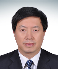 Changsheng Liu (刘昌胜)合作论文数Shanghai University;Chongqing University13
 David Kaplan合作论文数Department of Biomedical Engineering, School of Engineering, Tufts University13
 Jie Wang (王杰)合作论文数International Peace Maternity & Child Health Hospital, Shanghai Jiao Tong University School of Medicine13
 Ling Xu (徐凌)合作论文数School of Medicine, Shanghai Jiao Tong University13Xiaojuan Sun合作论文数13KeFeng Pan (潘可风)合作论文数12
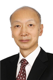 Jiang Chang (常江)合作论文数Shanghai Institute of Ceramics, Chinese Academy of Sciences12Xiaolin Wu (吴晓林)合作论文数Shanghai Jiao Tong University12Li Guanglong合作论文数12Yuezhi Lu合作论文数11
 Guifang Wang合作论文数11
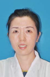 Yuqiong Wu (吴玉琼)合作论文数Zhongshan Hospital, Xiamen University11
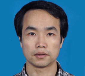 Kaili Lin (林开利)合作论文数National Clinical Research Center for Oral Diseases, Shanghai Ninth People’s Hospital, Shanghai Jiao Tong University School of Medicine10Kaige Lv合作论文数10Weiqiang Yu合作论文数10Xinchao Miao合作论文数9
 Longwei Hu合作论文数9
 Jiawei Yang合作论文数9




