OBJECTIVES:To investigate the differential effects of primary (PPT) and odontogenic (OPT) pathological Schneiderian membrane thickening on outcomes of transcrestal sinus floor elevation (TSFE) with simultaneous implant placement. MATERIAL AND METHODS:This prospective cohort study enrolled 89 patients (123 implants), divided into normal, PPT, and OPT groups. All patients underwent TSFE with simultaneous implant placement. The Schneiderian membrane perforation, early implant failure, postoperative complications, and implant stability quotient (ISQ) value were recorded. Cone beam computer tomography was obtained preoperatively, immediate postoperatively, and 6 months postoperatively to perform linear measurements including Schneiderian membrane thickness (SMT), residual bone height (RBH), sinus floor elevation height (SFEH), endo-sinus bone gain (ESBG), and graft material resorption height (GMRH). RESULTS:No early implant failure or postoperative complications were observed, and no significant difference in ISQ values was found among the three groups (p = 0.116). The OPT group had a significantly higher membrane perforation rate (30.4%) than the normal (9.5%) and PPT (7.7%) groups (p = 0.023), lower ESBG (2.88 ± 1.59 mm vs. normal 4.02 ± 1.63 mm, p = 0.015), and higher GMRH (2.98 ± 1.84 mm vs. normal 1.37 ± 1.15 mm/PPT 1.41 ± 1.26 mm, p < 0.001). PPT showed no significant differences in ESBG/GMRH versus normal (p > 0.05). CONCLUSIONS:Our results demonstrate that neither primary nor odontogenic pathological Schneiderian membrane thickening influences the early success rate of implants placed simultaneously with transcrestal sinus floor elevation. Nevertheless, odontogenic pathological Schneiderian membrane thickening significantly increases the incidence of Schneiderian membrane perforation and decreases the formation of new bone.
Periodontitis, a prevalent and progressive infectious inflammatory disease, is characterized by alveolar bone resorption and loss of periodontal attachment, presenting a formidable challenge for complete tissue regeneration. This study develops a biomimetic dual-layer scaffold (d-ACT@MIC/SIM) featuring synchronized minocycline (MIC) release and liposome-encapsulated simvastatin (SIM) delivery for optimal periodontal repair. The scaffold featured an asymmetric architecture, comprising a dense upper layer of chitosan (CS) and sodium alginate (AL) designed to prevent epithelial downgrowth, and a porous osteoconductive lower layer of CS, AL, and β-tricalcium phosphate (β-TCP) to support cell infiltration and bone ingrowth. Physicochemical characterization confirmed that d-ACT@MIC/SIM possessed favorable biocompatibility, robust antimicrobial efficacy against periodontal pathogens, and requisite mechanical integrity. In vivo studies, following the implantation of the biomimetic dual-layer drug-loading scaffold (d-ACT@MIC/SIM) into alveolar bone defects in a rat periodontitis model for 10 weeks, demonstrated significantly enhanced alveolar bone regeneration, with a bone volume/tissue volume (BV/TV) ratio of 80.6%. Furthermore, this multifunctional scaffold exhibited synergistic capabilities in promoting osteogenic differentiation, exerting sustained antibacterial activity, and mitigating local inflammatory responses. These findings highlight the substantial therapeutic potential of this asymmetrically structured, dual drug-loading biomimetic scaffold as a promising strategy for periodontal tissue engineering and the clinical management of periodontitis.
Periodontitis, a leading cause of adult tooth loss, requires effective plaque biofilm removal for successful treatment. Conventional mechanical debridement often fails to eradicate biofilms from deep root surfaces, leading to disease recurrence. Sonodynamic therapy (SDT) has emerged as a promising noninvasive antibacterial approach, owing to its capacity for deep tissue penetration. However, the efficacy of SDT is limited by the insufficient penetration of sonosensitizers through the dense extracellular polymeric substances of biofilms. Herein, a sonosensitizer-cationic polypeptide conjugate (PPa-cP) was developed via simple ring-opening reactions. The cationic design facilitated electrostatic binding to embedded bacteria within the biofilm, significantly enhancing sonosensitizer localization and the subsequent ultrasound-triggered bactericidal effect. PPa-cP achieved a reduction of >99.8% in viable bacteria within mature Porphyromonas gingivalis and Fusobacterium nucleatum biofilms. Transcriptomic analysis further revealed that PPa-cP disrupted Porphyromonas gingivalis at structural, metabolic, and functional levels. In a rat periodontitis model, PPa-cP significantly attenuated alveolar bone resorption and inflammatory responses. This work demonstrates a promising SDT conjugate with potential for treating periodontitis and other biofilm-associated infections.
Periodontitis is the leading cause of tooth loss in adults. Unfortunately, inflammation remains poorly controlled and prone to relapse, even after removing the initial plaque biofilm. The unique metabolic properties of mitochondria in the periodontal microenvironment provide a promising target for novel therapeutic strategies against periodontitis. Here, we integrate metabolomics and network biology to elucidate the potential role of nuclear factor E2-related factor 2/mitochondrial transcription factor (Nrf2/TFAM) in regulating mitochondrial metabolism in periodontitis. Based on this discovery, it is crucial to develop an innovative nanomedicine capable of effectively modulating the mitochondrial metabolism in periodontitis. Recently, itaconate (ITA), a key metabolite linking mitochondrial metabolism and inflammation, has emerged as a powerhouse in regulating immunity through Nrf2; however, its limited permeability hinders its application in biological systems. Therefore, we synthesize ITA-based nano cocktail (INC) with cell permeability and improved biological functions. At the cellular level, INC activates Nrf2/TFAM to remodel mitochondrial metabolism and regulate macrophage immune homeostasis. In mouse models of periodontitis, INC successfully reprograms mitochondrial metabolism within the gingiva, leading to an improved inflammatory microenvironment. Our study elucidates the role of INC in modulating mitochondrial metabolism, thereby offering an innovative therapeutic strategy for the management of periodontitis and other clinical conditions resulting from mitochondrial abnormalities.
Volumetric resorption of the alveolar ridge often occurs in both horizontal and vertical directions following tooth extraction. There is a specific lack of evidence for alveolar ridge reconstruction at molar and premolar sites with severe bone resorption. This randomized controlled trial used 3D and linear analyses to evaluate volumetric changes of the alveolar bone following alveolar ridge reconstruction (ARR) at molar and premolar sites with severe bone resorption as compared to un-assisted socket healing before implant placement. A total of 31 patients (15 men, 16 women) with > 50% hard tissue loss in one or more socket walls were recruited and randomized into either a test group (postextraction ARR using deproteinized bovine bone mineral with 10% collagen [DBBM-C] and platelet-rich fibrin [PRF] with a resorbable collagen membrane) or a control group (natural healing after extraction). The clinical, linear, and volumetric implant-related and patient-reported outcomes were analyzed after 4 months of healing. Linear bone assessments revealed significantly greater ridge width gains in the test group (25% in the mesial, midfacial, and distal aspects) and less reduction of vertical bone ridge than in the control group (P <.05). Further, volumetric bone remodeling was significantly higher in the test group (35.1% +/- 34.9% for ARR, 14.2% +/- 12.8% for control; P <.05). Patient-reported discomfort and keratinized mucosal changes were comparable between groups. ARR with a combination of DBBM-C, PRF, and a resorbable membrane at posterior sites with a severe socket wall deficiency (> 50% bone loss) is a safe and more capable therapeutic method when compared to natural healing and unassisted sockets. Collectively, the present analyses demonstrate that ARR represents an efficient method to maintain and augment crestal bone at posterior extraction sites with severe bone defects when assessed after 4 months of healing.
Bacterial infections can pose significant health risks as they have the potential to cause a range of illnesses. These infections can spread rapidly and lead to complications if not promptly diagnosed and treated. Therefore, it is of great significance to develop a probe to selectively target and image pathogenic bacteria while simultaneously killing them, as there are currently no effective clinical solutions available. This study presents a novel approach using near-infrared carbonized polymer dots (NIR-CPDs) for simultaneous in vivo imaging and treatment of bacterial infections. The core-shell structure of the NIR-CPDs facilitates their incorporation into bacterial cell membranes, leading to an increase in fluorescence brightness and photostability. Significantly, the NIR-CPDs exhibit selective bacterial-targeting properties, specifically identifying Staphylococcus aureus (S. aureus) while sparing Escherichia coli (E. coli). Moreover, under 808 nm laser irradiation, the NIR-CPDs exhibit potent photodynamic effects by generating reactive oxygen species that target and damage bacterial membranes. In vivo experiments on infected mouse models demonstrate not only precise imaging capabilities but also significant therapeutic efficacy, with marked improvements in wound healing. The study provides the dual-functional potential of NIR-CPDs as a highly effective tool for the advancement of medical diagnostics and therapeutics in the fight against bacterial infections.
因牙周疾病、肿瘤、外伤、先天畸形所致的口腔硬组织缺损给口腔功能重建带来极大困难.硅磷酸钙[Ca5(PO4)2SiO4,简称CPS]生物陶瓷因其良好的生物相容性、可降解性、骨传导性和骨诱导性,成为新一代骨缺损修复材料的研究热点.天然人体矿化组织中含有Cu2+、Fe3+、Zn2+、Mg2+、Sr2+等多种金属离子,具有其独特的生物特性,将这些金属离子加入到硅磷酸钙中对于材料改性有着巨大的潜力.本文概述了目前针对硅磷酸钙性能的研究进展,重点介绍了离子掺杂对生物陶瓷理化性质的影响及金属离子在抗菌、促进成骨和新血管生成等方面的作用及其可能机制,并对硅磷酸钙类材料今后的发展方向做一展望.
Being the most common cause of implant failure, peri-implantitis is defined as a pathological condition associated with the occurrence of peri-implant plaque, characterized by peri-implant mucosal inflammation and progressive loss of the supporting bone tissue attributed to the persistence of pro-inflammatory cytokines. Docosahexaenoic acid (DHA), which is a type of omega-3 polyunsaturated fatty acid, is generally used for the treatment of many inflammatory diseases. However, a suitable form for dosing and its therapeutic effect on peri-implantitis remain unclear. In this study, a novel nanostructured lipid carrier (NLC) loaded with squalene and DHA was fabricated (DHA-loaded NLC). The encapsulation efficiency and drug loading efficiency values of the DHA-loaded NLC were 78.13% ± 1.85% and 28.09% ± 0.48%, respectively. The release of DHA was gradual and steady until 144 h. In addition, the free-radical-scavenging rate of DHA-loaded NLC (0.57 ± 0.03) was much higher than that of sole DHA (0.17 ± 0.003). By inhibiting nuclear factor-κB p65 nuclear translocation, DHA-loaded NLC prevented the activation of nuclear factor-κB downstream inflammatory pathways and exerted anti-inflammatory effects on macrophages. Moreover, DHA-loaded NLC showed better effects on preventing alveolar bone resorption of rat peri-implantitis model than sole DHA. Hence, DHA-loaded NLC enhanced the anti-inflammatory bioavailability of DHA, offering a novel approach for the treatment of peri-implantitis.
Objectives Ferroptosis is associated with multiple inflammatory diseases. Periodontitis is an inflammatory disease mainly caused by oral opportunistic pathogens. However, the ferroptosis-periodontitis relationship has not been thoroughly described. We here analyzed whether ferroptosis is involved in periodontitis. Materials and methods Human gingival fibroblasts (HGFs) were stimulated with P. gingivalis-LPS and ferrostatin-1 (Fer-1, a ferroptosis inhibitor), and changes in mitochondrial morphology, ferroptosis-related factors, and inflammation levels were detected. After the rat experimental periodontitis model was established, changes in ferroptosis-related factors and inflammation levels were re-evaluated in the same manner. Results Porphyromonas gingivalis-LPS-induced mitochondrial shrinkage, an increase in mitochondrial membrane density, and upregulation of reactive oxygen species in HGFs. The expression of prostaglandin-endoperoxide synthase 2, transferrin receptor 1, and malondialdehyde and inflammation levels were upregulated, whereas the expression of solute carrier family seven member 11, glutathione peroxidase 4, superoxide dismutase, and glutathione were downregulated. Fer-1 attenuated these aforementioned changes and inflammation levels induced by P. gingivalis-LPS. The in vivo experiment results were consistent with the in vitro experiment results. Conclusions Ferroptosis is involved in inflammatory processes in HGFs upon P. gingivalis-LPS stimulation. Ferroptosis is observed in the gingival tissue of periodontitis rats.
To explore the application effect of the "medical record module"-oriented teaching mode in practice teaching of oral implant theory based on the textbook Modern Oral Implantology (second edition, edited by Su Yucheng) (referred to as Oral Implantology for short). 60 undergraduate students were selected as subjects, and real clinical cases were selected. After routine explanation, students were asked to write clinical medical records, which were recorded as pre-test medical records and scored according to the scoring table. After that, 30 students were randomly selected by lottery and divided into experimental group. According to the medical records of the whole process of oral implant therapy in clinical work, fifteen knowledge modules were decomposed, and the students in the experimental group were taught modularly according to the real clinical cases. After the teaching was completed, they were instructed to study Oral Implantology by themselves. The remaining students were divided into the control group, and they were taught in the classroom according to the traditional teaching plan. After the teaching was completed, they were instructed to learn Oral Implantology by themselves. A month later, the clinical real cases with the same difficulty were selected again. After routine explanation, students were asked to write clinical medical records, which were recorded as post-test medical records and scored according to the scoring table. The application effect of this teaching method in practice teaching of oral implant theory was evaluated through the score of medical records and satisfaction questionnaire. The average score of the medical records written by the students in the experimental group after the teaching intervention can reach 40.10 ± 1.80 points, which is significantly higher than the average score of 28.80 ± 1.32 points (P<0.001) in the control group and the average score of all students' pre-test medical records of 11.50 ± 2.27 points (P<0.001). 95% of students believe that this teaching mode can mobilize students' learning enthusiasm, exercise logical thinking and improve their comprehensive application ability. The "medical record module "-oriented teaching mode can achieve high quality and ideal teaching effect of oral implant theory, which can be considered for reference in the exploration of new oral implant teaching methods in the future.
The deficiency of keratinized mucosa is a common situation in the edentulous posterior mandibular area. This case report has rebuilt the sufficient width of keratinized mucosa in the peri-implant area with a modified free gingival graft surgery and kept the long-term health of the peri-implant tissue.
Vertical bone augmentation is one of the greatest challenges of bone regeneration in implant dentistry. When the bone defect is large, it is often necessary to open a second operative area for autologous bone block grafting or to use titanium mesh or non-absorbable membrane in combination with autologous bone blocks or particulates.The incidence of complications is high and the surgical trauma is significant. In this case, the patient had a severe vertical bone defect in the posterior maxillary region. BioOss Collagen combined with titanium tent poles and absorbable collagen membrane were used for vertical bone augmentation. The average bone height increased by 6.57 mm at 6months after bone grafting, which met the need of implant implantation. The bone tissue around the implant was relatively stable during the follow-up after restoration. Due to the predictable effect of bone augmentation, the simpler operation procedure and the less invasive results, this operation can provide a reference for the clinical selection of vertical bone augmentation.
牙齿缺失是老年群体常见的健康问题,种植修复能够有效恢复缺牙区的美观和功能.3~6个月的无负荷愈合期是实现种植体骨结合的基本前提,相对于传统无负荷愈合期,即刻负荷可显著缩短缺牙期,短期内恢复牙齿的美观和功能.但该方案对技术要求高,且后牙作为承担咀嚼力的主要受力区,对最终植入扭矩及上部修复体的咬合控制等因素都应有所考虑.本文就后牙区种植即刻负荷的理论依据、成功因素及临床评价作一综述.
Objective To investigate the mechanisms by which D-methionine (D-Met) eradicates Porphyromonas gingivalis biofilms by suppressing cyclic dimeric GMP (c-di-GMP) levels. Methods Cell viability, minimum inhibitory concentration (MIC) and minimum bactericidal concentration (MBC) were measured to determine the effective concentrations of D-Met, which were subsequently used in the following experiments. During the P. gingivalis biofilm formation inhibition experiment and the mature biofilm disassembly experiment, biofilm biomass, exopolysaccharide (EPS), biofilm morphology, integrity of the cell membrane, and the level of c-di-GMP were determined. Results D-Met < 40 mmol/L was biocompatible. During the biofilm formation inhibition and mature biofilm disassembly experiments, D-Met ≥ 20 mmol/L decreased the biofilm biomass and the production of EPS. SEM analysis showed that the extracellular matrix and bacterial density were drastically reduced by D-Met ≥ 20 mmol/L. TEM detection showed that 35 mmol/L D-Met ruptured the cell membrane during biofilm formation and increased the permeability of the cell membrane in the disassembly phase of mature biofilms. C-di-GMP levels decreased with increasing concentrations of D-Met in a concentration-dependent manner. Conclusion D-Met ≥ 20 mmol/L could eradicate P. gingivalis biofilms by suppressing c-di-GMP levels.
In recent years, dental implantation has become the preferred protocol for restoring dentition defects. Being the direct contact between implant and bone interface, osseointegration is the basis for implant exerting physiological functions. Nevertheless, biological complications such as insufficient bone volume, poor osseointegration, and postoperative infection can lead to implant failure. Emerging antibacterial-osteogenic multifunctional implant surfaces were designed to make up for these shortcomings both during the stage of forming osseointegration and in the long term of supporting the superstructure. In this mini-review, we summarized the recent antibacterial-osteogenic modifications of the dental implant surface. The effects of these modifications on biological performance like soft tissue integration, bone osteogenesis, and immune response were discussed. In addition, the clinical findings and prospects of emerging antibacterial-osteogenic implant materials were also discussed.
Objective: This study investigated the main mechanism and role of caspase-11/4 as a pattern recognition receptor (PRR) in periodontitis through caspase-11 inhibition.Design: Clinical tissue samples were collected from patients with periodontitis and healthy volunteers and evaluated through hematoxylin-eosin (HE) staining, immunohistochemical (IHC) staining, and real-time quan-titative PCR (RT-qPCR). In the rat periodontitis model, both these staining procedures, RT-qPCR, and western blotting were used to evaluate the histological, mRNA, and protein levels of caspase-11, interleukin-1 beta (IL-1 beta), and tumor necrosis factor-alpha (TNF-alpha). In vitro, the role of caspase-11, inhibited by siRNA, was investigated by analyzing the mRNA and protein levels of IL-1 beta and TNF-alpha in Porphylinomonas gingivalis (P. gingivalis) lipo-polysaccharide (LPS)-stimulated Raw264.7 macrophages.Results: Histological and molecular biological results of clinical and experimental animal periodontitis samples indicated that caspase-11/4 mRNA and protein levels significantly increased in inflammatory tissues. Caspase-11 is mainly distributed in leukocytes, which are labeled by CD45 in the submucosa. In vitro results further confirmed that the expression of caspase-11/4, IL-1 beta, and TNF-alpha significantly increased in LPS-stimulated macrophages, and these changes were significantly attenuated by inhibiting caspase-11/4 expression.Conclusions: The function of caspase-11 in rat periodontitis models is similar to that of caspase-4 in human clinical periodontitis. IL-1 beta and TNF-alpha release in periodontitis depends on the recognition of P. gingivalis LPS by caspase-11/4.
随着种植修复技术的不断应用与发展,种植相关的感染已经成为威胁种植长期效果的重要因素.细菌在种植体表面形成菌斑生物膜,其独特结构可以抵抗宿主防御功能,加速破坏其周围组织,导致种植体周围炎的发生,最终导致种植体周围骨进行性丧失、种植体松动、脱落.因此,抑制种植体表面微生物的黏附和生物膜的形成是防治种植体周围炎的主要策略.近年来,种植体表面的抗菌处理已逐渐成为研究热点.然而,表面的抗菌改性同样会影响表面的其他生物学性能,如涂层对上皮组织边缘封闭作用的影响以及涂层对结缔组织附着作用的影响,涂层促进良好的软组织结合对于维持种植体的物理屏障具有重要作用;骨结合是种植成功的基础,而不同的抗菌涂层处理将影响骨结合的进程,以及其作用机制依据材料的不同存在差异;抗菌涂层对局部免疫微环境的影响以及参与免疫平衡调节亦具有重要作用等.本文将从种植体表面抗菌设计的原理和抗菌表面产生的生物学性能影响两方面进行综述.
Peri-implantitis is the main cause of dental implant failure, which is associated with pyroptosis. The roles of D-aspartic acid (D-Asp) on pyroptosis and the mechanism of the protective effect of D-Asp on human gingival fibroblasts (HGFs) remain unknown. This study investigated the effects of D-Asp on the pyroptosis of HGFs induced by high mobility group box 1 protein (HMGB1). The cytotoxic effects of D-Asp on HGFs was detected by Cell Counting Kit-8 assay, the membrane permeability was investigated by propidium iodide/ Hoechst 33,342 double staining, flow cytometry analysis, and lactate dehydrogenase releasing, The gene and protein expression levels were detected by real-time quantitative PCR, enzyme-linked immunosorbent assay, and Western blot, respectively. Cell viability analysis showed that D-Asp ≤ 30 mM had no cytotoxicity to HGFs. HMGB1 drastically raised the membrane permeability of HGFs, while 1/10/30 mM D-Asp suppressed the permeability and remained the integrity of the membrane. HMGB1 promoted the mRNA expression of NLRP3, caspase-1, GSDMD, IL-1β, and IL-18, and the protein expression of IL-1β, IL-18, caspase-1, GSDMD, and NLRP3. With the pretreatment of HGFs with D-Asp of 1/10/30 mM for 24 h, the cell membrane permeability was reduced and the expression of NLRP3, caspase-1, GSDMD, IL-1β, and IL-18 was significantly decreased compared with the HMGB1 group, indicating the competitive antagonism of D-Asp against HMGB1 on the binding with toll-like receptors. Hence, this study may provide a novel insight into preventing pyroptosis and propose a new strategy for the treatment of peri-implantitis.
Abstract Objective: This randomized controlled trial was primarily aimed at evaluating the volumetric changes of the alveolar bone after alveolar ridge reconstruction (ARR)in molar sites with severe bone resorption as compared with unassisted socket healing by means of three dimensional and linear analyses.Material and methods: A total of 31 patients (15 males, 16 females) with more than 50% of hard tissue loss in one or more socket walls were recruited and randomized into either the test group(ARR after extraction using deproteinized bovine bone mineral with 10% collagen (DBBM-C) and platelet-rich fibrin (PRF) with a resorbable collagen membrane) or the control group (natural healing after extraction).The clinical, linear, volumetric implant-related and patient-reported outcomes were analyzed following a 4-months healing process. This single blinded randomized clinical trial was approved by Chinese Clinical Trial Registry (Identifier: ChiCTR2100047321).Results: Linear bone assessments presented significantly more gains of ridge width presented at 25% level in mesial, mid-facial and distal aspects in the test group and less reduction of vertical bone ridge than in the control group from (P<0.05).Similarly, volumetric bone remodeling was significant higher in the test group (ARR=35.1±34.9%, control=14.2±12.8%, P<0.05). Assessment of patient-reported discomfort and keratinized mucosa changes were comparable between groupsConclusion: Alveolar ridge reconstruction with DBBM-C in combination with PRF and resorbable membrane in posterior sites with severe socket walls deficiency (>50% bone loss) is a safe and more capable method when compared with natural healing unassisted socket.Clinical Relevance: The presented study have demonstrated that alveolar ridge reconstruction could be an efficient method to maintain and augment crestal bone at posterior extraction sites with severe bone defect during 4 months of healing.
牙周炎是一种牙齿支持组织的慢性炎症性疾病,能引起结缔组织和牙槽骨的破坏而导致渐进性附着丧失,最终可能导致牙齿脱落.革兰氏阴性厌氧菌牙龈卟啉单胞菌是慢性牙周炎的重要致病菌,具有包括脂多糖、菌毛、血细胞凝集素和胞外蛋白酶在内的多种毒力因子,其中牙龈蛋白酶提供的蛋白水解活性占该生物体产生的一般蛋白水解活性的大部分(85%),已被认为是必需的毒力因子.本文就牙龈卟啉单胞菌牙龈蛋白酶的分类、分泌、致病作用及其抑制剂的研究作一综述.
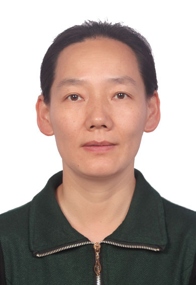 Qing Cai (蔡晴)合作论文数College of Materials Science and Engineering, Beijing University of Chemical Technology41
 Baosheng Li (李保胜)合作论文数40
 Yanmin Zhou (周延民)合作论文数Implant Center,Stomatology Hospital,Jilin University39
 He-Ling Wang (王鹤龄)合作论文数Akershus Univ Hosp14Lisheng ZHANG (张理生)合作论文数11HuiYan Zhang (张慧彦)合作论文数11ShunLi Chu (储顺礼)合作论文数10Yun-xia CUI (崔云霞)合作论文数8Yanqun Liu合作论文数8Hua Qi (齐华)合作论文数8Zuntai Li合作论文数7Zhenyang ZHANG (张震阳)合作论文数7
 Zhao Jinghui合作论文数7xu quan (全旭)合作论文数7Chunyu Han合作论文数6Shuwei Qiao合作论文数6haoyang wang (王昊阳)合作论文数6Jin Zhuohua (金卓华)合作论文数6Yanzhen OU (欧燕珍)合作论文数6Chun-yang WANG (王春阳)合作论文数6XU Tongtong (许彤彤)合作论文数6Yanjing Zhang合作论文数6Zixuan Wang合作论文数6
 Chengcheng Yin合作论文数6XiaoShuang Qi (齐晓爽)合作论文数6Jiang Jiayang合作论文数6Yu Haina (余海纳)合作论文数5Yang Li-ming合作论文数5LI Xiaoyu (李效宇)合作论文数5LI Zhen (李珍)合作论文数5LI Birong (李碧榕)合作论文数5Qiyue FU (付齐月)合作论文数5TongBin Liu (刘童斌)合作论文数5Zhaoyi Yin合作论文数4Wang Dan (汪丹)合作论文数4Xie Lili (谢丽丽)合作论文数4Mengxiao He (何梦晓)合作论文数4Yuyang Li合作论文数4
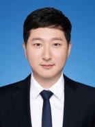 Baixiang Wang (王柏翔)合作论文数The Stomatology Hospital, Zhejiang University School of Medicine4WU Songtao (吴松涛)合作论文数4YanHai Che (车彦海)合作论文数4YanQiu Li (李艳秋)合作论文数4JingJing Wang (王晶晶)合作论文数4YuJie Liu (刘玉洁)合作论文数4Yuyan SUN (孙毓言)合作论文数3zhenping zhou (周振平)合作论文数3Yue YUAN (袁月)合作论文数3
 Shoujun Zhu (朱守俊)合作论文数College of Chemistry, Jilin University3




