Statement of problem. Information regarding the effect of tooth color under different light conditions on the accuracy of intraoral complete arch scanning is limited. Purpose. The purpose of this in vitro study was to evaluate the effect of color and ambient light conditions on the accuracy of mandibular complete arch scanning with an intraoral scanner (IOS) using a zirconia restoration model with different shades. Material and methods. Five mandible dentition models with zirconia restorations of different shades were fabricated by computer-aided design and computer-aided manufacturing (CAD-CAM). The spectral reflectance and transmittance curves were collected with a spectrophotometer to determine color parameters (Rb, T, S+A, L*, a*, b*, C*, and h). Under 4 different lighting conditions: no light (ZL), natural light (NL), room light (RL), and chair light (CL), each model was scanned 10 times by using an IOS (TRIOS 3). Three-dimensional (3D) deviation analysis and a linear deviation analysis were performed for an accurate quantitative measurement of intraoral scanning. The multivariate test was used to determine significant differences in 3D deviation and linear deviation among groups. The multiple linear regression test was conducted to investigate the relevant independent factors of mean absolute 3D deviation. Results. The 3D deviation analysis showed that the mean absolute 3D deviation of 3M2 model scanning was the lowest (P<.001). Moreover, under CL and RL, the accuracy results from the 3M2 model scan were demonstrated as significantly better than the tested scans under other light conditions (P=.021). The result of the linear deviation analysis indicated that the variation in distance was only significant between the bilateral canines (P=.032). Ambient light conditions, C*, and h were factors influencing mean absolute 3D deviation (R2=0.593, P<.001). Conclusions. Color change influenced the accuracy of intraoral mandibular complete arch scanning under different light conditions. This effect may be attributable to the interaction between the ambient light condition and color parameters such as C* and h. (J Prosthet Dent 2024;131:145.e1-e8)
Objectives: This study aims to investigate different treatments on new bone formation around immediate implants in the canine posterior mandible with varying sized mesial-distal gap. Materials and methods: The 4 th premolar and the 1 st molar of six Labrador dogs were extracted from the mandible, and 4 dental implants were placed 1 mm below the level of the buccal bone crest. Moderate/large mesial-distal gaps between the implants and the sockets were treated with one of four methods and divided into the following groups: (1) the blank group, (2) the collagen membrane (CM) group, (3) the deproteinized bovine bone mineral (DBBM) group, and (4) the DBBM+CM group. Sequential fluorescent labeling was performed at 4, 8, and 10 weeks after the operation. After 12 weeks, the dogs were euthanized, and specimens were collected for micro-CT scanning and histological analysis. Results: The survival rate of immediate implant was 100%. Micro-CT showed significant differences in bone mineral density (BMD) and bone volume fraction (BVF) among groups ( P = 0.040, P = 0.009); other indicators were not significantly different among groups. Histological analysis showed the proportion of new bone formation and bone-to-implant contact were not significantly different among groups. No significant difference in bone reduction height around dental implant among four groups and varied mesial-distal gap size. Conclusion: Owing to the restricted sample size, this pilot study lacks conclusive findings. Within the limitation, this study demonstrated that although DBBM significantly increase BMD and BVF, the use of DBBM/CM didn’t significantly improve bone formation and healing in extraction sockets around the implants in both moderate and large mesial-distal gap. Clinical Relevance: The use of deproteinized bovine bone in conjunction with collagen is a common practice in immediate implantation procedures in the posterior mandible. However, there is a lack of conclusive evidence regarding the timing and circumstances under which they should be employed.
Background The purpose of this study was to investigate the socket healing outcome after alveolar ridge preservation at infected molar sites using an erbium-doped yttrium aluminium garnet (Er:YAG) laser. Methods Eighteen patients who needed molar extraction and exhibited signs of infection were included and allocated into either the laser group or the control group. Er:YAG laser irradiation for degranulation and disinfection was performed with alveolar ridge preservation (ARP) in the laser group. Traditional debridement with a curette was performed in the control group. Two months after ARP, bone tissue samples were harvested at the time of implant placement for histological analysis. Assessment of dimension changes in alveolar bone was conducted by superimposing two cone-beam computed tomography (CBCT) scans taken at baseline and two months after extraction. Results Histologically, after two months of healing, Er:YAG laser treatment resulted in more newly formed bone (laser: 17.75 ± 8.75, control: 12.52 ± 4.99, p = 0.232). Moreover, greater osteocalcin (OCN) positive expression and lower runt-related transcription factor 2 (RUNX-2) positive expression were detected in the laser group. However, no statistically significant difference was observed between the two groups. The difference in the vertical resorption of the buccal bone plate was statistically significant between groups (laser: -0.31 ± 0.26 mm, control: -0.97 ± 0.32 mm, p < 0.05). Major changes in ridge width were observed at 1 mm below the bone crest. However, the differences between groups were not significant (laser: -0.36 ± 0.31 mm, control: -1.14 ± 1.24 mm, p = 0.171). Conclusions ARP with Er:YAG laser irradiation seemed to improve bone healing by regulating osteogenesis-related factor expression in the early stage at infected sites. Trial registration The trial was registered on the Chinese Clinical Trial Registry Platform ( https://www.chictr.org.cn/ ) (registration number: ChiCTR2300068671; registration date: 27/02/2023).
Abstract Aim This study aimed to explore new bone formation around implants in extraction sockets after immediate implantation in the canine posterior mandible in response to different treatments. Materials and methods The 4th premolar and the 1st molar of six Labrador dogs were extracted from the mandible, and then, 4 dental implants were implanted. The mesial and distal gaps between the implants and the sockets were treated with one of four methods and divided into the following groups: (1) the blank group (group BG), (2) the collagen membrane (CM) group (group C), (3) the deproteinized bovine bone mineral (DBBM) group (group D), and (4) the DBBM+CM group (group DC). Sequential fluorescent labeling was performed at 4, 8, and 10 weeks after the operation. After 12 weeks, the dogs were euthanized, and specimens were collected for micro-CT scanning and histological analysis. Results The survival rate was 100%. Micro-CT showed significant differences in bone mineral density (BMD) and bone volume fraction (BVF) among the four groups (P = 0.040, P = 0.009); other indicators were not significantly different among the groups. Histological analysis showed that the proportion of new bone formation and bone-to-implant contact (BIC) were not significantly different among the groups. Conclusion The current study demonstrated that although DBBM can significantly increase BMD and BVF, the use of DBBM and CM did not significantly improve bone formation in extraction sockets around the implants.Clinical relevance According to the current animal experiment, DBBM and CM may not have a significant impact on bone formation in the extraction socket of IIP in the posterior mandible, suggesting that there was no sufficient reason to recommend the application of these two kinds of materials in mesiodistal extraction sockets around IIP in mandibular molar sites.
Efficient initiation and resolution of inflammation are crucial for wound repair. However, with using tissue adhesives for wound repair, patients occasionally suffered from delayed healing process because slow elimination of those exogenous adhesives generally leads to chronic inflammation. As the demand for minimal invasive therapy continues to rise, desire for adhesive materials that can effectively reconnect surgical gaps and promote wound regeneration becomes increasingly urgent. Herein, by exploiting the inherent porous structure and performance of adhesion to tissue of mesoporous silica nanoparticles (MSNs), we demonstrate a tissue adhesive that can elicit acute inflammatory response and get eliminated after tissue reformation. With formation of nanocomposites in wound gaps, the injured tissues can get reconnected conveniently. The resultant accelerated healing process verify that the strategy of exploiting unique properties of nanomaterials can effectively promote inflammation resolution and wound repair. This design strategy will inspire more innovative tissue adhesives for clinical applications.
BackgroundDifferent substances are combined to compensate for each other's drawbacks and create an appropriate biomaterial. A novel Polyvinyl alcohol (PVA)/chitosan (CS) porous hydrogel was designed and applied to the treatment of osteochondral defects.MethodsHydrogels of various PVA/CS ratios were tested for physiochemical and mechanical properties in addition to cytotoxicity and biocompatibility. The hydrogels with the best PVA/CS ratio were used in the animal study. Osteochondral defects were created at the articular cartilage of 18 rabbits. They were assigned to different groups randomly (n=6 per group): the osteochondral defect only group (control group), the osteochondral defect treated with hydrogel group (HG group), and the osteochondral defect treated with hydrogel loaded with bone marrow mesenchymal stem cells (BMSCs) group (HG-BMSCs group). The cartilage was collected for macro-observation and histological evaluation at 12weeks after surgery.ResultsThe Hydrogel with PVA/CS ratio of 6:4 exhibited the best mechanical properties; it also showed stable physical and chemical properties with porosity and over 90% water content. Furthermore, it demonstrated no cytotoxicity and was able to promote cell proliferation. The HG-BMSCs group achieved the best cartilage healing.ConclusionsThe novel PVA/CS porous composite hydrogel could be a good candidate for a tissue engineering material in cartilage repair.
Background Functional reconstruction of maxillofacial bone defects is a considerable clinical challenge. Many studies have emphasized the osteogenic and angiopoietic abilities of stem cells for tissue regeneration. We previously showed that microRNA-21 (miRNA-21) can promote angiogenesis in human umbilical cord blood-derived mesenchymal stem cells (UCBMSCs). In the present study, the role of miRNA-21 in osteogenic differentiation of bone marrow-derived stem cells (BMSCs) was investigated. Methods Western blotting and qPCR were performed to investigate the influences of miRNA-21 on osteogenic differentiation of BMSCs. The effects of miRNA-21 on PTEN/PI3K/Akt/HIF-1α pathway were also assessed using western blotting. To further evaluate the roles of miRNA-21 in osteogenesis in vivo, we conducted animal experiments in rat and canine. New bone formation was assessed using micro-CT and histological methods. Results In the present study, we found that miRNA-21 promotes the migration and osteogenic differentiation of bone marrow-derived stem cells (BMSCs) in vitro. Using gain- and loss-of-function studies, we found that miRNA-21 promoted the osteogenic ability of BMSCs by increasing P-Akt and HIF-1α activation. Finally, we verified the essential role of miRNA-21 in osteogenesis by implanting a miRNA-21-modified BMSCs/β-tricalcium phosphate (β-TCP) composite into critical size defects. Radiography, micro-CT, and histology revealed significantly greater volume of new bone formation in the miRNA-21 group than in the control group. Conclusion In conclusion, our study demonstrated an essential role of miRNA-21 in promoting maxillofacial bone regeneration via the PTEN/PI3K/Akt/HIF-1α pathway.
A considerable amount of research has focused on improving regenerative therapy strategies for repairing defects in load-bearing bones. The enhancement of tissue regeneration with microRNAs (miRNAs) is being developed because miRNAs can simultaneously regulate multiple signaling pathways in an endogenous manner. In this study, we developed a miR-210-based bone repair strategy. We identified a miRNA (miR-210-3p) that can simultaneously up-regulate the expression of multiple key osteogenic genes in vitro. This process resulted in enhanced bone formation in a subcutaneous mouse model with a miR-210-3p/poly-L-lactic acid (PLLA)/bone marrow-derived stem cell (BMSC) construct. Furthermore, we constructed a model of critical-sized load-bearing bone defects and implanted a miR-210-3p/beta-tricalcium phosphate (beta-TCP)/bone mesenchymal stem cell (BMSC) construct into the defect. We found that the load-bearing defect was almost fully repaired using the miR-210-3p construct. We also identified a new mechanism by which miR-210-3p regulates Sclerostin protein levels. This miRNA-based strategy may yield novel therapeutic methods for the treatment of regenerative defects in vital load-bearing bones by utilizing miRNA therapy for tissue engineering. Statement of Significance The destroyed maxillofacial bone reconstruction is still a real challenge for maxillofacial surgeon, due to that functional bone reconstruction involved load-bearing. Base on the above problem, this paper developed a novel miR-210-3p/beta-tricalcium phosphate (TCP)/bone marrow-derived stem cell (BMSC) construct (miR-210-3p/beta-TCP/BMSCs), which lead to functional reconstruction of critical-size mandible bone defect. We found that the load-bearing defect was almost fully repaired using the miR-210-3p construct. In addition, we also found the mechanism of how the delivered microRNA activated the signaling pathways of endogenous stem cells, leading to the defect regeneration. This miRNA-based strategy can be used to regenerate defects in vital load-bearing bones, thus addressing a critical challenge in regenerative medicine by utilizing miRNA therapy for tissue engineering. (C) 2018 Acta Materialia Inc. Published by Elsevier Ltd. All rights reserved.
Periodontal ligament stem cells (PDLSCs) are a major source of mesenchymal stem cells (MSCs) in adults and are effective for tissue engineering, like promoting angiogenesis and bone regeneration. CXCL12 has been reported to be involved in the recruitment and engraftment of MSCs in wound sites. However, whether CXCL12 potentiates the angiogenesis of PDLSCs is not clear. In this experiment, we transduced PDLSCs with CXCL12, and evaluated the angiogenesis potential of CXCL12-modified PDLSCs through in vitro and in vivo studies. The results showed that CXCL12 overexpression significantly stimulated the gene and protein expressions of bFGF, VEGF, SCF and PLGF in PDLSCs; CXCL12 gene modified PDLSCs formed longer capillary‐like structure; Moreover, in vivo transplanted PDLSCs transduced with CXCL12 could significantly promote bone tissue repair and angiogenesis in a rat critical-sized calvarial bone defect model. Taken together, our study confirmed that CXCL12 can enhance the angiogenesis potential of PDLSCs, which are crucial in the repair and regeneration of bone tissue.
BACKGROUND: As is known to all, cartilage tissue engineering has three elements, seed cells,scaffold materials, and cytokines. Scaffold materials play an important role in cartilage tissue engineering.OBJECTIVE: To search and review the literatures about tissue-engineered cartilage scaffold materials in the last decade,and to discuss problems and the development direction of the scaffolds.METHODS: A computer-based search of relevant articles published from 2006 to 2016 was conducted in PubMed and CNKI using the key words of "cartilage tissue engineering, natural scaffold materials, synthetic scaffold materials,composite scaffolds,nanometer materials" in English and Chinese, respectively.RESULTS AND CONCLUSION:Scaffold materials in cartilage tissue engineering have different sources and kinds.Natural materials, synthetic materials and nanometer materials have their own advantages and disadvantages. Any single material is difficult to meet the clinical requirements of the cartilage. Material compounds with different proportions can be used to prepare scaffolds with good mechanical properties, high porosity, good compatibility and degradation. But there are still some problems, such as excessive speed of scaffold degradation and excessive cell growth. The current research is in in vitro experimental stage mostly, and the application in clinic has not been enforced yet due to small size and lack of a long-term follow up.
Iron-oxide-based contrast agents for magnetic resonance imaging (MRI) had been clinically approved in the United States and Europe, yet most of these nanoparticle products were discontinued owing to failures to meet rigorous clinical requirements. Significant advances have been made in the synthesis of magnetic nanoparticles and their biomedical applications, but several major challenges remain for their clinical translation, in particular large-scale and reproducible synthesis, systematic toxicity assessment, and their preclinical evaluation in MRI of large animals. Here, we report the results of a toxicity study of iron oxide nanoclusters of uniform size in large animal models, including beagle dogs and the more clinically relevant macaques. We also show that iron oxide nanoclusters can be used as T 1 MRI contrast agents for high-resolution magnetic resonance angiography in beagle dogs and macaques, and that dynamic MRI enables the detection of cerebral ischaemia in these large animals. Iron oxide nanoclusters show clinical potential as next-generation MRI contrast agents.
BACKGROUND:By mixing technology, various materials are mixed and complemented each other to enhance heat sensitivity, mechanical properties, viscoelasticity of the materials and improve their biocompatibility, biodegradability and other biomedical properties.OBJECTIVE:To prepare polyvinyl alcohol (PVA)/chitosan (CS) porous hydrogel and to observe its effects on the repair of articular cartilage defects.METHODS: PVA, CS and polysorbate-80 were used as raw materials to prepare the hydrogels at the PVA/CS ratio of 5:5, 6:4, 7:3, 8:2 and 9:1 by freezing-thawing cycle and emulsification-frostice phase separation, and the physical and chemical properties, mechanical properties, and biocompatibility of the hydrogels were detected to find out the best PVA/CS ratio. Eighteen New Zealand rabbits were enroled to make models of bilateral articular cartilage defects, and then randomized into three groups: blank control group; control group; experimental group. The prepared hydrogel was compounded with rabbit bone marrow mesenchymal stem cells (BMSCs) and then implanted into the rabbit articular cartilage defect in the experimental group, the PVA/CS hydrogel was implanted in the control group, and nothing was implanted in the blank control group. After 12 weeks, the animals were killed and the repairing effect was observed by gross observation and histological examination.RESULTS AND CONCLUSION:The PVA/CS porous composite hydrogel was successfully prepared, and PVA:CS=6:4 was the best in the presence of good mechanical properties, stable physical and chemical properties, and water content≥ 90%. In additional, scanning electron microscopy showed a porous network structure, with the porosity≥ 90%. The results of cell counting kit-8 assay and the results of cell death and survival showed that the hydrogel was non-cytotoxic and beneficial to the cell proliferation. In the blank control group, knee articular cartilage defects were not repaired within 12 weeks after surgery, showing significant granulation tissue filling. In the control group, knee articular cartilage defects were full of cartilage-like tissues with no smooth surface, but there were a great amount of chondrocyte-like cells. In the experimental group, knee articular cartilage defects were well repaired and full of a great amount of chondrocytes with the smooth surface. To conclude, the PVA/CS porous composite hydrogel could repair articular cartilage defects as an ideal tissue-engineered cartilage material.
Scaffolds for tissue engineering (TE) which closely mimic the physicochemical properties of the natural extracellular matrix (ECM) have been proven to advantageously favor cell attachment, proliferation, migration and new tissue formation. Recently, as a valuable alternative, a bottom-up TE approach utilizing cell-loaded micrometer-scale modular components as building blocks to reconstruct a new tissue in vitro or in vivo has been proved to demonstrate a number of desirable advantages compared with the traditional bulk scaffold based top-down TE approach. Nevertheless, micro-components with an ECM-mimicking nanofibrous structure are still very scarce and highly desirable. Chitosan (CS), an accessible natural polymer, has demonstrated appealing intrinsic properties and promising application potential for TE, especially the cartilage tissue regeneration. According to this background, we report here the fabrication of chitosan microspheres with an ECM-mimicking nanofibrous structure for the first time based on a physical gelation process. By combining this physical fabrication procedure with microfluidic technology, uniform CS microspheres (CMS) with controlled nanofibrous microstructure and tunable sizes can be facilely obtained. Especially, no potentially toxic or denaturizing chemical crosslinking agent was introduced into the products. Notably, in vitro chondrocyte culture tests revealed that enhanced cell attachment and proliferation were realized, and a macroscopic 3D geometrically shaped cartilage-like composite can be easily constructed with the nanofibrous CMS (NCMS) and chondrocytes, which demonstrate significant application potential of NCMS as the bottom-up cell-carrier components for cartilage tissue engineering.
Critical limb ischemia (CLI) is a severe blockage in the arteries of the lower extremities. However, the effective and optimal treatment for CLI remains to be elucidated. Previous therapeutic research is mainly focused on proangiogenic growth factors administrations. Recently, miR-21 has been revealed to play a crucial role in angiogenesis. Thus, we hypothesize that miR-21 over-expression in human umbilical cord blood-derived mesenchymal stem cells (UCBMSCs) can effectively treat CLI. Herein, UCBMSCs were transduced with lentivirus-miR-21-Luciferase (Lenti-miR-21) or lentivirus- LacZ-Luciferase (Lenti-LacZ). The results indicated that miR-21 induced UCBMSCs proliferation, migration, and angiogenesis in vitro. Subsequently, general observation and laser Doppler perfusion imaging were introduced to detect perfusion in muscles of CLI-nude mice on 1, 4, 7, 14, and 28 day postoperation. There was a significant improvement in blood vessels of the ischemic limb in Lenti-miR-21 group at 7 day compared with the saline or Lenti-LacZ groups. At 28 day, histological analysis confirmed that UCBMSCs over-expressing miR-21 increased neovascularization in CLI. Furthermore, carboxyl terminus of Hsc70-interacting protein (CHIP) was found to be the target gene for miR-21-mediated activation of hypoxia-inducible factor 1α (HIF-1α) in UCBMSCs. In summary, our study demonstrated that over-expressing miR-21 in UCBMSCs could improve neovascularization in CLI through enhancing HIF-1α activity by targeting CHIP, which may hold great therapeutic promise in treating CLI.
In this study, a high‐performance T1–T2 dual‐model contrast agent by gadolinium‐doped iron oxide nanoparticle (GION) is developed. Following its development, the application of this agent in vivo by combining doxorubicin (DOX) and folic acid (FA) (FA–GION–DOX) for targeted drug delivery to monitor cancer treatment is explored. GION showed transverse and longitudinal relaxivities up to 182.7 × 10⁻³ and 7.87 × 10⁻³m−1 s−1, respectively, upon Gd/Fe ratio in GION at 1/4. DOX released from FA–GION–DOX is pH dependent and only kills cancer cell after FA receptor‐mediated internalization into the acidic environment of endosomes and lysosomes. Systemic delivery of FA–GION–DOX significantly inhibits the growth of tumors and shows good magnetic resonance enhancement in a human cervical cancer xenograft model. Thus, FA–GION–DOX has a potential application for the targeted and magnetic resonance imaging guided therapy of cervical cancer.
For artificial nerve conduits, great improvements have been achieved in mimicking the structures and components of autologous nerves. However, there are still some problems in conduit construction, especially in terms of mechanical properties, biomimetic surface tomography, electrical conductivity and sustained release of neurotrophic factors or cells. In this study, we designed and fabricated a novel electrospun nerve conduit enhanced by multi-walled carbon nanotubes (MWNTs) on the basis of a collagen/poly(ε-caprolactone) (collagen/PCL) fibrous scaffold. Our aim was to provide further knowledge about the mechanical effects and efficacy of MWNTs on nerve conduits as well as the biocompatibility and toxicology of MWNTs when applied in vivo. The results showed that as one component, carboxyl MWNTs could greatly alter the composite scaffold’s hydrophilicity, mechanical properties and degradability. The electrospun fibers enhanced by MWNTs could support Schwann cell adhesion and elongation as a substrate in vitro. In vivo animal studies demonstrated that the MWNT-enhanced collagen/PCL conduit could effectively promote nerve regeneration of sciatic nerve defect in rats and prevent muscle atrophy without invoking body rejection or serious chronic inflammation. All of these results showed that this MWNT-enhanced scaffold possesses good biocompatibility and MWNTs might be excellent candidates as engineered nanocarriers for further neurotrophic factor delivery research.
BACKGROUND:Pediatric aggressive fibromatosis (AF) is a rare, benign tumor with locally infiltrative growth. Therefore, how to prevent reoccurrence while maintaining the mandible contour and continuity as much as possible is very important when the mandible is involved. PROCEDURE:We selected 10 pediatric patients with AF involving the mandible in our department between January 2001 and December 2011. Patient clinical data, including patient characteristics, symptoms at presentation, management, and treatment outcome, were reviewed. RESULTS:Patients' ages ranged from 3 to 16 years with six males and four females. According to imaging, there were three cases where the tumor adhered to the mandible; the periosteum was resected with the tumor, followed by cryotherapy or cauterization. Another five cases involved the destruction of the periosteum and the cortical plate. Resection of the involved mandible with the tumor was performed, and the margin was trimmed. In the last two cases, the tumor had invaded the whole ramus, and the immediate iliac graft was operated on after resecting the lesions. Neither radiotherapy (RT) nor chemotherapy was used. There was no recurrence or contour defect of the face. The function of the mandible was not affected, and only one case showed a slight limitation when opening the mouth. CONCLUSIONS:For the treatment of pediatric AF, we recommend complete tumor resection. As for the involved mandible, preserving the mandible contour and continuity as much as possible and providing adjunctive therapy, such as cryotherapy or cauterization, are vital. RT is not recommended.
The successful clinical outcome of the implanted tissue-engineered bone is dependent on the establishment of a functional vascular network. A gene-enhanced tissue engineering represents a promising approach for vascularization. Our previous study indicated that hypoxia-inducible factor-1α (HIF-1α) can up-regulate the expression of vascular endothelial growth factor (VEGF) and stromal-derived factor 1 (SDF-1) in bone mesenchymal stem cells (BMSCs). The angiogenesis is a co-ordinated process that requires the participation of multiple angiogenic factors. To further explore the angiogenic effect of HIF-1α mediated stem cells, in this study, we systematically evaluated the function of HIF-1α in enhancing BMSCs angiogenesis in vitro and in vivo. A constitutively active form of HIF-1α (CA5) was inserted into a lentivirus vector and transduced into BMSCs, and its effect on vascularization and vascular remodeling was further evaluated in a rat critical-sized calvarial defects model with a gelatin sponge (GS) scaffold. The expression of the key angiogenic factors including VEGF, SDF-1, basic fibroblast growth factor (bFGF), placental growth factor (PLGF), angiopoietin 1 (ANGPT1), and stem cell factor (SCF) at both mRNAs and proteins levels in BMSCs were significantly enhanced by HIF-1α overexpression compared to the in vitro control group. In addition, HIF-1α-over expressing BMSCs showed dramatically improved blood vessel formation in the tissue-engineered bone as analyzed by photography of specimen, micro-CT, and histology. These data confirm the important role of HIF-1α in angiogenesis in tissue-engineered bone. Improved understanding of the mechanisms of angiogenesis may offer exciting therapeutic opportunities for vascularization, vascular remodeling, and bone defect repair using tissue engineering strategies in the future.
Background To cope with the limitations faced by autograft acquisitions particularly for multiple nerve injuries, artificial nerve conduit has been introduced by researchers as a substitute for autologous nerve graft for the easy specification and availability for mass production. In order to best mimic the structures and components of autologous nerve, great efforts have been made to improve the designation of nerve conduits either from materials or fabrication techniques. Electrospinning is an easy and versatile technique that has recently been used to fabricate fibrous tissue-engineered scaffolds which have great similarity to the extracellular matrix on fiber structure. Results In this study we fabricated a collagen/poly(ε-caprolactone) (collagen/PCL) fibrous scaffold by electrospinning and explored its application as nerve guide substrate or conduit in vitro and in vivo . Material characterizations showed this electrospun composite material which was made of submicron fibers possessed good hydrophilicity and flexibility. In vitro study indicated electrospun collagen/PCL fibrous meshes promoted Schwann cell adhesion, elongation and proliferation. In vivo test showed electrospun collagen/PCL porous nerve conduits successfully supported nerve regeneration through an 8 mm sciatic nerve gap in adult rats, achieving similar electrophysiological and muscle reinnervation results as autografts. Although regenerated nerve fibers were still in a pre-mature stage 4 months postoperatively, the implanted collagen/PCL nerve conduits facilitated more axons regenerating through the conduit lumen and gradually degraded which well matched the nerve regeneration rate. Conclusions All the results demonstrated this collagen/PCL nerve conduit with tailored degradation rate fabricated by electrospinning could be an efficient alternative to autograft for peripheral nerve regeneration research. Due to its advantage of high surface area for cell attachment, it is believed that this electrospun nerve conduit could find more application in cell therapy for nerve regeneration in future, to further improve functional regeneration outcome especially for longer nerve defect restoration.
 Duohong Zou合作论文数12
 Zhiyuan Zhang (张志愿)合作论文数School of Medicine, Shanghai Jiaotong University;Shanghai Ninth People’s Hospital9
 Jiacai He (何家才)合作论文数Anhui Medical University7
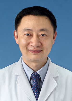 Xin-quan Jiang (蒋欣泉)合作论文数Shanghai Stomatological Hospital4
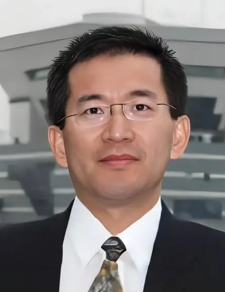 Wenjie Zhang (张文杰)合作论文数Shanghai Ninth People’s Hospital, Shanghai Jiao Tong University School of Medicine4Chao Zhu合作论文数3Tao Wu合作论文数3YouMing Zhu (朱友明)合作论文数3
 Weimin Zhu (朱伟民)合作论文数Affiliated Stomatological Hospital of Nanchang University3
 Daping Wang (王大平)合作论文数Shenzhen Second People's Hospital;Guangzhou Medical University3Mohan Wang合作论文数3Liangquan Peng合作论文数3
 Chi Yang (杨驰)合作论文数Shanghai Ninth People’s Hospital, School of Medicine, Shanghai Jiao Tong University3Zhoujing Zhang合作论文数2
 Wei Liu合作论文数2Huai-Ling Gao合作论文数2Honghai Fu合作论文数2Yujie Ning合作论文数2
 Shuhong Yu (俞书宏)合作论文数Department of Chemistry, School of Chemistry and Materials Science, University of Science and Technology of China2
 Wang Daming合作论文数2
 Liang Dong合作论文数2
 Wen Zhao (赵雯)合作论文数School of Life Sciences, Northwestern Polytechnical University2
 Wei Han (韩伟)合作论文数Nanjing Stomatological Hospital, Medical School of Nanjing University2
 Jian Zhou (周坚)合作论文数北京协和医院2
 Shaoyi Wang (王绍义)合作论文数Shanghai Ninth People’s Hospital, Shanghai Jiao Tong University School of Medicine2Bin Hu (胡斌)合作论文数2Xiuli Zhang合作论文数2
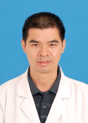 Yuanyin Wang (王元银)合作论文数College of Stomatology, Anhui Medical University2
 Yiqun Wu (吴轶群)合作论文数Shanghai Ninth People’s Hospital, Shanghai JiaoTong University School of Medicine2Wenwen Yu合作论文数2
 Yuanliang Huang合作论文数2Dongxia Ye合作论文数2Qiutong Yang合作论文数1Xinxiu Duan合作论文数1Kai Zhang合作论文数1Baiyan Sui合作论文数1Kai-Run Zhang合作论文数1Jian Chen合作论文数1Li Zhang合作论文数1Faming Pan合作论文数1Xianyan Long合作论文数1Sulan You合作论文数1Fengying Sun合作论文数1Siheng Zhu合作论文数1Lili Shen合作论文数1Lizhen Wang合作论文数1Kang Chen合作论文数1
 Greg Zhang合作论文数1




