Representation learning is all about discovering the hidden modular attributes that generate the data faithfully. We explore the potential of Denoising Diffusion Probabilistic Model (DM) in unsupervised learning of the modular attributes. We build a theoretical framework that connects the diffusion time-steps and the hidden attributes, which serves as an effective inductive bias for unsupervised learning. Specifically, the forward diffusion process incrementally adds Gaussian noise to samples at each time-step, which essentially collapses different samples into similar ones by losing attributes, e.g., fine-grained attributes such as texture are lost with less noise added (i.e., early time-steps), while coarse-grained ones such as shape are lost by adding more noise (i.e., late time-steps). To disentangle the modular attributes, at each time-step t, we learn a t-specific feature to compensate for the newly lost attribute, and the set of all {1,...,t}-specific features, corresponding to the cumulative set of lost attributes, are trained to make up for the reconstruction error of a pre-trained DM at time-step t. On CelebA, FFHQ, and Bedroom datasets, the learned feature significantly improves attribute classification and enables faithful counterfactual generation, e.g., interpolating only one specified attribute between two images, validating the disentanglement quality. Codes are in https://github.com/yue-zhongqi/diti.
Prompt tuning methods have achieved remarkable success in parameter-efficient fine-tuning on large pre-trained models. However, their application to dual-modal fusion-based visual-language pre-trained models (VLPMs), such as GLIP, has encountered issues. Existing prompt tuning methods have not effectively addressed the modal mapping and aligning problem for tokens in different modalities, leading to poor transfer generalization. To address this issue, we propose Synchronous Dual Prompt Tuning (SDPT). SDPT initializes a single set of learnable unified prototype tokens in the established modal aligning space to represent the aligned semantics of text and image modalities for downstream tasks. Furthermore, SDPT establishes inverse linear projections that require no training to embed the information of unified prototype tokens into the input space of different modalities. The inverse linear projections allow the unified prototype token to synchronously represent the two modalities and enable SDPT to share the unified semantics of text and image for downstream tasks across different modal prompts. Experimental results demonstrate that SDPT assists fusion-based VLPMs to achieve superior outcomes with only 0.04% of model parameters for training across various scenarios, outperforming other single- or dual-modal methods.
Accurate segmentation in histopathology images at pixel-level plays a critical role in the digital pathology workflow. The development of weakly supervised methods for histopathology image segmentation liberates pathologists from time-consuming and labor-intensive works, opening up possibilities of further automated quantitative analysis of whole-slide histopathology images. As an effective subgroup of weakly supervised methods, multiple instance learning (MIL) has achieved great success in histopathology images. In this paper, we specially treat pixels as instances so that the histopathology image segmentation task is transformed into an instance prediction task in MIL. However, the lack of relations between instances in MIL limits the further improvement of segmentation performance. Therefore, we propose a novel weakly supervised method called SA-MIL for pixel-level segmentation in histopathology images. SA-MIL introduces a self-attention mechanism into the MIL framework, which captures global correlation among all instances. In addition, we use deep supervision to make the best use of information from limited annotations in the weakly supervised method. Our approach makes up for the shortcoming that instances are independent of each other in MIL by aggregating global contextual information. We demonstrate state-of-the-art results compared to other weakly supervised methods on two histopathology image datasets. It is evident that our approach has generalization ability for the high performance on both tissue and cell histopathology datasets. There is potential in our approach for various applications in medical images.
Electronic Health Records (EHRs), which contain patients' medical histories in various multi-modal formats, often overlook the potential for joint reasoning across imaging and table modalities underexplored in current EHR Question Answering (QA) systems. In this paper, we introduce EHRXQA, a novel multi-modal question answering dataset combining structured EHRs and chest X-ray images. To develop our dataset, we first construct two uni-modal resources: 1) The MIMIC-CXR-VQA dataset, our newly created medical visual question answering (VQA) benchmark, specifically designed to augment the imaging modality in EHR QA, and 2) EHRSQL (MIMIC-IV), a refashioned version of a previously established table-based EHR QA dataset. By integrating these two uni-modal resources, we successfully construct a multi-modal EHR QA dataset that necessitates both uni-modal and cross-modal reasoning. To address the unique challenges of multi-modal questions within EHRs, we propose a NeuralSQL-based strategy equipped with an external VQA API. This pioneering endeavor enhances engagement with multi-modal EHR sources and we believe that our dataset can catalyze advances in real-world medical scenarios such as clinical decision-making and research. EHRXQA is available at https://github.com/baeseongsu/ehrxqa.
This work presents a RISC-V system-on-chip (SoC) with eight application cores containing programmable-precision vector accelerators. The SoC is built by using a generator-based design methodology, which enables the integration of open-source and project-specific building blocks to develop differentiated functionality. The digital component generators use Chisel, the analog component generators use the Berkeley Analog Generator (BAG), and the physical design flow is implemented with Hammer. The chip totals 125 M gates and is implemented in a 16-nm finFET process. The vector accelerator achieves peak energy efficiency per task of 209 half-precision, 92 single-precision, and 56 double-precision GFLOPs/W for a matrix multiplication kernel at 0.55 V and 339 MHz.
Potential radioactive hazards in full-dose positron emission tomography (PET) imaging remain a concern, whereas the quality of low-dose images is never desirable for clinical use. So it is of great interest to translate low-dose PET images into full-dose. Previous studies based on deep learning methods usually directly extract hierarchical features for reconstruction. We notice that the importance of each feature is different and they should be weighted dissimilarly so that tiny information can be captured by the neural network. Furthermore, the synthesis on some regions of interest is important in some applications. Here we propose a novel segmentation guided style-based generative adversarial network (SGSGAN) for PET synthesis. (1) We put forward a style-based generator employing style modulation, which specifically controls the hierarchical features in the translation process, to generate images with more realistic textures. (2) We adopt a task-driven strategy that couples a segmentation task with a generative adversarial network (GAN) framework to improve the translation performance. Extensive experiments show the superiority of our overall framework in PET synthesis, especially on those regions of interest.
Hispathological image segmentation algorithms play a critical role in computer aided diagnosis technology. The development of weakly supervised segmentation algorithm alleviates the problem of medical image annotation that it is time-consuming and labor-intensive. As a subset of weakly supervised learning, Multiple Instance Learning (MIL) has been proven to be effective in segmentation. However, there is a lack of related information between instances in MIL, which limits the further improvement of segmentation performance. In this paper, we propose a novel weakly supervised method for pixel-level segmentation in histopathology images, which introduces Transformer into the MIL framework to capture global or long-range dependencies. The multi-head self-attention in the Transformer establishes the relationship between instances, which solves the shortcoming that instances are independent of each other in MIL. In addition, deep supervision is introduced to overcome the limitation of annotations in weakly supervised methods and make the better utilization of hierarchical information. The state-of-the-art results on the colon cancer dataset demonstrate the superiority of the proposed method compared with other weakly supervised methods. It is worth believing that there is a potential of our approach for various applications in medical images.
MRI brain structure segmentation plays an important role in neuroimaging studies. Existing methods either spend much CPU time, require considerable annotated data, or fail in segmenting volumes with large deformation. In this paper, we develop a novel multi-atlas-based algorithm for 3D MRI brain structure segmentation. It consists of three modules: registration, atlas selection and label fusion. Both registration and label fusion leverage an integrated flow based on grayscale and SIFT features. We introduce an effective and efficient strategy for atlas selection by employing the accompanying energy generated in the registration step. A 3D sequential belief propagation method and a 3D coarse-to-fine flow matching approach are developed in both registration and label fusion modules. The proposed method is evaluated on five public datasets. The results show that it has the best performance in almost all the settings compared to competitive methods such as ANTs, Elastix, Learning to Rank and Joint Label Fusion. Moreover, our registration method is more than 7 times as efficient as that of ANTs SyN, while our label transfer method is 18 times faster than Joint Label Fusion in CPU time. The results on the ADNI dataset demonstrate that our method is applicable to image pairs that require a significant transformation in registration. The performance on a composite dataset suggests that our method succeeds in a cross-modality manner. The results of this study show that the integrated 3D flow-based method is effective and efficient for brain structure segmentation. It also demonstrates the power of SIFT features, multi-atlas segmentation and classical machine learning algorithms for a medical image analysis task. The experimental results on public datasets show the proposed method's potential for general applicability in various brain structures and settings.
Whole brain segmentation is an important neuroimaging task that segments the whole brain volume into anatomically labeled regions-of-interest. Convolutional neural networks have demonstrated good performance in this task. Existing solutions, usually segment the brain image by classifying the voxels, or labeling the slices or the sub-volumes separately. Their representation learning is based on parts of the whole volume whereas their labeling result is produced by aggregation of partial segmentation. Learning and inference with incomplete information could lead to sub-optimal final segmentation result. To address these issues, we propose to adopt a full volume framework, which feeds the full volume brain image into the segmentation network and directly outputs the segmentation result for the whole brain volume. The framework makes use of complete information in each volume and can be implemented easily. An effective instance in this framework is given subsequently. We adopt the 3D high-resolution network (HRNet) for learning spatially fine-grained representations and the mixed precision training scheme for memory-efficient training. Extensive experiment results on a publicly available 3D MRI brain dataset show that our proposed model advances the state-of-the-art methods in terms of segmentation performance.
Numerous task-specific variants of conditional generative adversarial networks have been developed for image completion. Yet, a serious limitation remains that all existing algorithms tend to fail when handling large-scale missing regions. To overcome this challenge, we propose a generic new approach that bridges the gap between image-conditional and recent modulated unconditional generative architectures via co-modulation of both conditional and stochastic style representations. Also, due to the lack of good quantitative metrics for image completion, we propose the new Paired/Unpaired Inception Discriminative Score (P-IDS/U-IDS), which robustly measures the perceptual fidelity of inpainted images compared to real images via linear separability in a feature space. Experiments demonstrate superior performance in terms of both quality and diversity over state-of-the-art methods in free-form image completion and easy generalization to image-to-image translation. Code is available at https://github.com/zsyzzsoft/co-mod-gan.
Modern workloads, such as deep neural networks (DNNs), increasingly rely on dense arithmetic compute patterns that are ill-suited for general-purpose processors, leading to a rise in domain-specific compute accelerators [1]. Many of these workloads can benefit from varying precision during computation, e.g. different precisions among layers and between training and inference for DNNs has been shown to improve energy efficiency [2].
We present a bang-bang phase-locked loop (PLL) generator that encapsulates design methodologies for its circuit blocks and the complete PLL system. The generator is fully automated and parameterized, producing the layout and schematic based on process characterization and top-level specifications. Three 14GHz PLLs are instantiated in TSMC 16nm, GF 14nm and Intel 22nm technologies, demonstrating the process portability. The rapid generation time of less than four days enables fast PLL design and technology porting. The PLL design fabricated in TSMC 16nm shows RMS jitter of 565.4fs and power of 6.64mW from a 0.9V supply.
Fine-grained object recognition and classification in biomedical images poses a number of challenges. Images typically contain multiple instances (e.g. glands) and the recognition of salient structures is confounded by visually complex backgrounds. Due to the cost of data acquisition or the limited availability of specimens, data sets tend to be small. We propose a simple yet effective attention based deep architecture to address these issues, specially to improve background suppression and recognition of important instances per image. Attention maps per instance are learnt in an end-to-end fashion. Microscopic images of fungi (new data) and a publicly available Breast Cancer Histology benchmark dataset are used to demonstrate the performance of the proposed approach. Experimental results suggest that the proposed approach advances the state-of-the-art.
We present a cross-modality generation framework that learns to generate translated modalities from given modalities in MR images. Our proposed method performs Image Modality Translation (abbreviated as IMT) by means of a deep learning model that leverages conditional generative adversarial networks (cGANs). Our framework jointly exploits the low-level features (pixel-wise information) and high-level representations (e.g. brain tumors, brain structure like gray matter, etc.) between cross modalities which are important for resolving the challenging complexity in brain structures. Our framework can serve as an auxiliary method in medical use and has great application potential. Based on our proposed framework, we first propose a method for cross-modality registration by fusing the deformation fields to adopt the cross-modality information from translated modalities. Second, we propose an approach for MRI segmentation, translated multichannel segmentation (TMS), where given modalities, along with translated modalities, are segmented by fully convolutional networks (FCN) in a multichannel manner. Both of these two methods successfully adopt the cross-modality information to improve the performance without adding any extra data. Experiments demonstrate that our proposed framework advances the state-of-the-art on five brain MRI datasets. We also observe encouraging results in cross-modality registration and segmentation on some widely adopted brain datasets. Overall, our work can serve as an auxiliary method in medical use and be applied to various tasks in medical fields.
Automatic Non-rigid Histological Image Registration (ANHIR) challenge was organized to compare the performance of image registration algorithms on several kinds of microscopy histology images in a fair and independent manner. We have assembled 8 datasets, containing 355 images with 18 different stains, resulting in 481 image pairs to be registered. Registration accuracy was evaluated using manually placed landmarks. In total, 256 teams registered for the challenge, 10 submitted the results, and 6 participated in the workshop. Here, we present the results of 7 well-performing methods from the challenge together with 6 well-known existing methods. The best methods used coarse but robust initial alignment, followed by non-rigid registration, used multiresolution, and were carefully tuned for the data at hand. They outperformed off-the-shelf methods, mostly by being more robust. The best methods could successfully register over 98% of all landmarks and their mean landmark registration accuracy (TRE) was 0.44% of the image diagonal. The challenge remains open to submissions and all images are available for download.
Feature warping is a core technique in optical flow estimation; however, the ambiguity caused by occluded areas during warping is a major problem that remains unsolved. In this paper, we propose an asymmetric occlusion-aware feature matching module, which can learn a rough occlusion mask that filters useless (occluded) areas immediately after feature warping without any explicit supervision. The proposed module can be easily integrated into end-to-end network architectures and enjoys performance gains while introducing negligible computational cost. The learned occlusion mask can be further fed into a subsequent network cascade with dual feature pyramids with which we achieve state-of-the-art performance. At the time of submission, our method, called MaskFlownet, surpasses all published optical flow methods on the MPI Sintel, KITTI 2012 and 2015 benchmarks. Code is available at https://github.com/microsoft/MaskFlownet.
Deep learning (DL) has achieved state-of-the-art performance in many digital pathology analysis tasks. Traditional methods usually require hand-crafted domain-specific features, and DL methods can learn representations without manually designed features. In terms of feature extraction, DL approaches are less labor intensive compared with conventional machine learning methods. In this paper, we comprehensively summarize recent DL-based image analysis studies in histopathology, including different tasks (e.g., classification, semantic segmentation, detection, and instance segmentation) and various applications (e.g., stain normalization, cell/gland/region structure analysis). DL methods can provide consistent and accurate outcomes. DL is a promising tool to assist pathologists in clinical diagnosis.
To determine inter-lab reliability in sleep stage scoring using the 2014 American Academy of Sleep Medicine (AASM) manual. To understand in-depth reasons for disagreement and provide suggestions for improvement. This study consisted of 40 all-night polysomnographys (PSGs) from different samples. PSGs were segmented into 37,642 30-s epochs. Five doctors from China and two doctors from America scored the epochs following the 2014 AASM standard. Scoring disagreement between two centers was evaluated using Cohen’s kappa (κ). After visual inspection of PSGs of deviating scorings, potential disagreement reasons were analyzed. Inter-lab reliability yielded a substantial degree (κ = 0.75 ± 0.01). Scoring for stage W (κ = 0.89) and R (κ = 0.87) achieved the highest agreement, while stage N1 (κ = 0.45) reflected the lowest. Considering the relative disagreement ratio, N2-N3 (22.09%), W-N1 (19.68%), and N1-N2 (18.75%) were the most frequent combinations of discrepancy. American and Chinese doctors showed certain characteristics in the scoring of discrepancy combination W-N1, N1-N2, and N2-N3. There are seven reasons for disagreement, namely “on-threshold characteristic” (29.21%), “context influence” (18.06%), “characteristic identification difficulty” (8.81%), “arousal-wake confusion” (7.57%), “derivation inconsistence” (2.15%), “on-borderline characteristic” (0.92%), and “misrecognition” (33.27%). This study demonstrated the sleep stage scoring agreement of the 2014 AASM manual and explored potential sources of labeling ambiguity. Improvement measures were suggested accordingly to help remove ambiguity for scorers and improve scoring reliability at the international level.
3D medical image registration is of great clinical importance. However, supervised learning methods require a large amount of accurately annotated corresponding control points (or morphing), which are very difficult to obtain. Unsupervised learning methods ease the burden of manual annotation by exploiting unlabeled data without supervision. In this article, we propose a new unsupervised learning method using convolutional neural networks under an end-to-end framework, Volume Tweening Network (VTN), for 3D medical image registration. We propose three innovative technical components: (1) An end-to-end cascading scheme that resolves large displacement; (2) An efficient integration of affine registration network; and (3) An additional invertibility loss that encourages backward consistency. Experiments demonstrate that our algorithm is 880x faster (or 3.3x faster without GPU acceleration) than traditional optimization-based methods and achieves state-of-the-art performance in medical image registration.
Background: Quantitative areas is of great measurement of wound significance in clinical trials, wound pathological analysis, and daily patient care. 2D methods cannot solve the problems caused by human body curvatures and different camera shooting angles. Our objective is to simply collect wound areas, accurately measure wound areas and overcome the shortcomings of 2D methods. Results: We propose a method with 3D transformation to measure wound area on a human body surface, which combines structure from motion (SFM), least squares conformal mapping (LSCM), and image segmentation. The method captures 2D images of wound, which is surrounded by adhesive tape scale next to it, by smartphone and implements 3D reconstruction from the images based on SFM. Then it uses LSCM to unwrap the UV map of the 3D model. In the end, it utilizes image segmentation by interactive method for wound extraction and measurement. Our system yields state-of-the-art results on a dataset of 118 wounds on 54 patients, and performs with an accuracy of 0.97. The Pearson correlation, standardized regression coefficient and adjusted R square of our method are 0.999, 0.895 and 0.998 respectively. Conclusions: A smartphone is used to capture wound images, which lowers costs, lessens dependence on hardware, and avoids the risk of infection. The quantitative calculation of the 3D wound area is realized, solving the challenges that 2D methods cannot and achieving a good accuracy.
 Yan Xu (许燕)合作论文数School of Biological Science and Medical Engineering, Beihang University49
 Yubo Fan (樊瑜波)合作论文数School of Biological Science and Medical Engineering, Beihang University;Key Laboratory for Biomechanics and Mechanobiology of Ministry of Education, Beihang University;Beijing Key Lab for Design & Evaluation Technology of Implantable & Interventional Medical Devices, Beihang University15
 Chao Huang合作论文数Sigma Ctr, Microsoft Res China13
 Maode Lai (来茂德)合作论文数Sir Run Run Shaw Hospital, School of Medicine, Zhejiang University;Department of Pathology, School of Medicine, Zhejiang University13Frankie Liu合作论文数11
 Junichi Tsujii合作论文数University of Tokyo;National Institute of Informatics;Microsoft Research Asia;University of Manchester10
 Min Chu合作论文数AISpeech Co., Ltd.10
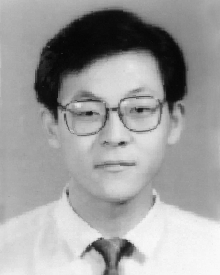 Jian-Lai Zhou合作论文数10
 Ron Ho合作论文数Meta10
 Ashok V. Krishnamoorthy合作论文数Axalume Inc.9
 Elad Alon合作论文数Department of Electrical Engineering and Computer Sciences, University of California, Berkeley;Berkeley Wireless Research Center, University of California, Berkeley9Philip Amberg合作论文数9
 Zhuowen Tu合作论文数Department of Computer Science and Engineering, Jacobs School of Engineering, University of California, San Diego;Department of Cognitive Science, University of California, San Diego;Amazon9Jon Lexau合作论文数9
 Xuezhe Zheng合作论文数Oracle Labs9Ivan Shubin合作论文数8Yu Shi合作论文数7
 John Cunningham合作论文数School of Electronic and Electrical Engineering, Faculty of Engineering and Physical Sciences, University of Leeds7
 Hiren Thacker合作论文数Oracle Labs7
 Borivoje Nikolic合作论文数Department of Electrical Engineering and Computer Sciences, University of California at Berkeley;Berkeley Wireless Research Center, University of California at Berkeley7
 Jin Yao合作论文数oracle corporation6
 Kannan Raj合作论文数6Xingyu Fan合作论文数5tian bai合作论文数5michael tsang合作论文数5Hu Peng合作论文数5Yipei Wang合作论文数State Key Laboratory of Software Development Environment and Key Laboratory of Biomechanics and Mechanobiology of Ministry of Education and Research Institute of Beihang University in Shenzhen, Beihang University, Beijing 100191, China5
 Frank Seide合作论文数Facebook AI5
 Shengyu Zhao合作论文数Pony.ai5
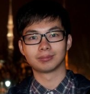 Jiahua Liu合作论文数Naturali5
 Yu Zheng (郑宇)合作论文数JD.COM;Zhiyuan College, Shanghai Jiao Tong University5
 Zhipeng Jia合作论文数5
 Jun-Yan Zhu合作论文数Robotics Institute, School of Computer Science, Carnegie Mellon University5Ying Luo合作论文数5
 Richard Lippmann合作论文数Lincoln Laboratory, Massachusetts Institute of Technology5Raman Chandrasekar合作论文数Microsoft Research5Jin-Hyoung Lee合作论文数4jackson hsieh合作论文数4Joseph Wu合作论文数4jichen wu合作论文数4bruce chen合作论文数4
 Zhongkai Wang合作论文数Univ Calif Berkeley, Berkeley, CA 94720 USA4
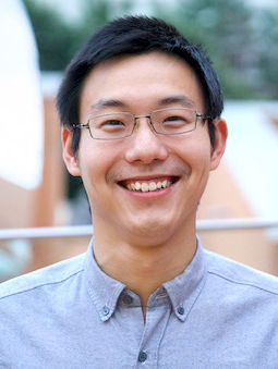 Jiajun Wu (吴佳俊)合作论文数Computer Science Department, Stanford University4Lin Shiyun合作论文数4
 Guoliang Li合作论文数Oracle Labs4
 Tao Chen合作论文数Department of Chemical and Process Engineering, School of Chemistry and Chemical Engineering, University of Surrey4
 Jae-Duk Han合作论文数Sch Elect Elect Engn, Yonsei Univ4
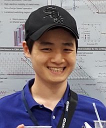 Woorham Bae合作论文数4
 Stevan S. Djordjevic合作论文数Department of Electrical and Computer Engineering;University of California , Davis;Department of Electrical and Computer Engineering, University of California , Davis4




