BACKGROUND:Clinical application of mesenchymal stromal cells (MSCs) for cartilage regeneration has been hampered by their poor retention of chondrogenic potential during in vitro culture, which has led to highly variable cartilage repair outcomes. Consequently, there is an urgent need for a reliable assay to predict MSC chondrogenic potential during cell manufacturing. METHODS:In this study, we developed a nondestructive MSCs iron flux monitoring methodology using spent culture media, utilizing micromagnetic resonance relaxometry (µMRR). We demonstrated that the dynamics of iron uptake and release by MSCs in culture can be reliably inferred from iron concentration changes in culture medium, with an unprecedented temporal resolution (<1 hour). RESULTS:Analysis of 3 MSC donors across 6 independent culture batches revealed a significant correlation between iron homeostasis (iron flux during culture) and chondrogenic differentiation outcomes, while a departure from iron homeostasis (significant iron uptake and accumulation) was correlated with impaired chondrogenesis. By contrast, cell proliferation, although essential for manufacturing to achieve sufficient cell numbers, did not reliably correlate with chondrogenic capacity. Furthermore, ascorbic acid supplementation during culture, which is known to promote MSCs proliferation and chondrogenic quality, regulated iron homeostasis by limiting iron flux. CONCLUSION:Our findings identify iron homeostasis as a potential chondrogenic-associated critical quality attribute of MSCs. This rapid, nondestructive monitoring strategy offers a promising approach to improve manufacturing efficiency and consistency by providing real-time insight into cellular iron flux. In addition, the methodology here impacts broader iron biology by providing a time-resolved iron flux measurement that is not currently available.
Early detection of harmful algal blooms remains challenging due to the low abundance of target microalgae and the limited sensitivity of conventional detection methods. Here, we present a high-throughput microfluidic cell concentrator that enables rapid and efficient pre-concentration of microalgae from large sample volumes. The system integrates a multiplexed inertial microfluidic device with a recirculation system, allowing repeated concentration cycles without manual sample transfer. Using the concentration system, we demonstrated the concentration of suspensions of a normal microalga (C. vulgaris) and a harmful cyanobacterium (M. aeruginosa). Starting from an initial density of 1 × 10⁵ cells/mL and a volume of 1 L, three concentration cycles increased the cell density to the 107 cells/mL range within 90 min. The increased cell density resulted in more than a 100-fold enhancement in fluorescence signal measured using a plate reader. Excitation-emission matrix analysis further demonstrated that pre-concentration significantly improved species-specific fluorescence signatures. These results indicate that the proposed microfluidic concentrator enables rapid, scalable, and effective enrichment of microalgae and offers a promising platform for early detection and monitoring of harmful algal blooms.
Lentiviral vectors (LVVs) are widely used as gene-delivery vehicles in cell and gene therapy, enabling stable integration of transgenes into target cells. However, efficient clarification of LVV harvest remains a major bottleneck in large-scale manufacturing, as conventional depth filtration often results in significant product loss. Here, we introduce a fouling-free microfluidic cell clarification (MCC) system that enables high-efficiency LVV recovery. By precisely controlling the harvest flow rate and adjusting the recirculated cell density by adding buffer, we established a quantitative relationship between clarification efficiency and throughput. Under optimized conditions, the MCC achieved > 95% product recovery while maintaining > 95% cell removal efficiency. The MCC process closely followed the theoretical recovery model assuming negligible product loss within the device. Across three independent batch operations, apparent average LVV recovery remained substantially higher than that of a benchmark process employing a cellulose depth filter-based process. Scalability tests demonstrated consistent performance at flow rates up to 1.5 L/h without loss of clarification efficiency, demonstrating its potential for large-scale biomanufacturing. Beyond LVVs, the MCC platform is broadly applicable to cell-based modalities that secrete therapeutic products, offering a continuous, low-shear, and fouling-free alternative to conventional filtration processes.
The rapid increase in global plastic consumption has intensified microplastic contamination, raising concerns regarding ecological and human health impacts. Conventional purification methods, such as membrane filtration and coagulation-flocculation, face limitations including fouling, clogging, and bulky instrumentation, restricting their suitability for compact or point-of-use (POU) systems. Here, we present a fully automated benchtop microplastic removal system integrating a compact spiral microchannel module with an air-driven self-cleaning mechanism, designed to produce microplastic-free water without manual intervention. The platform employs a diaphragm pump, a non-contact water level sensor, and an Arduino-based controller to autonomously alternate between purification and air-flushing modes, maintaining stable focusing and preventing particle accumulation. Using 20-30 mu m polystyrene microbeads, the device achieved continuous membrane-free purification with a purified-water collection rate of similar to 100 mL/min and an average removal efficiency of 97.6%. High-speed imaging confirmed reliable clog-free operation, and a techno-economic analysis showed clear cost and sustainability advantages over conventional POU membrane units. These results highlight the potential of the proposed benchtop platform as a compact, energy-efficient, and maintenance-free microplastic purification solution for practical POU water purification.
Scalable production of cell therapy doses relies on inexpensive, efficient production of gene delivery vectors, such as lentiviral vectors, in HEK293 cell culture. Intensified perfusion processes improve the volumetric productivity of cell culture by continuously supplying nutrients, oxygen, and media to cells while removing harmful metabolites, thereby enabling higher producer cell densities. Membrane filter-based cell retention devices commonly used in perfusion bioprocessing can experience significant clogging and fouling over long-term processes, which leads to the undesired retention of lentiviral vectors in the filter matrix. In this work, we used spiral microfluidic technology as a cell retention device to continuously harvest lentiviral vectors and remove metabolic waste from HEK293 cells in a bioreactor running high cell density perfusion cultures. With the spiral microfluidic device, we performed four perfusion culture runs with maximum cell densities between 15 × 106 and 25 × 106 cells per mL, achieving up to seven days of continuous lentiviral vector production and lossless harvesting with maximum, unconcentrated, functional titers on the order of 108 transducing units (TU) per mL. These production titers are competitive with other bioprocessing approaches in industry and academia. The highest cell-specific productivity (over 50 TU per cell per day) and cell-specific yields of our study (over 80 TU per cell) were achieved when using spiral device-mediated perfusion bioprocessing to cultivate cells and then transferring the cells to a shake flask environment with daily media replacement to generate lentiviral vectors.
The clinical adoption of emerging cell and gene therapies relies on the efficient upstream bioprocessing and high-recovery harvesting of viral vectors. AAV purification from cell culture begins with the removal of cells, through a process called cell clarification, which is typically performed with a membrane filter. Cells and debris accumulate on the filter matrix during clarification, resulting in filter clogging and significant product loss. Here, we implement spiral inertial microfluidic technology as an alternative, membraneless clarification strategy to separate AAVs from the host cells. We assemble a parallelized plastic spiral microfluidic system that divides an input flow rate of cell culture across 100 spirals to harvest clarified material at 20 mL min-1. The microfluidic clarification platform achieves a viral vector recovery of 85%, with the additional 15% of recoverable AAVs remaining in the system. However, up to 20% of cells were also found in the harvest, signifying that cell removal by inertial clarification is incomplete. We incorporate spiral microfluidics with the 3M Harvest RC filter (0.2 μm) (3M, USA) into a two-step harvest process, where spiral microfluidic clarification is used first to remove most of the cells from the culture. Performing spiral microfluidic clarification before depth filtration improves the filter throughput by 56.25%, from 44.8 L m-2 to 70 L m-2 and leads to a two-step AAV recovery of ∼74%, exceeding the 40 to 70% clarification recoveries generally reported. Our results support that further optimization and scale-out of the spiral inertial microfluidic system could increase the overall clarification recovery of AAV production processes.
Nuclear magnetic resonance relaxometry measurements have a diverse array of applications in medicine, biology, and chemistry, and they are valuable for monitoring cell cultures, diagnosing diseases, and detecting defects in pharmaceutical products. However, relaxometry workflows typically require laborious procedures and large sample volumes, which are both slow and destructive. This makes it difficult to use relaxometry in applications such as drug screening and cell culture development, where it has significant potential to be useful. In this work, we describe and demonstrate a low-cost benchtop system that can perform relaxometry measurements on 16 wells of a 96-well culture plate in one shot, enabling rapid, nondestructive scanning of multiple groups without any sample preparation. We demonstrate the application of this multiwell scanner for iron measurement and parallel measurements.
BACKGROUND:The zonal organization of articular cartilage is critical for the biphasic mechanical properties of the tissue. Current treatments for articular cartilage have yet to regenerate this zonal architecture, compromising the functional efficacy of the repaired tissue, which could account for tissue failure in the long term. Autologous chondrocyte implantation (ACI) still suffers from inconsistent efficacy and a long recovery period stemming from implantation of a heterogeneous chondrocyte mixture. HYPOTHESIS:Stratified implantation of zonal chondrocytes would facilitate the recapitulation of articular cartilage zonal properties and improve the repair efficacy of ACI treatment. STUDY DESIGN:Controlled laboratory study. METHODS:Autologous chondrocytes extracted from porcine articular cartilage were subjected to dynamic microcarrier expansion followed by size-based segregation using a spiral microfluidic device for the enrichment of zonal chondrocytes. Zonal chondrocytes were implanted into a chondral defect as a bilayered hydrogel construct consisting of superficial zone chondrocytes overlaying middle/deep zone chondrocytes (n = 6). Twelve months after implantation, the repair efficacy was compared against implantation of full-thickness cartilage-derived heterogeneous chondrocytes expanded on tissue culture plates (n = 5) or microcarriers (n = 6). RESULTS:Quantitative assessment of the repair tissues, including gross morphology, histological analysis, micro-computed tomography (micro-CT), compression modulus, and surface lubrication analysis, at 12 months demonstrated statistically significant improvement in cartilage and subchondral bone repair with zonal chondrocyte bilayered implantation. Magnetic resonance imaging (MRI) T2 mapping indicated progressive improvement in graft maturation as early as 3 months, reaching normalcy at 9 months. CONCLUSION:This study demonstrates that with appropriate expansion and isolation of zonal chondrocytes, stratified zonal chondrocyte implantation is able to facilitate restoration of articular cartilage zonal architecture and significantly enhance the functional repair as compared with current ACI treatment. CLINICAL RELEVANCE:With appropriate expansion and enrichment of zonal chondrocytes, stratified zonal chondrocyte implantation could represent a significant advancement over current ACI-based cartilage repair, with the potential to support quicker and better recovery.
Conventional iron determination methods, such as colorimetric assays, often lack sensitivity at low iron levels and are susceptible to interference from complex compositions in serum and plasma. In this study, we developed a rapid, sensitive, and accurate method for iron determination in 1 min utilizing an inexpensive benchtop 0.5-T micromagnetic resonance relaxometry (μMRR) system with acidification treatment of samples (pH < 1). The method yielded a highly linear calibration curve (R2 > 0.999) between the transverse relaxation rate R2 and iron concentration from 0.5 to 1000 μM, with a limit of detection (LOD) of 0.25 μM and a minimal assay volume of 5 μL. The accuracy of this method was validated by inductively coupled plasma mass spectrometry (ICP-MS) across a diverse range of biological samples. This μMRR-based approach offers a rapid and convenient alternative for serum and plasma iron measurements, which can substantially reduce clinical diagnostic time and support real-time iron monitoring for patients.
Recent regulatory guidance now encourages the use of sequencing as an alternative adventitious agent testing assay to lengthy compendial in vivo assays used for cell line qualification. Most short-read sequencing assays, however, still require over a week to obtain a final test result since the sequencing must be completed before bioinformatic analysis can begin, which is still too long for some cell and gene therapy products that must be released as soon as possible to reach critically ill patients. Oxford Nanopore sequencing can address these issues, as it provides real-time basecalling and sequence alignment, which can reduce the overall assay time. Still, as with any sequencing platform, the abundance of background nucleic acid from the human or mammalian host can mask the signal from a low-level viral contaminant. To address this, we have developed a sensitive sample preparation workflow using concentration, nuclease treatment, and agnostic PCR methods to eliminate background signals and amplify viral contaminant reads, leading to a 3-log improvement in the limit of detection that is comparable to or better than short-read sequencing approaches. This approach will lead to more rapid and improved detection of viral contaminants in cell and gene therapy manufacturing.
Understanding the biophysical properties of cells is essential for biological research, diagnostics, and therapeutics. Microfluidics enhances biophysical cell characterization by enabling precise manipulation and real-time measurement at the microscale. However, the high-throughput nature of microfluidic systems generates vast amounts of data, complicating analysis. Integrating artificial intelligence (AI) methods, including machine learning and deep learning, with microfluidic technologies addresses these challenges. AI excels at analyzing large, complex datasets, improving the accuracy and efficiency of microfluidic experiments and facilitating new biological discoveries. This review examines the synergy between microfluidics and machine learning for biophysical cell characterization, categorizing existing methods based on the types of input data used for machine learning analysis, highlighting recent advancements, and discussing challenges and future directions in this interdisciplinary field.
Live microbial contamination poses high risks to cell and gene therapies, threatening manufacturing processes and patient safety. Rapid, sensitive detection of live microbes in complex environments, such as CAR-T cell cultures, remains an urgent need. Here, an innovative sample-to-result workflow is introduced using digital loop-mediated isothermal amplification (dLAMP), enhanced by Electrostatic Microfiltration (EM)-based enrichment, for rapid sterility testing. By rationally designing primers targeting 16S and 18S rRNA, dLAMP assay enables both universal detection (covering >80% of known species) and strain-specific identification of bacterial and fungal contaminants in CAR-T cell spent medium and final products, directly from microorganism lysates. Enhanced by EM-based enrichment of low-abundance live microbes, the workflow achieves unparalleled sensitivity and speed, detecting contamination levels as low as 1 CFU/mL in complex CAR-T cell cultures within 6 h. Compared to qPCR and 14-day compendial methods, the approach demonstrates superior accuracy and significantly faster turnaround times. This workflow holds transformative potential for real-time monitoring in cell therapy manufacturing and rapid safety assessments of CAR-T cell products prior to patient infusion. Beyond cell therapy, the method is broadly applicable to infectious disease diagnostics, biomanufacturing monitoring, food safety, and environmental surveillance.
Nuclear magnetic resonance (NMR) measurements of moving samples are limited by the depletion of the sample during long experiments, making it difficult to apply NMR relaxometry techniques to moving fluid samples for various manufacturing and reaction monitoring applications. Traditional techniques for compensating for flow-induced artificial relaxation in postprocessing do not work for samples with unknown or variable flow rates, which is a significant barrier to the widespread adoption of NMR monitoring systems for existing processes. In this work, we present a compact NMR coil geometry that removes the effect of sample depletion during long experiments on moving samples, making it possible to perform flow-agnostic measurements on samples with variable or uncontrolled flow rates. We evaluate this coil geometry using relaxometry experiments on fast-flowing water samples and demonstrate its potential for process monitoring with real-time measurements of the dairy concentration.
The rapid increase in plastic consumption has accelerated microplastic pollution, raising concerns about potential ecological and health risks. Despite the development and application of various microplastic removal technologies, they exhibit inherent limitations, such as membrane fouling/clogging and low removal efficiency. In this study, we introduce a high-throughput membrane-free microplastic removal system utilizing a plastic spiral inertial microfluidic device. The continuous and clogging-free operational capabilities of spiral inertial microfluidics, coupled with a robust scaling-up of a mass-producible plastic device, allow us to overcome the limitations of conventional microplastic removal methods while meeting the throughput requirements for practical water treatment applications. Utilizing a multiplexed plastic spiral unit, we successfully demonstrated 10-liter-scale high-throughput microplastic removal with a high microparticle removal efficiency (up to 99%, depending on particle size) at a harvesting rate of purified water of 125 mL/min (not limited and can be further increased by utilizing multiple units in parallel) without any fouling/clogging issue.
Acute myocardial infarction (AMI), a leading cause of death globally, triggers complex inflammatory responses critical to patient outcomes. However, rapid tools for profiling immune responses at the single-cell level are lacking. The integrated Single-cell Enzyme and Antigen Quantification (iSEAQ) system addresses this gap by enabling high-throughput, single-cell analysis of immune cell activity using just 20 μL of blood. This novel tool processes live CD66b and CD3 cells to quantify the secretion of Granzyme B, Neutrophil Elastase, and CD31 within minutes. Longitudinal studies on nine AMI patients revealed that CD66b+ cells are major contributors (up to 95%) to key inflammatory enzymes, including the unexpected secretion of Granzyme B. iSEAQ achieves unparalleled sensitivity (0.4 fg/cell) and predictive accuracy (>90%) for patient profiling across AMI onset, treatment, and discharge. This innovation provides clinicians with a rapid, precise method to monitor immune responses, unveiling new insights into AMI inflammation and therapy.
Chimeric Antigen Receptor (CAR) T cell therapy is a pivotal treatment for hematological malignancies. However, CAR T cell products exhibit batch-to-batch variability in cell number, quality, and in vivo efficacy due to donor-to-donor heterogeneity, and pre/post-manufacturing processes, and the manufacturing of such products necessitates careful testing, both post-manufacturing and pre-infusion. Here, we introduce the Cell Trajectory Modulation (CTM) assay, a microfluidic, label-free approach for the rapid evaluation of the functional attributes of CAR T cells based on biophysical features (i.e., size, deformability). CTM assay correlates with phenotypic metrics, including CD4:CD8 ratio, memory subtypes, and cytotoxic activity. Validated across multiple donors and culture platforms, the CTM assay requires fewer than 10,000 cells and delivers results within 10 minutes. Compared to labeled flow cytometry processing, the CTM assay offers real-time data to guide adaptive manufacturing workflows. Thus, the CTM assay offers an improvement over existing phenotypic assessments, marking a step forward in advancing CAR T cell therapy manufacturing.
BACKGROUND:There is a current, absence of reliable, blood-sparing, diagnostic tools to measure and trend real-time changes in the levels of inflammation and its effects on the immune cells in the infant. METHODS:We deployed the BiophysicaL Immune Profiling for Infants (BLIPI) system in the neonatal intensive care unit to describe immune cell biophysical profiles using 50 microliters of blood per sample from term and preterm infants. RESULTS:A total of 19 infants (8 term, 11 preterm) were recruited and 24 blood samples were collected in their first month. Based on the profiles of immune cells' size and deformation, there was a clear distinction between term and preterm infants, with 48/50 markers significantly different. A preterm infant with late-onset bacterial sepsis had notable size and deformability differences compared to the rest of the preterm cohort. There was a significant correlation between immune cell biophysical profiles and clinical markers such as C-reactive protein, white blood cell counts, and immature-to-total neutrophil (I:T) ratios, with Pearson correlation coefficients for linear regression models of 0.98, 0.97 and 0.94 respectively. CONCLUSION:This study highlights the potential for the biophysical immune cell profiling system to provide an overview of the infant's current immune activation and response. IMPACT:We present a novel, minimally invasive diagnostic system that leverages the physical properties of immune cells to provide a rapid and direct assessment of the immune status, requiring 20 times less blood volume than standard tests. This study demonstrates the potential of a compact, deployable system that is capable of performing biophysical profiling to assess immune cell activation in term and preterm infants, by revealing distinct differences in cell size and deformation between groups. The system's sensitive, quantitative measures were correlated with routine clinical biomarkers, highlighting its ability to provide a rapid, minimally invasive, real-time monitoring of neonatal immune status.
Monitoring immune cell function is increasingly being recognized as a more relevant biomarker than traditional white blood cell counts, yet the need for repeated relatively large blood volumes still continues to pose a significant challenge. To overcome this, we developed a sample-sparing platform using inertial microfluidics that can process as little as 10 μL of blood to isolate leukocytes for downstream functional analysis. Our platform isolates leukocytes with ∼80% purity, >90% in-device recovery, and >95% viability. Neutrophils were our primary focus due to their sensitivity to external stimuli and their critical role in immune responses. Neutrophils isolated through our new method did not show inadvertent activation, as evidenced by unchanged expression of activation markers CD62L and CD11b, with phenotypes comparable to control cells in whole blood. We conducted a range of functional assays, including phagocytosis, ROS production, and NETosis with all tests confirming that neutrophils maintained their functionality after isolation. These assays were performed using standard laboratory workflows, demonstrating the platform's compatibility with techniques such as flow cytometry and cell culture assays. Furthermore, we showed the versatility of our platform by successfully isolating leukocytes from challenging samples, including mouse blood from the vena cava or tail vein, as well as human capillary blood obtained by fingerstick. This adaptability highlights the potential of this platform for clinical and research applications, particularly in frequent immune monitoring or cases where sample volume is limited.
Rapid and sensitive detection of pathogens in various samples is crucial for disease diagnosis, environmental surveillance, as well as food and water safety monitoring. However, the low abundance of pathogens (<10 CFU) in large volume (1 mL-1 L) samples containing vast backgrounds critically limits the sensitivity of even the most advanced techniques, such as digital PCR. Therefore, there is a critical need for sample preparation that can enrich low-abundance pathogens from complex and large-volume samples. This study develops an efficient electrostatic microfiltration (EM)-based sample preparation technique capable of processing ultra-large-volume (>= 500 mL) samples at high throughput (>= 10 mL min(-1)). This approach achieves a significant enrichment (>8000x) of extremely-low-abundance pathogens (down to level of 0.02 CFU mL(-1), i.e., 10 CFU in 500 mL). Furthermore, EM-enabled sample preparation facilitates digital amplification techniques sensitively detecting broad pathogens, including bacteria, fungi, and viruses from various samples, in a rapid (<= 3 h) sample-to-result workflow. Notably, the operational ease, portability, and compatibility/integrability with various downstream detection platforms highlight its great potential for widespread applications across diverse settings.
BACKGROUND:The functional heterogeneity of culture-expanded mesenchymal stem cells (MSCs) has hindered the clinical application of MSCs. Previous studies have shown that MSC subpopulations with superior chondrogenic capacity can be isolated using a spiral microfluidic device based on the principle of inertial cell focusing.HYPOTHESIS:The delivery of microfluidic-enriched chondrogenic MSCs that are consistent in size and function will overcome the challenge of the functional heterogeneity of expanded MSCs and will significantly improve MSC-based cartilage repair.STUDY DESIGN:Controlled laboratory study.METHODS:A next-generation, fully automated multidimensional double spiral microfluidic device was designed to provide more refined and efficient isolation of MSC subpopulations based on size. Analysis of in vitro chondrogenic potential and RNA sequencing was performed on size-sorted MSC subpopulations. In vivo cartilage repair efficacy was demonstrated in an osteochondral injury model in 12-week-old rats. Defects were implanted with MSC subpopulations (n = 6 per group) and compared with those implanted with unsegregated MSCs (n = 6). Osteochondral repair was assessed at 6 and 12 weeks after surgery by histological, micro-computed tomography, and mechanical analysis.RESULTS:A chondrogenic MSC subpopulation was efficiently isolated using the multidimensional double spiral device. RNA sequencing revealed distinct transcriptomic profiles and identified differential gene expression between subpopulations. The delivery of a chondrogenic MSC subpopulation resulted in improved cartilage repair, as indicated by histological scoring, the compression modulus, and micro-computed tomography of the subchondral bone.CONCLUSION:We have established a rapid, label-free, and reliable microfluidic protocol for more efficient size-based enrichment of a chondrogenic MSC subpopulation. Our proof-of-concept in vivo study demonstrates the enhanced cartilage repair efficacy of these enriched chondrogenic MSCs.CLINICAL RELEVANCE:The delivery of microfluidic-enriched chondrogenic MSCs that are consistent in size and function can overcome the challenge of the functional heterogeneity of expanded MSCs, resulting in significant improvement in MSC-based cartilage repair. The availability of such rapid, label-free enriched chondrogenic MSCs can enable better cell therapy products for cartilage repair with improved treatment outcomes.
 Chwee Teck Lim合作论文数Mechanobiology Institute, National University of Singapore;Department of Biomedical Engineering, College of Design and Engineering, National University of Singapore45
 Sung Jae Kim合作论文数College of Medicine, Yonsei University34
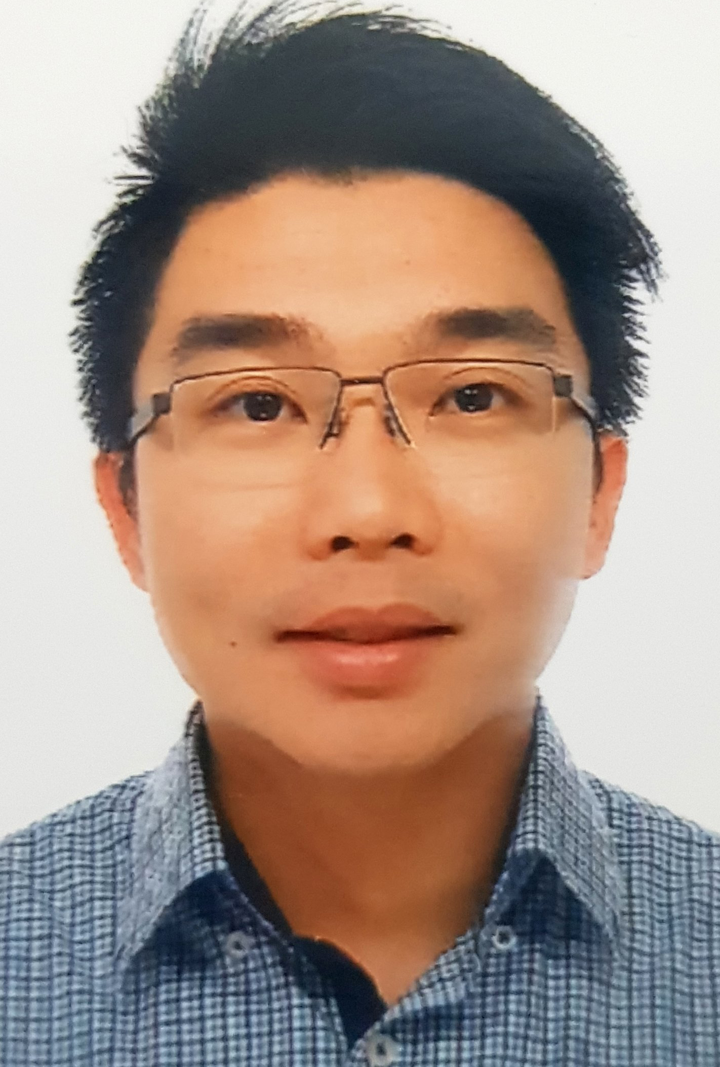 Han Wei Hou合作论文数School of Mechanical and Aerospace Engineering, College of Engineering, Nanyang Technological University28
 Yong-Ak Song合作论文数CAD/CAM Center, Korea Institute of Science and Technology28
 Ali Asgar S. Bhagat合作论文数Department of Electrical and Computer Engineering, University of Cincinnati24
 Bee Luan Khoo (邱美孌)合作论文数Department of Biomedical Engineering, City University of Hong Kong;Hong Kong Centre for Cerebro-Cardiovascular Health Engineering;CityU Shenzhen-Futian Research Institute22
 Zirui Li (李子瑞)合作论文数School of Mechanical Engineering, Hebei University of Technology21
 Sung Hee Ko合作论文数20
 Jeon Hyungkook合作论文数Department of Mechanical Engineering, Pohang University of Science and Technology (POSTECH),19
 Majid E. Warkiani合作论文数School of Biomedical Engineering, University of Technology Sydney19
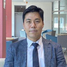 Kim Bumjoo合作论文数18Rhokyun Kwak合作论文数Sch Mech & Aerosp Engn, Seoul Natl Univ18
 Aniruddh Sarkar合作论文数Wallace H. Coulter Department of Biomedical Engineering, College of Engineering, Georgia Institute of Technology17
 Taehong Kwon合作论文数Dept Elect Engn & Comp Sci, MIT16
 Wei Ouyang合作论文数Department of Electrical Engineering and Computer Science,MIT16
 Jianping Fu合作论文数Department of Biomedical Engineering, College of Engineering, University of Michigan-Ann Arbor15Cheow Lih Feng合作论文数Department of Biomedical Engineering, National University of Singapore14Wu Lidan合作论文数14
 Pham Van Sang合作论文数Singapore-MIT Alliance, National University of Singapore14
 Mao Pan (潘懋)合作论文数School of Earth and Space Sciences, Peking University14
 Sha Huang (黄沙)合作论文数Chinese PLA General Hospital14
 Weng Kung Peng合作论文数Graduate School of Engineering Science, Osaka University14
 Guofeng Guan合作论文数Shenzhen Genflow Technol Co Ltd12
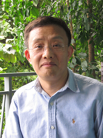 Jian-Sheng Wang合作论文数Department of Physics, Faculty of Science, National University of Singapore12
 Kwon Hyukjin J合作论文数12
 Douglas A. Lauffenburger合作论文数Department of Chemical Engineering, School of Engineering, Massachusetts Institute of Technology;Department of Biology, Massachusetts Institute of Technology12
 Ying-Chih Wang合作论文数Focus On Therapeutic Outcomes, Inc.12
 Ye Ai合作论文数11
 Yang Zheng合作论文数11
 Jeong Hoon Lee合作论文数Department of Electrical Engineering, School of Electronics and Information Technology, Kwangwoon University11
 Hugh I. Kim合作论文数Department of Chemistry, College of Science, Korea University11
 Geunbae Lim合作论文数11
 Hansen Bow合作论文数Department of Electrical Engineering and Computer Science, Massachusetts Institute of Technology11Leon D Li合作论文数10Zeming Kerwin Kwek合作论文数Faculty of Engineering, National University of Singapore10
 Gui-Rong Liu (刘桂荣)合作论文数Department of Aerospace Engineering and Engineering Mechanics, University of Cincinnati10
 Yuzong Chen (陈宇综)合作论文数Institute of Biomedical Health Technology and Engineering, Shenzhen Bay Laboratory;Shenzhen International Graduate School, Tsinghua University10
 Harold G. Craighead合作论文数School of Applied and Engineering Physics, Cornell University10
 Kyungyong Choi合作论文数Dept Biol Engn, MIT9
 David J. Collins合作论文数Department of Biomedical Engineering, The University of Melbourne9
 Hyunryul Ryu合作论文数Seoul National University, Seoul National University9
 Yehua Gan (甘业华)合作论文数Hospital of Stomatology, Peking University9
 Yoon Junghyo合作论文数School of Mechanical Engineering, Korea University9
 Chia-Hung Chen (陳家宏)合作论文数Department of Biomedical Engineering, City University of Hong Kong9monica diez silva合作论文数8Ching Ann Tee合作论文数8
 Eng Hin Lee合作论文数Yong Loo Lin School of Medicine, National University of Singapore8
 Tae Su Choi合作论文数Department of Chemistry, Pohang University of Science and Technology (POSTECH),8
 Peter Preiser合作论文数School of Biological Sciences, Nanyang Technological University8




