Introduction La bronchopneumopathie chronique obstructive (BPCO) est une maladie pulmonaire induite par la fumée de cigarette et caractérisée par un trouble ventilatoire obstructif irréversible. Cependant, un tiers des patients atteints de BPCO meurent d’un cancer pulmonaire, sans que les mécanismes conduisant au développement de ce cancer chez ces patients ne soient complétement élucidés. L’inflammation chronique, commune à ces deux pathologies est entretenue par la présence de cellules sénescentes. Ces cellules sont caractérisées par un arrêt de prolifération cellulaire associé à la synthèse de médiateurs inflammatoires qui participent au développement des tumeurs. Le rôle des fibroblastes sénescents (issus de tissus non cancéreux) est encore débattu, mais active la progression tumorale via leur sécrétome et notamment les phospholipases (sPLA2). Nous avons montré dans une étude précédente que les fibroblastes de patients BPCO exprimaient la sPLA2 XIIA, mais son rôle dans le processus cancéreux est inconnu. Le but de ce travail est de déterminer si cette protéine pourrait participer à la progression tumorale. Méthodes Les fibroblastes proviennent d’explants de patients fumeurs (TF, n=10) et de patients avec BPCO modérée (BPCO, n=11). Les groupes sont comparables en âge et pour la consommation tabagique. Deux lignées cellulaires cancéreuses (A549 et NCIH1563) ont été traitées soit avec du milieu conditionné (MCD) issus de ces fibroblastes déprivés ou non en sPLA2 XIIA soit avec différentes doses de sPLA2 XIIA recombinante. L’effet prolifératif a été déterminé par un dosage MTT et un marquage au Ki67. La migration a été évaluée par test sur blessure. Les voies de signalisation ont été analysées par western blot, et en utilisant différents inhibiteurs pharmacologiques. Les effets de la sPLA2 XIIA ont été étudiés dans des organoïdes. Résultats Les lignées cancéreuses expriment les récepteurs PLA2R1 et les protéoglycannes à héparanes sulfates (PHS). L’exposition des lignées à des MCDs issus des fibroblastes sénescents provenant de fumeurs BPCO induit une prolifération plus marquée que pour les MCDs de fibroblastes de TF. Une diminution de la prolifération des cellules cancéreuses a été observée après déplétion des MCD en sPLA2 XIIA confirmant le rôle de cette protéine, L’exposition des lignées cancéreuses à des concentrations de sPLA2 XIIA identiques à celles détectées dans les MCDs induit la prolifération, la migration des deux lignées cellulaires avec des effets plus marqués chez la lignée NCIH1563. L’utilisation d’inhibiteurs des PHS et de la voie des MAPK bloque les effets induits par la sPLA XIIA. Conclusion La sPLA2 XIIA induit la progression tumorale chez les patients BPCO via PHS/MAPK et pourrait constituer une cible thérapeutique.
Introduction Ces dernières années, de nouvelles options thérapeutiques ont été étudiées en périopératoire dans le cancer bronchique non à petites cellules (CBNPC). L’étude rétrospective FALCONS vise à améliorer la compréhension des caractéristiques et du parcours de soins des patients atteints de CBNPC, de stade précoce opéré en complétant les données issues des essais cliniques avec des données de vie réelle. FALCONS s’appuie sur le registre national français EPITHOR, qui collecte des données épidémiologiques et cliniques sur les chirurgies thoraciques. Méthodes FALCONS analyse les données de patients ayant bénéficiés d’une chirurgie thoracique entre 2016 et 2018 (cohorte A, n=9429, 27 sites), ce qui inclut les bénéficiaires d’un traitement néoadjuvant (cohorte A1, n=758) et d’un traitement adjuvant (cohorte A2, n=993). Les caractéristiques à l’inclusion, la prise en charge thérapeutique et la survie globale (SG) ont été analysées dans chaque cohorte avec un suivi minimal de 5 ans. Une collecte de données à partir des dossiers patients complètera prochainement les analyses (cohorte B, n=517, 11 sites). Résultats La répartition des patients par sexe et par habitudes tabagiques était similaire dans les trois cohortes. Les patients des cohortes A1 et A2 étaient plus jeunes que ceux de la cohorte A. Dans la cohorte néoadjuvante A1, il y avait moins de patients présentant un score ECOG PS 0 (A1 : 42 % ; A2 : 53 % ; À : 49 %), plus de patients présentant un score de dyspnée de 0 (A1 : 63 % ; A2 : 59 % ; À : 58 %), plus de tumeurs épidermoïdes (A1 : 31 % ; A2 : 26 % ; À : 24 %), plus de pneumonectomies (A1 : 16 % ; A2 : 11 % ; À : 6 %), moins de lobectomie (A1 : 68 % ; A2 : 74 % ; À : 72 %), moins de segmentectomie (A1 : 4 % ; A2 : 6 % ; À : 11 %) et moins de patients atteints de comorbidités (A1 : 86 % ; A2 : 92 % ; À : 90 %). Dans la cohorte A1, les stades tumoraux TNM III-IV étaient plus fréquents avant chirurgie (A1 : 67 % ; A2 : 32 % ; À : 19 %) qu’après chirurgie (A1 : 46 % ; A2 : 48 % ; À : 24 %). Au 31 décembre 2023, les taux de SG à 5 ans étaient estimés à 53 % dans la cohorte A1, 52 % dans la cohorte A2 et 65 % dans la cohorte A. La SG médiane était respectivement de 65,6 mois (IC95 % : 58,2–77,6), 64,8 mois (IC95 % : 58,9–70,6) et non atteinte. Conclusion Cette étude nationale menée en vie réelle confirme une amélioration significative récente des résultats en termes de survie, ce qui reflète probablement la mise en œuvre progressive en France, de l’obligation réglementaire de décisions thérapeutiques multidisciplinaires pour les CBNPC et l’amélioration continue de la qualité de la chirurgie. Malgré ces résultats encourageants, la mortalité reste élevée aux stades précoces, ce qui souligne la nécessité de nouveaux traitements pour le CBNPC aux stades opérables ; à l’instar des immunothérapies, récemment introduites dans un contexte périopératoire.
IntroductionEmphysema is a respiratory disease characterized by chronic alveolar destruction. Lipofibroblasts (LIF) play a key role in the stem cell niche surrounding alveolar type II (AT2) cells and may contribute to alveolar regeneration. We have previously shown that senescent cell elimination induces alveolar regeneration, increased LIF numbers and activation of the sterol regulatory binding protein (SREBP) and peroxisome proliferator-activated receptor gamma (PPARG) pathways [1]. However, it remains unclear whether the activation of these pathways can increase fibroblast stem cell niche properties and promote alveolar regeneration during emphysema.MethodsHuman lung tissue slides were obtained from patients with or without emphysema and immunofluorescent staining was performed to identify LIF (ADRP+Vimentin+). Both human primary fibroblasts and primary AT2 cells were isolated from lobectomies through the explant method and magnetic sorting (HT2-280+) respectively. Fibroblasts were treated with Rosiglitazone and T0901317 for 72hours. LIF phenotype acquisition was evaluated through IF staining, qPCR and lipidomic analysis. Stem cell niche properties were evaluated by performing alveolar organoid formation assay by co-culturing treated fibroblasts with H-441 cells or primary AT2 cells. Adult C57BL6 mice received intra-tracheal injection of either Elastase or PBS. From D21 mice were treated by intraperitoneal injections of Rosiglitazone (5μg/g/d), T0901317 (10μg/g/d) or vehicule, 5/7days. Lungs were collected at D90. Left lung was fixated for morphological analysis.ResultsOur study showed a decrease in LIF populations among patients with emphysema compared to controls. Furthermore, Rosiglitazone, a PPARG agonist, and T0901317, a SREBP agonist, can induce lipogenic differentiation in human lung fibroblasts. Activation of both pathways increased the expression of ADRP and the activation of the SREBP pathway induced the accumulation of neutral lipids in the fibroblasts. Using an organoid model of alveolar regeneration, we show that activating these pathways increases the stem cell niche properties of fibroblasts and enhances the number of organoids formed with either H441 cells or primary AT2. Lastly, in a murine mode of elastase-induced emphysema, we show that Rosiglitazone partially reverts emphysema.ConclusionActivation of PPARG and SREBP pathways promotes lipogenic differentiation of fibroblasts, enhances human alveolar organoid formation and partially reverts emphysema in vivo. These results provide insight into potential therapeutic strategies for promoting alveolar regeneration in patients with emphysema.
IntroductionLung alveoli are essential for gas exchanges between air and blood. Alveoli can be destroyed as in emphysema, a disease in which an accumulation of senescent cells has been observed. No curative treatment is yet able to create new alveoli and regenerative treatments described in mice failed to be transferred successfully to patients with emphysema. This emphasizes the need to develop intermediate steps between mouse models and clinical trials for drug development in emphysema. Our aim was to develop 3D alveolar cell culture that could be used to screen senotherapeutic strategies, first organoids derived from alveolar epithelial and mesenchymal cell lines (H441and MRC5) and then patient-derived alveolospheres from epithelial cells.MethodsH441 and MRC5 were cocultured in a drop of Matrigel in 24 trans-well and 96-well plates. AT1 (HT1-56) and AT2 (HT2-280) markers were assessed by immunofluorescence. The variability of organoid sizes and shapes were assessed over time to determine the optimal number of cells (MRC5 and H441). We first used 24 trans-well plates, and next miniaturized with 96-well plates. These seeding conditions were compared between day 8 and day 16 of culture. Several senescence inducers were tested to trigger reproducible senescence in H441: Doxorubicin (5nM to 50μM) and Etoposide (500nM to 100μM). Secondly, patient-derived alveolospheres were developed by isolating HT2-280 positive cells (AT2 cells) from human lung explants (Tata - Star protocol) and β-galactosidase was assessed to measure cell senescence.ResultsCulturing H441 in Matrigel led to the formation of spheroidal 3D structures expressing both AT1 and AT2 markers. Co-culturing H441 with MRC5 in Matrigel increased by 150% the organoid number compared to H441 alone but did not affect their size. In all culture conditions the organoid size increased between day 8 and day 16 of culture. Increasing the amount of H441 initially seeded with MRC5 increased the organoid number (230% between 625 and 1250 H441 and 770% between 625 and 5K H441) but does not influence their size. The best seeding condition for the 24 trans-well plate was 1250 H441 with 50K MRC5 and for the 96-well plate is 250 H441 with 2500 MRC5. The maximum non-toxic dose was 25μM for etoposide and 5μM for doxorubicin. Both treatments induced senescence in H441 cells. When the alveolospheres have been passed, the percentage of β-galactosidase positive organoids was 40% at first passage and 52% at second passage.ConclusionWe developed two senescent alveolar organoids assay that could be used to test senotherapeutic drugs.
Sickle Cell anemia (SCA) patients carry an increased risk of stroke and an important left atrial (LA) remodeling. Some healthcare data base analysis revealed an increased risk of arrhythmia in SCA but the reality of atrial arrhythmia (AA) has never been prospectively. The aim of this study was to identify predictors of atrial arrhythmia in SCA patients. From January 2019 to November 2022, consecutive adult patients with SCA referred to ambulatory cardiology department for possible cardiac involvement were prospectively included (Drepacoeur cohort). All patients that had 24-hours ECG monitoring (24h-holter), transthoracic echocardiography (TTE) and biology workup on the same day were analyzed in this study. The primary end point was the occurrence of AA, defined by the presence of excessive supraventricular ectopic activity (ESVEA) on 24h-holter (i.e. > 720 premature atrial contractions [PACs] or any run ≥ 20 PACs), or recent history of paroxysmal atrial fibrillation (AF). Patients in persistent AF were excluded from the analysis. Overall, 130 patients were included and 125 were analyzed (5 exclusions for persistent AF). Mean age was 44 ± 12 years (range 18–69), 48% were male. Heart function was mainly preserved with a mean left ventricular ejection fraction (LVEF) of 57.5 ± 5.9% and LA was dilated (56 ± 19 mL/m2). Atrial arrhythmia was observed in 28 (22%) patients (17 with more than 720 PACs/day, 7 with at least a run ≥ 20 PACs and 10 with recent history of paroxysmal AF). In univariate analysis, AA was associated with a multitude of clinical, echocardiographic and biological parameters (Figure 1). In multivariate analysis, age and LA volume were independently associated with AA (OR = 1,07 CI95% [1,01 ; 1,14], P = 0,02 and OR = 1,04 CI95% [1,01 ; 1,07], P = 0,03 respectively). They both were correlated with PAC load on 24h-holter (R = 0,55, P < 0,001 and R = 0,31, P < 0.001 respectively). Interestingly, an age over 46 years old or a LA volume > 53 mL/m2 could predict AA with a PPV of 32% and a NPV of 98%. Atrial arrythmia is common in adult SCA patients and is associated with ageing and LA remodeling. This study provides tools for early detection of AA in this highly vulnerable population.
IntroductionLe vieillissement vasculaire entraîne une raréfaction des microvaisseaux. Le mécanisme proposé est une insuffisance de signalisation du facteur de croissance endothélial vasculaire (VEGF) dont l’action est perturbée par une production accrue d’une forme soluble de son récepteur VEGFR1 (sVEGFR1) qui l’empêche de se fixer à son récepteur actif, le VEGFR1 exprimé par les CEs. Les CEs des microvaisseaux pulmonaires (PCEs) sont nécessaires à la fonction des poumons et nécessitent le signal de survie du VEGF, en l’absence duquel elles peuvent rentrer en sénescence, caractérisée par un arrêt de la division cellulaire et la libération de facteurs nocifs pour les poumons. Nous avons montré une accumulation de PCEs sénescentes au cours de l’hypertension (HTP) et de l’emphysème pulmonaires.MéthodeÉvaluer l’insuffisance de signalisation du VEGF dans la sénescence des PCEs liée à deux pathologies, l’hypertension (HTP) et l’emphysème pulmonaires.RésultatsLes poumons des souris âgées par rapport aux jeunes souris ont une densité capillaire plus faible se manifestant par une diminution du nombre de PCEs identifiées en immunohistochimie par marquage de l’ICAM1 ou de l’isolectine B4. Le traitement des souris par l’inhibiteur du récepteur du VEGF Sugen amplifie ces effets et augmente le nombre de PCEs sénescentes marquées p16, en diminuant le réseau capillaire pulmonaire, en détériorant l’hémodynamique pulmonaire et en induisant de l’emphysème. L’expression pulmonaire de sVEGFR1 chez les souris développant une HTP hypoxique est augmentée et aggravée par le Sugen, de même que l’expression pulmonaire de deux facteurs modulant l’épissage alternatif du VEGFR1 et la formation de sVEGFR1, le domaine Jumonji contenant la protéine 6 (Jmjd6) et le TNFSF15. Nous montrons in vitro qu’un traitement par le sVEGFR1 de PCEs en culture issues de patients (étude Costemcells) entraîne une augmentation de la sénescence cellulaire. Pour contrer la forte susceptibilité des PCEs à la sénescence dans l’HTP et l’emphysème, nous avons généré un nouveau modèle murin qui surexprime le VEGF selon un système Tet On inductible et spécifique au tissu. Ces souris sont en cours d’étude, croisées avec des souris exprimant la Cre-recombinase dans le foie (souris Alb-Cre), afin d’assurer un apport contrôlé de VEGF du foie vers les poumons.ConclusionL’augmentation du sVEGFR1 circulant au cours du vieillissement et des pathologies pulmonaires tels que l’HTP et l’emphysème pourrait être responsable de la sénescence des PCEs. Contrecarrer l’insuffisance de signalisation du VEGF devrait permettre de réduire la sévérité de ces pathologies.
Lipofibroblasts form a sub -population of fibroblasts located in the mesenchymal alveolar stem cell niche. They show close proximity with alveolar epithelial type 2 cells and play a key role in alveolar development and lung homeostasis. Their role in various diseases such as acute respiratory distress syndrome, pulmonary fibrosis and emphysema is progressively better understood. Through the activation of signaling pathways such as PPARg lipofibroblasts may help to induce endogenous alveolar regeneration. (c) 2024 Published by Elsevier Masson SAS on behalf of SPLF.
IntroductionAlveolar type 2 (AT2) to type 1 (AT1) transition is a key process in lung repair after injury. It involves transitional cell states such as activated AT2 and Krt8+ alveolar differentiation intermediate cells (ADI). The dysfunction of this transition impairs the repair process and is described in lung fibrosis. However, how AT2 to AT1 transition is affected in emphysema and the role of this dysfunction in this disease remains to be determined. We aimed to characterize the AT2 to AT1 transition in elastase-induced emphysema in mice and emphysematous patients, and to determine the impact of alveolar differentiation impairment on emphysema development.MethodsMurine emphysema is established in the elastase model 21days after elastase instillation (established emphysema) and persists at late time points (150days). We performed single cell RNAseq of EpCAM+ cells in established and late emphysema and clustered the different alveolar epithelial cells populations. We next assessed the functionality of the emphysematous epithelial cells compared to controls by analyzing their ability to form organoids obtained by co-culturing epithelial cells (EpCAM+ from elastase or control lung) with control lung fibroblasts. Single cell RNAseq was next performed on elastase and control organoids. In human lung tissue section, we quantified the number of SpC+ Krt8+ and Krt8+/Krt17+ cells by immunofluorescence in emphysematous or non-emphysematous lung.ResultsIn the elastase model, we profiled 91.094 cells and identified 6 alveolar cell states: AT2 (Lyz2+/Etv5+), inflammatory AT2 (Lcn2+/IL33+/Lrg1+), Sprr1a-pre-ADI and Sppr1a+ Krt8-ADI (Cldn4+, Itgb6+, Lgals3+), proliferating AT2 (Top2a+/Mki67+) and AT1 (Hopx+/Ager+). Among these populations, we identified only the subset of inflammatory AT2 to be increased in frequency in established and late emphysema. Cell cycle was reduced in each emphysema epithelial cells compared to controls (phase G2M). The gene set SenMayo to evaluate senescent cells was slightly increase in the alveolum of elastase mice vs. control (pooled time points). Transcirptomic analysis in epithelial cells reveal an upregulation of P53 and TNF signaling and a downregultation of E2F and mitotic spindle pathways. Pseudotime trajectory analysis of alveolar lineages revealed that inflammatory AT2 cells located at the branches from the middle of AT2 cells. Regarding organoids formation, they were fewer and smaller when they were obtained from elastase-treated WT epithelial cells than control mice. In organoids, we identified the same clusters than in the lung but did not find any increase in frequency of one of thess clusters in elastase mice derived organoids compared to controls. In human, no difference was observed in frequency of ADI cells (SpC-Krt8+ and Krt8+ Krt17+ cells) compared to non-emphysematous lungs.ConclusionEmphysema is associated with an increased of activated AT2 that may be in competition with the AT2 to AT1 transition and limit the ability of AT2 to repair the alveolar destruction.
IntroductionCellular senescence – defined as a stable cell-cycle arrest combined with stereotyped phenotypic changes – plays a causal role in a variety of aged-associated lung diseases. Previous work by our groups and others has shown that cellular senescence contributes to the pathobiology of both lung emphysema and lung fibrosis. However, it remains a conundrum on how these different pathological entities share a common pathogenic mechanism, i.e. lung senescent cell accumulation. The mTOR (mechanistic target of rapamycin) pathway has been validated as o,ne of a cell-senescence inducer. We reasoned here that activation of mTORC1 could either promote lung emphysema or fibrosis depending on the specific lung cell types undergoing senescence.MethodsWe generated mice with conditional deletion of the tuberous sclerosis complex heterodimer TSC1 (a negative mTORC1 regulator) in selected lung cells including fibroblast and SMCs (SM22-TSC1–/–mice), endothelial cells (P-ECs; PDGF-TSC1–/–mice), and alveolar epithelial cells (AECs; SPC-TSC1–/–mice) Mice with conditional and cell-specific TSC1 deletion were generated using the cre-lox strategy.ResultsCompared with controls, SM22-TSC1–/–mice developed lung fibrosis as assessed by the Ashcroft score but without lung emphysema as assessed by the absence of changes in the mean linear intercept. In contrast, PDGF-TSC1–/–mice developed lung emphysema with no significant associated lung fibrosis whereas SPC-TSC1–/–mice developed both lung emphysema and lung fibrosis, although to a lower extent than SM22TSC1 mice. This occurred together with an increase of the lung protein levels of the senescence markers p16 and p21as well as of the DNA damage marker gH2AX in the three mice strains, with p16 being higher in SM22-TSC1–/–mice than in the two other mutant mice. By the phenotypes of the mutant mice, the fibrosis markers Col1A, PAI1, and vimentin Col3, were increased in both SM22 and SPC-TSC1–/–mice whereas Col3 and pSmad3 were increased only in SM22-TSC1–/–mice. As expected TSC1 deletion in targeted cells leads to higher lung levels of p-AktSer-473, p-GSK-3, p-S6K, and p–4E-BP1 after 3 months. To question whether mice with TSC1 deletion in selected cells developed lung alterations due to the induction of cell senescence, we treated the three mutant mouse strains with the senolytic compound ABT263 (50mg/kg/day) 3 times a week during the 3 weeks following tamoxifen treatment. Treatment with ABT263 efficiently reduced the phenotypic alterations in the three mouse strains.ConclusionLung mTOR activation may lead to either lung fibrosis or emphysema via senescence of selected lung cells.
IntroductionObstructive sleep apnoea (OSA) syndrome is a common sleep disorder in which recurrent apnoea and hypopnoea produce repeated episodes of hypoxemia followed by reoxygenation. We previously demonstrated that OSA was associated with telomere shortening and increased susceptibility to cell senescence [1]. The cyclin-dependent kinase inhibitor p21, the main effector of p53, is activated in response to DNA damage and telomere dysfunction but is down-regulated by certain clock genes. Sleep abnormalities also disrupt endogenous circadian clock genes, more specifically Rev-Erbα, a central transcriptional repressor of the molecular clock that downregulates p21 expression.MethodsHere we investigated the effect of chronic intermittent hypoxia (CIH), a model of OSA, on lung cell senescence induction in mice. To this end, we exposed 2-month-old c57/bl6 mice to CIH (8hours a day) for one to 21 days and harvested lungs for histological and biological analyses, the day after CIH was stopped.ResultsWe found that the rhythmicity of the canonical clock genes Rev-Erbα, Rev-Erbß and Bmal-1 was associated with a rhythmicity of lung p21 during normoxia, with opposite variations of Rev-Erbα and p21. Exposure to CIH lead to an increase in Rev-Erbα at ZT 2-4 (morning) compared to normoxia, together with a downregulation of p21, observed either after 1.2, 7, or 21 days of CIH exposure, and lasting longer than 48hours after return to normoxia. No changes were observed at later time of the day. Consistent with this, senescence markers, including p16 and SASP components, were not affected by exposure to CIH. Similar changes were observed in aged (18-month-old) mice. Bulk RNA sequencing performed 3 weeks after CIH confirmed dysregulation of circadian clock and clock-controlled genes (Dbp, Rev-Erbα, Hlf) together with an increase in several collagen transcripts. Treatment of the mice with a Rev-Erbα antagonist (SR8278), partially restored the reduction in p21 expression after one day CIH, pointing toward a causal link between changes in lung Rev-Erbα and p21 during CIH. Interestingly, mice exposed to 21 days and even to 35 days CIH did not develop lung structural alterations, potentially as a consequence of a protection afforded by p21 downregulation and protection against cell senescence. Studies are underway to assess the consequences of CIH exposure in Rev-Erbα and Bmal-1 deficient mice.ConclusionOur data indicate that CIH has a major impact on clock genes expression, which may in turn play a protective role against cell senescence.
Given the unique hemodynamic features of sickle cell anemia (SCA), the assessment of diastolic function is particularly challenging. Left Atrial (LA) dilation is common in these patients, but unlike other populations, several studies did not find an association with diastolic function parameters. We hypothesize that LA reservoir function using strain analysis (LASr) could be used to evaluate diastolic function in SCA and provide prognostic information. From January 2019 to December 2022, consecutive adult patients with SCA referred to ambulatory cardiology department for possible cardiac involvement were prospectively included (Drepacoeur cohort). All patients that had thransthoracic echocardiography (TTE) with sufficient image quality to perform atrial strain were analysed in this study. LA volume and LASr were correlated with diastolic function parameters: lateral e'wave using Doppler tissue imaging and E/e' ratio, but also with two known prognostic markers: NT-pro BNP and tricuspid regurgitation velocity (TRV). Forty-three patients were included. Mean age was 43 ± 11, 51% were male. Heart function was mainly preserved with a mean left ventricular ejection fraction (LVEF) of 57 ± 5%. Left atrial volume was increased 38 ± 6 mL/m2 (range 22–47 mL/m2). Regarding prognostic markers, 3 (7%) had a TRV ≥ 3 m/s and 12 (28%) had a NT-pro BNP ≥ 160 pg/mL. While left atrial volume did not correlate with the usual diastolic and prognostic markers, LASr showed correlation with e' (r = 0.46, P = 0.003), E/e' (r = 0.37, P = 0.02), and NT-proBNP serum level (r = −0.47, P = 0.002) but not with TRV (P = 0.2). Interestingly LASr could predict a NT-proBNP value ≥ 160 pg/mL with a sensitivity of 70% and a specificity of 95%. In SCA patients, unlike LA volume, LASr using echocardiography correlates with diastolic function parameters as well as NT-pro BNP. This new tool could help further define the complex diastolic dysfunction in these patients.
Abstract Background Sickle cell anemia (SCA) is associated with both cardiac and systemic vascular disorders, which are the leading cause of morbi-mortality. Although these patients cumulate multiple cardiovascular (CV) risk factors, along with chronic inflammation and endothelial activation, some pre-clinical studies underlined a paradoxical protection against atherosclerosis. Purpose The aim of this study was to evaluate the prevalence of coronary artery disease in a high-risk SCA population. Methods From January 2019 to December 2023, consecutive adult patients with SCA were prospectively included in the DREPACOEUR registry to specifically analyze cardiac structure and function. They all had a comprehensive CV evaluation at steady-state in day-hospital including clinical exam, cardiac imaging, rhythm monitoring along with biology analysis. Only patients that underwent coronary imaging (CT coronary angiogram) were included in this study. Results Ninety patients were included. Mean age was 44±12, 50% were male with a high prevalence of systemic hypertension (57%), obstructive sleep apnea (21%) and 10% smokers. Only one patient had history of diabetes while no dyslipidemia (or lipid lowering therapy) was reported. Heart function was mainly preserved with a mean left ventricular ejection fraction (LVEF) of 58±5%. Expectedly, patients showed low hemoglobin level 8,7±1.4g/dL with high hemolysis markers. Glomerular filtration rate was 109 [62; 122] mL/min/m² with 21% having chronic kidney disease. Plasmatic LDLc level was low (0.7±0.3g/L, with 20% <0.5g/l) while triglycerides (TG) were highly variable (1.1±0.7mg/dL). Strikingly, no patient showed signs of significant coronary artery disease, with 87% presenting a calcium score of 0, 77% had a CAD-RADS of 0 while no patient was higher than 2. Correlation and linear regression analysis were performed to find independent parameters associated with LDLc and TG. Total bilirubin was the only variable that correlated with both LDLc and TG (R= -0.37, p<0.001 and R= -0.33, p=0.001 respectively). Conclusion Despite systemic vasculopathy and chronic inflammation, ageing SCA patients showed no significant coronary artery disease. These data are in line with pre-clinical studies suggesting a lipid-lowering role of bilirubin and its protective effect against atherosclerosis.
Les lipofibroblastes constituent une sous-population de fibroblastes présents dans la niche mésenchymateuse alvéolaire. Du fait de leur proximité étroite avec les pneumocytes de type 2 ils jouent un rôle clé dans le développement alvéolaire et le maintien de l’homéostasie alvéolaire. Leur rôle, dans différentes pathologiques telles que le syndrome de détresse respiratoire aiguë, la fibrose pulmonaire et l’emphysème, est progressivement mieux compris. Via l’activation des voies de signalisations telles que PPARg, les lipofibroblastes semblent pouvoir participer à l’induction d’une régénération alvéolaire endogène.
La réhabilitation préopératoire associée au sevrage tabagique permet de réduire la morbi-mortalité après chirurgie de cancer bronchopulmonaire (CBNPC). Son organisation est complexe. L’objectif de cette étude exploratoire est d’évaluer la faisabilité et les résultats d’un programme de réhabilitation préopératoire par exercices à haute intensité, à domicile, guidée à distance à l’aide d’une montre connectée sous la supervision d’enseignants en activité physique. Le programme consiste en un entraînement à haute intensité quotidien sur 10 à 30 jours avant chirurgie pour des patients atteints de CBNPC ayant un VEMS et/ou une DLCO < 80 %, avec indication de lobectomie ou segmentectomie sans chimiothérapie néoadjuvante. Les patients ne pouvant utiliser un smartphone sont exclus. Le critère principal est l’augmentation moyenne de 15 % des capacités cardiorespiratoires évaluées par un test d’effort (VO2 max) avant et après réhabilitation. Les critères secondaires sont: observance du programme, nombre de séances et intensité des entraînements, délais à la chirurgie, complications postopératoires, mortalité, durée d’hospitalisation. Vingt-neuf patients ont été inclus, 3 n’ont pas pu effectuer le programme. Quatre-vingt-dix-sept pour cent (n = 25/26) ont réalisé l’intégralité du programme. La durée moyenne de la réhabilitation était de 14,5 jours (n = 25 ≥ 10 jours, 22 séances/patient d’une durée moyenne de 37,6 minutes et 27,2 % du temps au-dessus de la fréquence cardiaque cible). Le pic moyen de VO2 était de 19,6 mL/kg/min avant le programme, avec une amélioration moyenne de 7,7 ± 3,5% soit 1,2 mL/kg/min. Pour les patients avec un pic de VO2 initial inférieur à 16 mL/kg/min (n = 4), le gain était de 13,1 % soit 1,9 mL/kg/min. La durée moyenne d’hospitalisation était de 12 jours. Les complications postopératoires de grade ≥ 2 selon les critères de Clavien–Dindo étaient de 33 %, avec une mortalité postopératoire nulle. Deux patients ont eu un report de chirurgie d’une semaine pour pouvoir réaliser la réhabilitation. Le programme d’entraînement préopératoire à haute intensité guidé à distance a été réalisé par la grande majorité des patients (86 %) avec une bonne adhésion sans retarder la chirurgie. Il permet une augmentation des capacités cardiorespiratoires.
La régénération alvéolaire implique des modifications dynamiques d’un type de cellules progénitrices alvéolaires, les pneumocytes de type 2 (pII), pouvant être altérés dans les maladies respiratoires tel que l’emphysème. Cibler p16INK4a (p16), un inhibiteur du cycle cellulaire, dans les pII pourrait restaurer leur plasticité et promouvoir la régénération alvéolaire. In vivo, la quantification de l’emphysème (mesure du mean linear intercept), le nombre de pII (pro-SpC+) et de celllules KRT8+ ont été réalisés dans le modèle de destruction alvéolaire par instillation d’élastase (analyse à j21, j90 et j150) chez des souris sauvage (WT) et p16-/-. In vitro, le nombre et la taille d’organoïdes alvéolaires (EpCAM+) ont été quantifiés chez les souris WT et p16-/-, ayant été traitées par de l’élastase ou du PBS (contrôle), à j14 de culture avec une mesure en parallèle de la senecence (ßgalactosidase). Des senolytiques (Dasasinib et Quercetin) ont été utilisés in vivo de j21 à j90 (5/7 jours) après l’injection d’élastase et in vitro sur les organoïdes alvéolaire. In vivo, p16 était surexprimée dans les pII de souris WT avec emphysème en comparaison au contrôle. La délétion de p16 n’était pas protectrice après l’injection d’élastase, mais les souris p16-/- avaient moins de destruction alvéolaire que les souris WT à j90 et j150, mettant en évidence un processus de régénération débutant après j21. À j21, la délétion de p16 chez les souris traitées à l’élastase était associée à une augmentation du nombre de pII, de cellules pro-SPC + Krt8+ et une diminution de la sénescence cellulaire. Le nombre et la taille des organoïdes était plus important avec des cellules EpCAM + p16-/- que EpCAM + WT, venant de poumons avec emphysème. L’utilisation de sénolytiques sur les organoïdes diminue le marquage de ßgalactosidase associé à la sénescence et augmente leur nombre. De plus, l’utilisation de sénolytiques in vivo entraînait un processus de régénération alvéolaire également associé à une augmentation des pII et des cellules pro-SpC + KRT8+. L’analyse en single cell RNA sequencing montrait une augmentation, uniquement chez les p16-/- avec emphysème, d’un sous-groupe de pII exprimant Krt8. Cibler p16 pourrait corriger la dysfonction des cellules épithéliales alvéolaires et permettre la régénération alvéolaire endogène en augmentant une nouvelle sous-population de pII caractérisée par le marqueur KRT8+.
 Jorge Boczkowski合作论文数37M. Zysman合作论文数hôpitaux de Brabois, CHU de Nancy18Bernard Maitre合作论文数17B. Ribeiro Baptista合作论文数17Francois Chabot合作论文数17Serge Adnot合作论文数16S. Lanone合作论文数14Geneviève Derumeaux合作论文数14Elisabeth Marcos合作论文数12
 Christos Chouaid合作论文数Centre de Recherche Clinique, Centre Hospitalier Intercommunal de Créteil;Department of Pneumology and Thoracic Oncology, CHI Créteil;Department of Pneumology and Thoracic Oncology, Intermunicipal Hospital Centre of Créteil12M. Toigo合作论文数10A. Covali-Noroc合作论文数10
 Grégoire Justeau合作论文数Serv Pneumol, CHU Angers10
 Serpe Adnot合作论文数Institut Mondor de Recherche Biomédicale, Universit Paris-Est Creteil10
 Bruno Housset合作论文数9
 Amal Houssaini合作论文数7Carole Planès合作论文数Department of Physiology, Université de Versailles‐Saint Quentin7
 Laurent Savale合作论文数6C. Thibault De Menonville合作论文数6
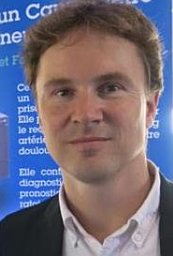 Laurent Plantier合作论文数CEPR, 7 Université de Tours6Le Corvoisier Philippe合作论文数6
 Charles Pilette合作论文数Christian de Duve Institute of Cellular Pathology, University of Louvain;and;Christian de Duve Institute of Cellular Pathology, University of Louvain6Frederic Schlemmer合作论文数6N Roche合作论文数5
 Thibaud Damy合作论文数5V. Bonnefoy合作论文数5M. Guecamburu合作论文数5Xavier Drouot合作论文数5
 Sobia Abid合作论文数Hôpital Henri Mondor, France and Université Paris-Est Créteil;University Hospitals of Leuven, University of Leuven;Hôpital Henri Mondor, Université Paris-Est Créteil;Telomeres and Telomerase Group, and Spanish National Cancer Research Centre (CNIO),;University Hospitals of Leuven, Université Paris-Est Créteil5Emmanuel Gomez合作论文数5
 Jean-Luc Dubois-Rande合作论文数Université Paris-Est Créteil;Université Numérique en Santé et Sport5L. Margarit合作论文数5C. Morelot合作论文数4B Mari合作论文数4Philippe Bonniaud合作论文数4
 Camille Taille合作论文数4
 Isabella Annesi-Maesano合作论文数4
 Pablo Bartolucci合作论文数4L-A Essari合作论文数4
 Pierre-Regis Burgel合作论文数4Randa Belgacemi合作论文数4Thomas d'Humières合作论文数AP HP, Hop Univ Henri Mondor4a boudjemaa合作论文数4
 Florence Canoui-Poitrine合作论文数4
 Si-Tahar Mustapha合作论文数Inst Pasteur, Unite Def Innee & Inflammat4Dagouassat Maylis合作论文数4
 Gregory Bouchaud合作论文数4
 Christophe Guignabert合作论文数IECB and INSERM U1045, University of Bordeaux4
 G. Deslee合作论文数Service des Maladies Respiratoires et Allergologie, Centre Hospitalier Universitaire de Reims4




