Background/Objectives: The survival of patients experiencing transformation of follicular lymphoma (tFL) is generally inferior to that of their non-transformed FL counterparts. While rituximab (R) has been shown to reduce transformation rates, data on Obinutuzumab (O), a third generation anti-CD20 monoclonal antibody, are limited. Methods: This retrospective study analyzed risk factors for tFL and evaluated outcomes following transformation in 1145 consecutive patients with FL (2010-2023). Results: Over a median follow-up of 70 months, 9% (n = 103) of FL patients developed tFL, with a median time of 36 months from FL diagnosis to transformation. In multivariate analysis, O-based, compared to R-based regimens and maintenance therapy (compared to no maintenance), were independently associated with reduced risk of histologic transformation (HR 0.40, 95% CI 0.19-0.90, p = 0.026 and HR 0.42, 95% CI 0.23-0.77, p = 0.005, respectively). Transformed FL was associated with shorter overall survival (OS) from FL diagnosis compared to non-transformed FL (median not reached, p < 0.001), while prior exposure to anti-FL chemoimmunotherapy predicted shorter OS following transformation (median 34.6 months vs. not reached, p = 0.001). Multivariate analysis confirmed prior exposure to anti-FL therapy (HR 5.27, p < 0.001), male sex (HR 2.36, p = 0.014), and age over 65 (HR 2.54, p = 0.019) to be associated with shorter OS among patients with tFL. Conclusions: These findings, although requiring further validation, suggest that O-based regimens may reduce the risk of transformation and support prior studies demonstrating adverse survival outcomes in previously treated tFL patients.
11109 Background: While recent FL therapy advances offer various treatment options, data are limited on FL treatment preferences in the shared decision-making process. A comprehensive survey with a DCE design was conducted to assess preferences of patients, caregivers, and physicians for different attributes that impact treatment choice. Methods: A web-based DCE survey available in English and Spanish was administered in Oct-Nov 2024 to patients with FL, caregivers, and physicians recruited in the US, the UK, Spain, Australia, and Canada through the Follicular Lymphoma Foundation (FLF). FL treatment attributes were selected based on targeted literature review, clinical inputs, and review with FLF patient and caregiver advisors. Attributes included efficacy (progression-free survival [PFS]), safety (impact of adverse events [AEs], including fatigue, cytokine release syndrome [CRS], and neurologic events [NE], on quality of life [QOL]), and convenience (mode of administration, treatment duration and frequency of visits, time needed to travel to treatment center). Survey responses were analyzed by patient, caregiver, and physician groups. Preference weights were generated from conditional logistic regression models and used to calculate the relative importance of attributes and willingness to trade off. Results: A total of 337 patients, 37 caregivers, and 29 physicians (median age: 59, 45, and 51 y, respectively) from 25 countries (>75% from US, UK, and Spain) responded to the DCE survey. The majority (93.7%) of patients reported having experienced ≥1 AE from previous treatment. Patients preferred treatments with longer PFS; mild or no impact of fatigue, CRS, and NEs on QOL during treatment; oral tablets vs infusions; a 3-mo duration with twice-weekly visits vs continuous duration with visits once every 3 mo; and <30 min of travel time vs >2 h (all P <.05). PFS was ranked as the most important attribute across patients, caregivers, and physicians. Following efficacy, treatment convenience attributes were ranked higher by patients and caregivers while safety attributes were more important to physicians. On average, patients were willing to accept reductions of 1 y of PFS for treatment requiring <30 min of travel vs >2 h, 0.7-1 y to receive treatments with less impact of AEs on QOL, 0.6 y for oral tablets vs blood collection and intravenous infusion, and 0.5 y for 3-mo treatment vs continuous duration. Conclusions: Efficacy is the most important attribute in treatment choice for patients, caregivers, and physicians. Following efficacy, patients and caregivers prioritize convenience and reduced impact of AEs, while physicians prioritize safety over convenience. Insights on differences between preferences highlight the importance of informed discussion and a balanced, individualized approach to treatment selection.
Background The prognostic utility of positron emission tomography/computer tomography (PET/CT) scan and bone marrow biopsy (BMB) as response assessments in mantle cell lymphoma (MCL) is incompletely explored. We sought to: 1) interrogate the prognostic impact of a mid-induction PET/CT scan, and 2) investigate whether end-of-induction (EOI) BMB results add prognostic significance to EOI PET/CT scan results in patients with MCL receiving frontline chemoimmunotherapy (CIT) in the context of the E1411 clinical trial. Methods Briefly, the E1411 randomized phase 2 study (NCT01415752) investigated whether addition of bortezomib to bendamustine/rituximab (BR) induction and/or addition of lenalidomide to rituximab maintenance improved progression-free-survival (PFS) in treatment-naïve MCL. Patient characteristics and results are as previously published (Smith et al., 2024). Neither addition of bortezomib to BR nor addition of lenalidomide to rituximab maintenance following BR-based induction impacted PFS. PET/CT scans, read locally, were completed at initial staging, after cycle 3, and at EOI. BMB was part of initial staging and was required at EOI for any patient with baseline BM involvement who otherwise met complete remission (CR) criteria. Response criteria were per International Working Group 2007. Survival distributions were estimated by Kaplan-Meier method. Univariable Cox proportional hazards models were used to evaluate the association between PET/CT or BMB and PFS or overall survival (OS). Results 37% (132/358) of patients who initiated induction therapy had interim PET/CT results available, with 42% (56/132) remaining PET-positive after 3 cycles of treatment. With median survival follow-up of 7.5 years (yrs), median PFS for interim PET-positive vs. negative patients was 4.9 vs. 7.4 yrs (HR 1.49; 95% CI: 0.89-2.49; p=0.13). Median OS for interim PET-positive vs. negative patients was 9.2 vs. 10.2 yrs (HR 1.40; 95% CI: 0.76-2.57; p=0.28). All patients who initiated induction therapy underwent baseline BMB, 82% of whom (293/358) had confirmed lymphoma involvement. 237/304 patients who completed 6 cycles of induction had EOI BMB results available, 96% (227/237) of which were negative for lymphoma. Rates of EOI BM involvement by PET response were as follows: 1/181 patients with CR, 6/50 with partial response (PR), 1/1 with stable disease, 0/1 with progressive disease, 2/3 with response unevaluable. Median PFS and OS of patients with CR and negative EOI BM was 6.8 yrs and 10.1 yrs, respectively (95% CI: 5.9-NR). Within patients who achieved PR and had EOI BM results available, the median PFS for EOI BM-positive vs. negative was 3.1 vs. 5.4 yrs (HR 2.38; 95% CI: 0.95-5.99; p=0.06). The median OS for EOI BM-positive vs. negative patients was 6.3 vs. 9.4 yrs (HR 2.39; 95% CI: 0.67-8.50; p=0.18). Conclusion Our results generate 3 key either conclusions or hypotheses for future studies. First, the trend to inferior PFS with PET-positive disease after 3 cycles of induction therapy provides foundation to evaluate whether patients may benefit from early adjustment of therapeutic strategy. Second, the extremely low rate of 0.6% of patients in a CR with EOI BM involvement suggests EOI BMB is not needed for patients achieving metabolic CR after frontline CIT. These results align with a recent study demonstrating baseline BMB adds little value to response assessment in follicular lymphoma, leading to recommendation for deferral in most cases (Rutherford et al., 2023). Patients with MCL could therefore similarly be spared a procedure that does not provide clear clinical utility. Third, the trend toward difference in PFS by EOI BMB status even in patients with a PR additionally identifies a group for whom escalated treatment, such as intensified maintenance treatment, may be beneficial to investigate. Limitations of our analyses include the unavailable interim PET/CT results for 63% of patients who initiated induction therapy. Additionally, none received frontline BTK inhibition. Future directions include combining minimal residual disease with interim and EOI PET/CT results. EOI BMB analyses should be performed in trials investigating frontline BTK inhibitors and particularly those without chemotherapy, to determine if a BMB is of similarly low clinical utility across treatment types. Such analyses will shed further light on the assessments with highest prognostic utility for MCL.
We performed a phase I/II trial to explore the safety and efficacy of carfilzomib (K) in combination with R-CHOP (KR-CHOP) in patients with diffuse large B cell lymphoma (DLBCL). A total of 48 patients were enrolled and 47 were treated. The overall response rate (ORR) was 89% (70% complete response). At a median follow-up of 31 months, 3-year Kaplan-Meier estimates of PFS and OS were 79% and 87%, respectively. Treatment with KR-CHOP for non-GC DLBCL was associated with a decreased risk of disease progression and death relative to standard of care treatment with R-CHOP with hazard ratios (HR) of 0.16 [95% confidence interval (CI) 0.04-0.58, p = 0.002] and 0.31 [(95% CI, 0.09 - 0.99), p = 0.02], respectively. The most common grade 3 or 4 adverse events (AEs) were anemia (13%), thrombocytopenia (9%) and febrile neutropenia (9%). KR-CHOP is safe and may have preferential activity in non-GC DLBCL.
Abstract Although initial therapy of mantle cell lymphoma (MCL) is not standardized, bendamustine plus rituximab (BR) is commonly used in older patients. Rituximab (R) maintenance after induction is often used. Thus, the open-label, randomized phase 2 ECOG-ACRIN Cancer Research Group E1411 trial was designed to test 2 questions: (1) does addition of bortezomib to BR induction (BVR) and/or (2) addition of lenalidomide to rituximab (LR) maintenance improve progression-free survival (PFS) in patients with treatment-naïve MCL? From 2012 to 2016, 373 previously untreated patients, 87% aged ≥60 years, were enrolled in this trial. At a median follow-up of 7.5 years, there is no difference in the median PFS of BR compared with BVR (5.5 vs 6.4 years; hazard ratio [HR], 0.90; 90% confidence interval [CI], 0.70-1.16). There were no unexpected additional toxicities with BVR treatment compared with BR, with no impact on total dose/duration of treatment received. Independent of the induction treatment, addition of lenalidomide did not significantly improve PFS, with median PFS in R vs LR (5.9 vs 7.2 years; HR, 0.84; 90% CI, 0.62-1.15). Most patients completed the planned 24 cycles of LR at the scheduled dose. In summary, adding bortezomib to BR induction does not prolong PFS in treatment-naïve MCL, and LR maintenance was not associated with longer PFS compared with R alone after BR. Nonetheless, the >5-year median PFS outcomes in this prospective cooperative group trial indicate the efficacy of BR followed by R maintenance as highly effective initial therapy for older patients with MCL. This trial was registered at www.clinicaltrials.gov as #NCT01415752.
INTRODUCTION: Lenalidomide and Rituximab (R2) is an effective frontline treatment regimen for patients (pts) with indolent B-cell lymphoma including follicular lymphoma (FL). Recent phase III data from the RELEVANCE trial comparing R2 to traditional chemoimmunotherapy showed that this regimen is generally well-tolerated and has favorable clinical efficacy [61% overall response, 53% CR rate, 77% 3-year progression free survival (PFS) (Morschhauser, et al)]. Proteasome inhibitors such as bortezomib disrupt NF-KB signaling and have shown clinical activity in indolent NHL. Although randomized trials have failed to demonstrate clinical benefit of adding bortezomib to standard chemoimmunotherapy regimen bendamustine + rituximab (BR) for frontline treatment of FL, the addition of proteasome inhibitors to lenalidomide is a mainstay of treatment for plasma cell neoplasms due to synergistic antitumor effect. The oral proteasome inhibitor ixazomib has less potential for dose-limiting neuropathy than bortezomib, making it an attractive option to incorporate into the R2 regimen. We sought to investigate the safety and efficacy of the addition of ixazomib to R2 for FL and indolent B-cell NHL through a phase I clinical trial of this combination for patients with high risk disease. METHODS: Adult (age ≥ 18) pts with untreated FL or other indolent lymphoma, adequate organ function and performance status were enrolled. To be enrolled, FL patients were required to have stage 2, 3 or 4 disease, with high tumor burden by GELF criteria and/or FLIPI score of 3-5. During 3 x 3 dose escalation, ixazomib was given at a dose of 2 mg (n=3), 3 mg (n=3) or 4 mg (n=12) PO on days 1, 8, and 15 with lenalidomide 20 mg PO on days 1-21 every 28 days. Rituximab was administered at standard dosing on days 1,8,15,21 for cycle 1, once every 28 days for cycles 2-6 and then once every 2 months for cycles 7-12. Treatment was continued for 12 cycles and no maintenance therapy was specified per protocol. All pts received low dose aspirin for venous thromboembolism prophylaxis and acyclovir for prevention of VZV reactivation. Response assessments by CT were performed after cycle 3 and 6 and by PET/CT at the end-of-treatment (cycle 12). RESULTS: 20 pts were enrolled and 18 were eligible for treatment [15 FL (14 grade 1-2, 1 grade 3A), 2 splenic marginal zone lymphoma and 1 nodal marginal zone lymphoma].The median age of treated pts was 61 (range 40-83) years old. 55% of patients were female. Stage at diagnosis was II (n =2), III (n = 4) and IV (n = 12). For FL pts, FLIPI scores at enrollment were low (n=2), intermediate (n = 5) and high risk (n=8) and FLIPI-2 scores were low (n=3), intermediate (n = 2) and high risk (n=10). There were no dose limiting toxicities during 3 x 3 dose escalation. Grade (G) 1/2 and G3/4 treatment-related hematologic adverse events (AEs) included neutropenia (6%, 28%), thrombocytopenia (16.7%, 5.6%) and anemia (16.7%, 0%). The most common treatment-related AEs included nausea/vomiting (44% G1, 11% G2), diarrhea, (50% G1, 22% G2, 5% G3), rash (33% G1, 6% G2, 11%G3), peripheral neuropathy (22% G1, 6% G2), myalgia/arthralgia (17% G1, 17% G2), and infection (33% G2, 17% G3). There was one pulmonary embolism and no cases of febrile neutropenia. As of June, 2020, median follow-up among living pts was 21 months. 4 pts discontinued treatment due to disease progression; 2 with transformation to aggressive lymphoma. Of the transformed cases, one subject died on study due to progression disease and one developed CNS disease on study treatment but proceeded to autologous stem cell transplant. The best overall response rate was 61.2% [55.6% CR, 5.6% PR): 22.2% had stable disease and 16.7% had disease progression. 18-month Kaplan-Meier estimates of PFS and overall survival were 71% and 94%, respectively (Figure). CONCLUSION: R2 can safely be combined with at the target dose of 4 mg of ixazomib for treatment-naïve indolent NHL patients. Non-hematologic AEs were generally consistent with known toxicity of each component of therapy. CR rate and PFS were was similar to the outcomes reported in the RELEVANCE trial despite enrolling high risk patient. R2 may serve as backbone for future studies of novel treatment combinations for high risk FL after thorough evaluation for occult transformation to aggressive lymphoma. Disclosures Hill: Abbvie: Consultancy, Honoraria, Research Funding; Genentech: Consultancy, Honoraria, Research Funding; Novartis: Consultancy, Honoraria; BMS: Consultancy, Honoraria, Research Funding; Celgene: Consultancy, Honoraria, Research Funding; Takeda: Research Funding; Karyopharm: Consultancy, Honoraria, Research Funding; AstraZenica: Consultancy, Honoraria, Research Funding; Kite, a Gilead Company: Consultancy, Honoraria, Research Funding; Beigene: Consultancy, Honoraria, Research Funding; Pharmacyclics: Consultancy, Honoraria, Research Funding. Jagadeesh:MEI Pharma: Research Funding; Regeneron: Research Funding; Seattle Genetics: Membership on an entity's Board of Directors or advisory committees, Research Funding; Debiopharm Group: Research Funding; Verastem: Membership on an entity's Board of Directors or advisory committees. Caimi:Celgene Corp: Other: Incyte Corporation - Ownership - Pharmacyclics, Inc. - Ownership - Celgene Corp. - Other, Speakers Bureau; ADC Therapeutics: Research Funding; Genentech: Research Funding. Smith:Takeda: Research Funding; Celgene: Research Funding. OffLabel Disclosure: Ixazomib is off-label for treatment of NHL
Cyclin E/Cdk2 kinase activity is frequently deregulated in human cancers, resulting in impaired apoptosis. Here, we show that cyclin E/Cdk2 phosphorylates and stabilizes the pro-survival Bcl-2 family protein Mcl-1, a key cell death resistance determinant to the small molecule Bcl-2 family inhibitors ABT-199 and ABT-737, mimetics of the Bcl-2 homology domain 3 (BH3). Cyclin E levels were elevated and there was increased association of cyclin E/Cdk2 with Mcl-1 in ABT-737-resistant compared to parental cells. Cyclin E depletion in various human tumor cell-lines and cyclin E-/- mouse embryo fibroblasts showed decreased levels of Mcl-1 protein, with no change in Mcl-1 mRNA levels. In the absence of cyclin E, Mcl-1 ubiquitination was enhanced, leading to decreased protein stability. Studies with Mcl-1 phosphorylation mutants show that cyclin E/Cdk2-dependent phosphorylation of Mcl-1 residues on its PEST domain resulted in increased Mcl-1 stability (Thr92, and Thr163) and Bim binding (Ser64). Cyclin E knock-down restored ABT-737 sensitivity to acquired and inherently resistant Mcl-1-dependent tumor cells. CDK inhibition by dinaciclib resulted in Bim release from Mcl-1 in ABT-737-resistant cells. Dinaciclib in combination with ABT-737 and ABT-199 resulted in robust synergistic cell death in leukemic cells and primary chronic lymphocytic leukemia patient samples. Collectively, our findings identify a novel mechanism of cyclin E-mediated Mcl-1 regulation that provides a rationale for clinical use of Bcl-2 family and Cdk inhibitors for Mcl-1-dependent tumors.
Supplementary Figures 1-5, Tables 1-4 from Aberrant Huntingtin Interacting Protein 1 in Lymphoid Malignancies
Representative images (20 x magnification) of formalin-fixed paraffin-embedded clots prepared from bone marrow aspirates.
Table A1. Prior therapies received (agents received by >5 patients); Table A2. Summary of pevonedistat plasma pharmacokinetic parameters on days 1 and 9 of cycle 1 on schedule A (administration on days 1, 2, 8, 9 of 21-day cycles); Table A3. Summary of pevonedistat plasma pharmacokinetic parameters on days 1 and 4 (or, alternatively, day 11) of cycle 1 on schedule B (administration on days 1, 4, 8, 11 of 21-day cycles); Table A4. Changes from baseline in gene expression levels for NAEregulated transcriptional targets on cycle 1, day 1 following pevonedistat administration at the MTDs on schedules A and B; Table A5. Patients achieving partial responses; Table A6. Patients achieving stable disease and receiving at least 5 cycles of pevonedistat.
<p>S2: Inactivation of bortezomib by liver homogenate; S3: CCF642 effect on survival of normal bone marrow, myeloid neoplasia, myeloma and lymphoma cells; S4: Biotin-stained Western blot after treatment of MM1.S cells with B-CCF642</p>
Despite improvements in indolent non-Hodgkin lymphoma (iNHL) therapy, they remain generally incurable and new treatments are needed. Will phosphatidylinositol 3-kinase (PI3K) inhibitors be among these? PI3K exists in four isoforms (α, β, γ, and δ). The δ isoform, preferentially expressed in hematopoietic cells, plays a key role in B-cell development and function [1]. PI3K, activated in B-cells by various upstream signals including the B-cell receptor, signals downstream to regulate B-cell survival, proliferation, and mobility [2] (Figure 1). As dysregulated PI3K signaling contributes to iNHL B cell survival and proliferation, PI3K became a rational therapeutic target for drug development, particularly PI3Kδ selective agents to promote lymphocyte specificity, reducing off-target toxicity [2]. For a comprehensive understanding of the role of PI3K in B-cell lymphoma and its therapeutic implications, we refer readers to a prior review on this topic, Gottfried von Keudell & Alison J. Moskowitz, 2019 [3]. Several PI3K inhibitors (PI3Ki) with varying isoform specificity but including δ demonstrated durable responses and improved progression-free survival (PFS) in iNHL and chronic lymphocytic leukemia (CLL), leading to various therapeutic approvals. While initially considered small-molecule inhibitors of B cells, accumulating data reveal additional immune and other off-target toxicity, highlighting several critical issues including suboptimal dosing regimens, limitations of single-arm trial interpretation, and ultimately concerns about adverse effects on overall survival. Moreover, the optimal approach to developing future PI3Kis for lymphoma remains a matter of debate. The Food and Drug Administration (FDA) has issued specific directives regarding the use of PI3Kis in hematologic malignancies and requires randomized studies for future PI3Kis approval decisions using overall survival (OS) as a clinical outcome endpoint [4]. This editorial review addresses existing data on PI3Ki in B-cell iNHL (not CLL or T cell lymphoma), and potential pathways to continue PI3Ki drug development in light of the US FDA discussion of the use of PI3Ki in hematological malignancies [5]. The role of PI3Ki in T-cell lymphoma was beyond the scope of the article, however this is an area of active investigation with evidence that PI3Ki may play a valuable role, especially as the risk:benefit calculation differs. 2. Efficacy vs toxicity of PI3Ki
The randomized, double-blind, phase 3 MAGNITUDE study evaluated NIRA+AAP vs placebo and AAP (PBO+AAP) as 1st line therapy for mCRPC that was HRR+ based on targeted next-generation sequencing of tumor tissue and/or plasma-based assays.1 Here we aimed to assess treatment efficacy by assay. Tumor tissue and/or plasma was collected from mCRPC patients (pts) who consented to the pre-screening protocol and tested using local tissue assays or centrally using the Foundation Medicine FoundationOne®CDx (F1CDX®) tissue assay or Agilent-Resolution Bioscience Resolution HRD™ Plasma assay. Pts HRR+ (BRCA1, BRCA2, FANCA, PALB2, CHEK2, BRIP1, HDAC2, ATM, CDK12) were randomized 1:1 to receive NIRA+AAP or PBO+AAP (NCT03748641). Primary (radiographic progression-free survival, rPFS) and secondary end-points (time to cytotoxic chemotherapy, TCC; time to symptomatic progression, TSP; and overall survival, OS) are reported for interim analysis 2 for each subgroup (events [NIRA+AAP/PBO+AAP], Hazard ratio and 95% CI). Overall, 423 pts were HRR+ with 225 BRCA+. Pts, 159 (96), were HRR+ (BRCA+) with both assays, the remaining pts were positive with either tissue/plasma (Table). Pts, 124 (62) HRR+ (BRCA+), were tissue positive but plasma negative, and 38 (17) HRR+ (BRCA+) pts were positive with plasma, but tissue negative. Hazard ratios for end-points, except OS, had p<0.05 for pts BRCA positive by either assay, with similar results for HRR+ pts. Table: 1806PPopulation, n (NIRA+APP / PBO+APP)rPFSTCCTSPOSBRCA+Overall 113/11257/78 0.55 (0.39-0.78)28/44 0.56 (0.35-0.90)31/51 0.54 (0.35-0.85)43/49 0.88 (0.58-1.34)Plasma 71/790.63 (0.42-0.96)0.49 (0.28-0.87)0.51 (0.30-0.89)0.88 (0.53-1.46)Tissue 76/860.51 (0.33-0.77)0.44 (0.24-0.82)0.50 (0.28-0.89)0.83 (0.49-1.40)All HRROverall 212/211124/140 0.76 (0.60-0.97)57/77 0.67 (0.47-0.94)54/83 0.60 (0.42, 0.84)90/89 1.01 (0.75-1.36)Plasma 129/1480.77 (0.57-1.04)0.60 (0.39-0.92)0.58 (0.38-0.88)1.04 (0.73-1.49)Tissue 144/1470.80 (0.59-1.08)0.62 (0.40-0.96)0.56 (0.36-0.86)0.97 (0.67-1.42) Open table in a new tab Clinically meaningful benefit from NIRA+AAP are similar for BRCA+ pts detected by either tissue or plasma-based assays. Positivity by either test is sufficient to guide treatment decisions. 1. Efstathiou E, et al. J Clin Oncol. 2023;41(suppl 6):170.
In ARASENS (NCT02799602), DARO + ADT + DOC significantly reduced risk of death by 32.5% (HR 0.68; 95% CI 0.57–0.80; P<0.0001) vs ADT + DOC in pts with mHSPC. In post hoc analyses, HRs for overall survival (OS) were similar for pts with high-volume (HV HR 0.69; 95%CI 0.57–0.82) and low-volume (LV HR 0.68; 95% CI 0.41–1.13) disease. We report PSA outcomes in these pt subgroups. Pts with mHSPC were randomized to DARO 600 mg twice daily or placebo (PBO), both with ADT + DOC. PSA was measured at screening and every 12 weeks. Analyses included the proportion of patients achieving undetectable PSA (<0.2 ng/mL) and time to PSA progression. The association between undetectable PSA at any time with time to PSA progression and OS was evaluated using Kaplan-Meier estimates with HRs (95% CI) based on a stratified Cox regression model. Of 1305 pts, 1005 (77%) had HV disease and 300 (23%) had LV disease. Rapid undetectable PSA response as early as 12 weeks was achieved in nearly twice as many patients receiving DARO vs PBO and continued to increase for both HV and LV subgroups over time (at 24 weeks: HV 43.1% vs 21.7%, LV 66.9% vs 31.5%; at 52 weeks, HV 54.7% vs 23.6%, LV 77.9% vs 34.9%; and at any time, HV 62.2% vs 25.8%, LV 83.8% vs 38.4%). Time to PSA progression was prolonged in pts receiving DARO vs PBO in HV (HR 0.30; 95% CI 0.24–0.37) and LV (HR 0.09; 95% CI 0.05–0.18) pts (4-yr PSA progression-free rates: HV 65.8% vs 27.0%; LV 91.7% vs 36.3%). Pts receiving DARO who achieved undetectable PSA had improved time to PSA progression (HV HR 0.11, 95% CI 0.07–0.16; LV HR 0.02, 95% CI 0.004–0.10) and improved OS (HV HR 0.20; 95% CI 0.15–0.27; LV HR 0.34; 95% CI 0.14–0.79) vs those who did not. Early treatment intensification with DARO + ADT + DOC allows pts with HV and LV mHSPC to achieve rapid, deep, and durable PSA responses that are associated with improved time to PSA progression and OS.
Background: Patients with metastatic castration-resistant prostate cancer (mCRPC) and BRCA alterations have poor outcomes. MAGNITUDE found patients with homologous recombination repair gene alterations (HRR+), particularly BRCA1/2, benefit from first-line therapy with niraparib plus abiraterone acetate and prednisone (AAP). Here we report longer follow-up from the second prespecified interim analysis (IA2).Patients and methods: Patients with mCRPC were prospectively identified as HRR+ with/without BRCA1/2 alterations and randomized 1:1 to niraparib (200 mg orally) plus AAP (1000 mg/10 mg orally) or placebo plus AAP. At IA2, secondaryendpoints [time to symptomatic progression, time to initiation of cytotoxic chemotherapy, overall survival (OS)] were assessed.Results: Overall, 212 HRR+ patients received niraparib plus AAP (BRCA1/2 subgroup, n = 113). At IA2 with 24.8 months of median follow-up in the BRCA1/2 subgroup, niraparib plus AAP significantly prolonged radiographic progression-free survival {rPFS; blinded independent central review; median rPFS 19.5 versus 10.9 months; hazard ratio (HR) = 0.55 [95% confidence interval (CI) 0.39-0.78]; nominal P = 0.0007} consistent with the first prespecified interim analysis. rPFS was also prolonged in the total HRR+ population [HR = 0.76 (95% CI 0.60-0.97); nominal P = 0.0280; median follow-up 26.8 months]. Improvements in time to symptomatic progression and time to initiation of cytotoxic chemotherapy were observed with niraparib plus AAP. In the BRCA1/2 subgroup, the analysis of OS with niraparib plus AAP demonstrated an HR of 0.88 (95% CI 0.58-1.34; nominal P = 0.5505); the prespecified inverse probability censoring weighting analysis of OS, accounting for imbalances in subsequent use of poly adenosine diphosphate-ribose polymerase inhibitors and other life-prolonging therapies, demonstrated an HR of 0.54 (95% CI 0.33-0.90; nominal P = 0.0181). No new safety signals were observed.Conclusions: MAGNITUDE, enrolling the largest BRCA1/2 cohort in first-line mCRPC to date, demonstrated improved rPFS and other clinically relevant outcomes with niraparib plus AAP in patients with BRCA1/2-altered mCRPC, emphasizing the importance of identifying this molecular subset of patients.
 Brian T. Hill合作论文数Department of Hematology and Medical Oncology, Cleveland Clinic;Department of Medicine, School of Medicine, Case Comprehensive Cancer Center, Case Western Reserve University34
 Robert M. Dean合作论文数Department of Medicine, School of Medicine, Case Western Reserve University;Case Comprehensive Cancer Center, Case Western Reserve University;Department of Hematology and Medical Oncology, Taussig Cancer Institute, Cleveland Clinic31
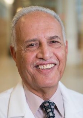 Tahseen I. Al-Saleem合作论文数Department of Pathology, Fox Chase Cancer Center, Temple University Health System, Inc.31
 Brad Pohlman合作论文数31
 Deepa Jagadeesh合作论文数Department of Hematologic Oncology and Blood Disorders, Cleveland Clinic;Department of Medicine, School of Medicine, Case Western Reserve University;Case Comprehensive Cancer Center, Case Western Reserve University29Indira Joshi合作论文数26
 Fred Saad合作论文数Urology Department, University Hospital of Montreal;Department of Surgery, Université de Montréal21
 Ranjana Advani合作论文数Division of Oncology, Department of Medicine, Stanford University;Stanford Cancer Institute20
 Hossein Borghaei合作论文数Department of Hematology, Fox Chase Cancer Center;Department of Oncology, Fox Chase Cancer Center19
 Eric Hsi合作论文数Department of Pathology, Wake Forest University School of Medicine18Frederic J. Reu合作论文数18Michael Millenson合作论文数17
 Russell J Schilder合作论文数17
 Elisabeth M. Paietta合作论文数Department of Oncology, Albert Einstein College of Medicine;Department of Medicine, Albert Einstein College of Medicine16Lisa A. Rybicki合作论文数16
 Karim Fizazi合作论文数Institut Gustave Roussy;Department of Oncology, University of Paris-Saclay15
 Eric Small合作论文数Department of Urology, University of California, San Francisco;Division of Hematology and Oncology, Department of Medicine, University of California, San Francisco13
 Paolo F. Caimi合作论文数Department of Medicine, Division of Hematology and Oncology, School of Medicine, Case Western Reserve University;Case Comprehensive Cancer Center, Case Western Reserve University;Department of Hematology and Medical Oncology, Cleveland Clinic13
 Luis E. Fayad合作论文数Department of Lymphoma/Myeloma, Division of Cancer Medicine, The University of Texas MD Anderson Cancer Center13Mm Millenson合作论文数12Czuczman Myron S合作论文数12
 Neal Shore合作论文数Carolina Urologic Research Center, Atlantic Urology Clinics12Martin S. Tallman合作论文数12Ayad Al-Katib合作论文数12
 John P. Leonard合作论文数Weill Cornell Medical College11
 Brad S. Kahl合作论文数Division of Oncology, John T. Milliken Department of Medicine, Washington University School of Medicine11
 Christy Samaras合作论文数Department of Hematology and Medical Oncology, Cleveland Clinic10
 Eduardo Martin Sotomayor合作论文数Cancer Institute, Tampa General Hospital10
 Dana E. Rathkopf合作论文数Sidney Kimmel Center for Prostate and Urologic Cancers, Memorial Sloan Kettering Cancer Center10Andres Forero合作论文数10
 Matt Kalaycio合作论文数Department of Hematology and Medical Oncology, Cleveland Clinic10Nam H. Dang合作论文数9
 Jason Valent合作论文数9Gordon Leo I合作论文数9Daniel J. Lindner合作论文数8T Xie合作论文数8
 Jane N Winter合作论文数Hematology and Oncologys, Department of Medicine, Feinberg School of Medicine, Northwestern University8
 Peter Martin合作论文数Division of Hematology/Oncology, Weill Cornell Medical College8
 Ann S. Lacasce合作论文数Department of Medical Oncology, Dana-Farber Cancer Institute;Harvard Medical School8
 Tombal Bertrand合作论文数Pôle de Chirurgie Expérimentale et Transplantation, Institut de Recherche Expérimentale et Clinique, Université Catholique de Louvain;Faculty of Medicine and Dentistry, Université Catholique de Louvain;Department of Surgery, Cliniques universitaires Saint-Luc;Division Of Urology, Cliniques universitaires Saint-Luc8
 Clark W. Distelhorst合作论文数Department of Pharmacology, School of Medicine, Case Western Reserve University;Department of Medicine, Division of Hematology and Oncology, School of Medicine, Case Western Reserve University;Department of Hematology and Oncology, University Hospitals Cleveland Medical Center8Jin F合作论文数8
 Nancy L. Bartlett合作论文数Divisions of Hematology & Oncology, Department of Medicine, Washington University School of Medicine in St. Louis8
 Anas Younes合作论文数AstraZeneca8
 John C. Byrd合作论文数College of Medicine, University of Cincinnati8
 Andre Rogatko合作论文数Departments of Medical Oncology and Biostatistics, Fox Chase Cancer Center8
 Randy Gascoyne合作论文数Centre for Lymphoid Cancers, BC Cancer Research Institute;Department of Pathology and Laboratory Medicine, Faculty of Medicine, University of British Columbia8
 Owen A O'Connor合作论文数8Dale R. Grabowski合作论文数Department of Translational Hematology and Oncology Research, Cleveland Clinic8




