Secondary lymphedema is a debilitating condition driven by impaired regeneration of lymphatic vasculature following lymphatic injury, surgical removal of lymph nodes in cancer patients, or infection. However, the extent to which collecting lymphatic vessels regenerate following injury remains unclear. Here, we employed a novel mouse model of lymphatic injury in combination with state-of-the-art lymphatic imaging to demonstrate that the implantation of an optimized fibrin gel following lymphatic vessel injury leads to the reconnection of the injured lymphatic vessel network through sprouting lymphangiogenesis of initial-like lymphatic vessels from the ends of the collecting lymphatic vessels, resulting in the restoration of lymph flow to the draining lymph node. Mechanistically, we found that fibrin implantation elevates the tissue levels of CCL5, a potent immune cell-recruiting chemokine. Notably, injured vessels in CCL5-KO mice made fewer connections following fibrin gel implantation. These novel findings shed light on the mechanisms underlying lymphatic regeneration and suggest that enhancing CCL5 signaling may be a promising therapeutic strategy for enhancing lymphatic regeneration.
Supplementary Fig. S9: Expression levels of Cxcr3 and its ligands are increased in ICC tissues after GC/dual ICB treatment in murine 425-ICC.
Stromal cells suppress CD8+ T cell proliferation via a contact independent mechanism.
Supplementary Fig. S8: Bulk tissue RNA sequencing analysis of ICC after GC/dual ICB combination therapy in orthotopic murine 425-ICC model.
Supplementary Fig. S12: Effect of ICB treatment scheduling on efficacy and toxicity.
Supplementary Fig. S2: Standard chemotherapy converts ICB-resistant ICCs to ICB-responsive tumors, significantly delays tumor progression and increases survival in mice.
Supplementary Fig. S6: CTLA-4 blockade mediates the efficacy of GC/ICB therapy in ICC and increases CD8+CTL frequency in murine ICC.
CAF exosomes alter TCR signaling, transcriptional regulation, and metabolic profiles in CD8+ T cells. A, Differentially expressed proteins were analyzed using volcano plots for comparisons between unstimulated murine CD8 T cells and T cells stimulated with anti-CD3/CD28 antibodies, after 48 hours, with GO Biological Process (GOBP) analysis shown in B. C, Differentially expressed proteins were analyzed using volcano plots for comparisons between murine CD8 T cells stimulated with anti-CD3/CD28 antibodies for 4 hours, after 24 hours in the presence of cancer cell or CAF exosomes, with GOBP analysis shown in D. E, Heatmap showing differentially expressed phosphorylated proteins in CD8 T cells exposed to cancer cells or CAF exosomes for 24 hours, followed by stimulated with anti-CD3/CD28 antibodies for 4 hours. F, Western blot of phosphorylated (p)ZAP-70 (Y352), total ZAP-70, pSLP-76 (S376), total SLP-76, pPLC-γ (Y783), total PLC-γ in unstimulated (-) CD8 T cells, and CD8 T cells stimulated with cancer cells or CAF exosomes (Exo.) and anti-CD3/CD28 antibodies for 15 minutes or 30 minutes.
PD-L1/PD-1 blockade fails to rescue CD8+ T cell proliferation in the presence of CAF exosomes.
Abstract Group 3 medulloblastomas (G3MB) carry the worst prognosis among medulloblastoma subtypes, yet molecularly targeted therapies remain elusive. Standard treatments cause severe long-term morbidity in survivors. Here, we identify tumor-derived sphingosine kinase 2 (SPHK2) as an essential driver of G3MB initiation and progression. SPHK2 exacerbates local immunosuppression by suppressing cytotoxic T-cell and NK-cell activity while promoting regulatory T-cell infiltration. Genetic or pharmacologic SPHK2 inhibition using Opaganib attenuates pro-survival tumor signaling and restores anti-tumor immunity, significantly improving survival in syngeneic G3MB mouse models. Combining Opaganib with fractionated low-dose radiation (f-LDRT) further enhances antigen presentation and reprograms tumor-associated myeloid cells toward an anti-tumor phenotype. This combination therapy markedly prolongs survival without inducing significant toxicity. Overall, our study establishes SPHK2 as a previously unrecognized therapeutic target and presents a safe, effective, microenvironment-reprogramming regimen for G3MB. One Sentence Summary Direct inhibition of tumor-derived SPHK2 overcomes local immunosuppression and downregulates pro-survival signaling in Group 3 medulloblastoma, while combination with fractionated low-dose radiation further enhances anti-tumor immunity and significantly improves survival.
Supplementary Fig. S11: Cxcr3 in CD8 T cells mediates the benefit of GC/ICB combination therapy in orthotopic murine 425-ICC model.
Density gradient ultracentrifugation-based isolation of serum reveals fractions characterized by exosomal markers.
Supplementary Fig. S4: IMC analysis of ICC after GC-based therapies in orthotopic murine 425-ICC model.
Cancer-associated fibroblasts (CAF), a major component of the breast tumor microenvironment, drive immune evasion in various cancers by promoting T-cell exclusion and dampening T-cell activation. Previous studies have implicated CAF-derived soluble factors in mediating these immunosuppressive effects. In this study, we investigated whether exosomes secreted by CAFs could suppress T-cell activity. Inhibition of global exosome secretion in breast tumor-bearing mice significantly reduced tumor growth and increased tumor-infiltrating T cells with lower exhaustion marker expression. Conversely, administration of CAF-derived exosomes into tumors produced the opposite effects. Moreover, CAF exosomes associated with T cells in vivo and impaired T-cell activation and cytotoxic potential in ex vivo assays. Proteomic and biochemical analyses of T cells exposed to CAF exosomes revealed dampened early T-cell receptor signaling. Mass spectrometry identified an extracellular matrix (ECM) signature on CAF exosomes. Depleting type I and type V collagens from CAF exosomes restored T-cell proliferation, whereas overexpression of collagen in cancer cells led to its incorporation into exosomes, which suppressed T-cell activation. These findings suggest that a signaling bridge between CAF exosomes and T cells, mediated by collagen, promotes T-cell dysfunction, contributing to immune evasion in breast cancer.Significance: Our data provide the first evidence that ECM proteins associate with mouse and human breast CAF-derived exosomes and directly impair T-cell activation and cytotoxicity. These findings suggest that signaling between collagen-rich CAF exosomes and T cells contribute to local and systemic T-cell dysfunction.
Supplementary Fig. S5: CTLA-4 blockade is critical for efficacy of combined chemotherapy with ICB in ICC by grafting 425 murine cells in C57Bl/6/FVB F1 mice.
 Peigen Huang合作论文数38
 Lance Munn合作论文数Department of Radiation Oncology, Mass General Research Institute, Massachusetts General Hospital;Harvard Medical School;Université de Technologie de Compiègne28
 Zohreh Amoozgar合作论文数Department of Radiation Oncology, Massachusetts General Hospital, Harvard Medical School28
 Rakesh K. Jain合作论文数Harvard Medical School, Harvard University;Edwin L. Steele Laboratories for Tumor Biology, Department of Radiation Oncology, Massachusetts General Hospital25
 Dan G. Duda合作论文数Houston Methodist Research Institute24Aya Matsui合作论文数21
 Dai Fukumura合作论文数Massachusetts General Hospital;Mass General Research Institute;Harvard Medical School, Harvard University21
 Meenal Datta合作论文数Edwin L. Steele Laboratory for Tumor Biology, Massachusetts General Hospital and Harvard Medical School;Department of Chemical and Biological Engineering, Tufts University;Harvard Medical School, Tufts University19Ze-Long Liu合作论文数Harvard Medical School, Massachusetts General Hospital18
 Timothy P. Padera合作论文数Edwin L. Steele Laboratories, Massachusetts General Hospital;Harvard Medical School17Xin Liu合作论文数16Koetsu Inoue合作论文数15
 Shuichi Aoki合作论文数Dept. of Surgery, Tohoku University Graduate School of Medicine15
 Nabeel M. Bardeesy合作论文数Center for Cancer Research, Massachusetts General Hospital;Harvard Medical School;KFCCR Faculty, Mass General Research Institute;Harvard Stem Cell Institute;OncoRev, LLC15Sarah Shin合作论文数14Daniel Staiculescu合作论文数14Zhiping Ruan合作论文数14Stefan Halvorsen合作论文数14Lingling Zhu合作论文数14Alexei Hernandez合作论文数14Zhangya Pu合作论文数14
 Lei Shi合作论文数Center for Cancer Research, Massachusetts General Hospital14
 Won Jin Ho合作论文数Department of Otolaryngology-Head and Neck Surgery, School of Medicine, Johns Hopkins University14Sassi Slim O合作论文数14Meghan J O'Melia合作论文数Emory Univ13
 Hengbo Zhou合作论文数Department of Pharmacology, University of Colorado–Denver, Aurora, USA12
 Chen Jiang合作论文数Sir Run Run Shaw Hospital, Zhejiang University12
 Min Wu (吴旻)合作论文数College of Life Sciences, Wuhan University;Taikang Center for Life and Medical Sciences, Wuhan University11
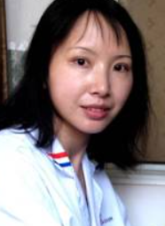 Lianyun Li (李联运)合作论文数College of Life Sciences, Wuhan University10
 Lutz Menzel合作论文数Translational Tumor Immunology, Max-Delbrück-Center for Molecular Medicine9
 James W. Baish合作论文数Department of Mechanical Engineering, Bucknell University9
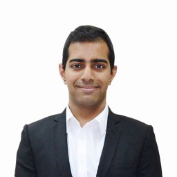 Ashwin S Kumar合作论文数Harvard-Massachusetts Institute of Technology Division of Health Sciences and Technology, Massachusetts Institute of Technology8
 Sonu Subudhi合作论文数Massachusetts General Hospital, Harvard Medical School8
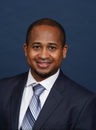 Dennis Hugh A Jones合作论文数Yale University, Yale Univ.8
 Patrik Andersson合作论文数Department of Medical Biochemistry and Biophysics;Karolinska Institute;Department of Medical Biochemistry and Biophysics, Karolinska Institute8Posada, Jessica M.合作论文数7Mark Duquette合作论文数7Shanmugarajan Krishnan合作论文数6
 Tatsuya Kobayashi合作论文数Massachusetts Gen Hosp, Endocrine Unit, Boston, MA 02114 USA6
 Gang Wei (魏刚)合作论文数Shanghai Institute of Nutrition and Health, Chinese Academy of Sciences6
 Jessalyn Ubellacker合作论文数6Lei Xu合作论文数6Jun Ren合作论文数Harvard Med Sch, Massachusetts Gen Hosp, Edwin L Steele Labs Tumor Biol, Boston, MA USA5Pereira Ethel R合作论文数Department of Genetics &Tumor Cell Biology, St. Jude Children's Research Hospital;Department of Genetics & Tumor Cell Biology, St. Jude Children's Research Hospital5
 William W Ho合作论文数5
 Somin Lee合作论文数5
 Neal Lindeman合作论文数Laboratory Medicine and Molecular Pathology, Weill Cornell Medicine5Sylvie Roberge合作论文数4
 Xiang Wang合作论文数4




