Abstract Group 3 medulloblastomas (G3MB) carry the worst prognosis among medulloblastoma subtypes, yet molecularly targeted therapies remain elusive. Standard treatments cause severe long-term morbidity in survivors. Here, we identify tumor-derived sphingosine kinase 2 (SPHK2) as an essential driver of G3MB initiation and progression. SPHK2 exacerbates local immunosuppression by suppressing cytotoxic T-cell and NK-cell activity while promoting regulatory T-cell infiltration. Genetic or pharmacologic SPHK2 inhibition using Opaganib attenuates pro-survival tumor signaling and restores anti-tumor immunity, significantly improving survival in syngeneic G3MB mouse models. Combining Opaganib with fractionated low-dose radiation (f-LDRT) further enhances antigen presentation and reprograms tumor-associated myeloid cells toward an anti-tumor phenotype. This combination therapy markedly prolongs survival without inducing significant toxicity. Overall, our study establishes SPHK2 as a previously unrecognized therapeutic target and presents a safe, effective, microenvironment-reprogramming regimen for G3MB. One Sentence Summary Direct inhibition of tumor-derived SPHK2 overcomes local immunosuppression and downregulates pro-survival signaling in Group 3 medulloblastoma, while combination with fractionated low-dose radiation further enhances anti-tumor immunity and significantly improves survival.
Cancer metastasis is a major contributor to patient morbidity and mortality1, yet the factors that determine the organs where cancers can metastasize are incompletely understood. Here we quantify the absolute levels of 124 metabolites in multiple tissues in mice and investigate how this relates to the ability of breast cancer cells to grow in different organs. We engineered breast cancer cells with broad metastatic potential to be auxotrophic for specific nutrients and assessed their ability to colonize different tissue sites. We then asked how tumour growth in different tissues relates to nutrient availability and tumour biosynthetic activity. We find that single nutrients alone do not define the sites where breast cancer cells can grow as metastases. In addition, we identify purine synthesis as a requirement for tumour growth and metastasis across many tissues and find that this phenotype is independent of tissue nucleotide availability or tumour de novo nucleotide synthesis activity. These data suggest that a complex interplay between multiple nutrients within the microenvironment dictates potential sites of metastatic cancer growth, and highlights the interdependence between extrinsic environmental factors and intrinsic cellular properties in influencing where breast cancer cells can grow as metastases.
Relationship between pathological response and circulating biomarker levels in the plasma of treated patients.
Immunofluorescence staining of residual PDAC in pathological responders and non-responders in FFX+CRT and losartan+FFX+CRT.
Heatmap showing differentially expressed genes (DEGs) and their expression in each patient in FFX+CRT and losartan+FFX+CRT-treated groups.
Group 3 Medulloblastoma (G3MB) is a pediatric brain cancer with poor prognosis, and a median 5-year overall survival of 45-58% in infants and young children, respectively. Moreover, the current standard-of-care therapy – which includes spinal radiation – causes tremendous morbidity. While immunotherapy with immune checkpoint blockade has transformed the treatment of many malignancies, it has yet to show benefit in the most common form pediatric brain tumors. Here we show that B cell depletion using Rituximab; anti-CD20: -- an FDA-approved antibody -- enhances anti-tumor immunity, promote tumor regression and improves overall survival of mice bearing orthotopic murine models of G3MB. By analyzing of human MB transcriptomics datasets, we discovered that pediatric MB patients with elevated B cell signatures have significantly reduced survival rates compared to signatures of other immune cell populations. Using immunohistochemistry and transcriptomic analysis of orthotopic murine models of G3MB, we found that intratumoral B cells are immunosuppressive – they directly inhibit anti-tumor CD8 T cells and reprogram the tumor microenvironment to an immunosuppressive milieu. Furthermore, systemic B cell depletion using murine equivalents of the FDA approved B cell depleting antibody (Rituximab; anti-CD20: 100µg/dose) significantly enhanced animal survival, and improved T cell infiltration and activation – estimated by flow cytometry and single cell RNA-sequencing. Furthermore, systemic B cell depletion enhanced the therapeutic efficacy of murine equivalents of standard-of-care radiation therapy (20.7Gy to tumor bed) and synergized with fractionated low-dose radiation therapy (1.81Gy x 6 daily doses) – which is likely to be less toxic to the developing pediatric brain. Finally, our mechanistic studies done revealed that tumor - derived placental growth factor (PlGF) reprograms B cells toward an immunosuppressive phenotype, characterized by increased IL-10 expression and suppression of CD8+ T cell function. These findings also suggest a potential benefit of anti-PlGF antibody treatment of MB in patient-derived models and its safe use in patients (Snuderl et al, Cell 2013; Sulnier-Sholler et al. Clinical Cancer Research 2022). In conclusion, our findings suggest that targeting B cells systemically represents a promising therapeutic strategy for G3MB treatment while preserving patients’ quality of life. The availability of FDA-approved B cell-targeting drugs for pediatric malignancies offers a path for rapid clinical translation, particularly crucial for this devastating disease. Citation Format: Ashwin S Kumar, Taylor P Uccello, Igor L Gomes dos Santos, Vasiliki Salameti, Sophie C Steinbuch, Sonu Subudhi, Rakesh K Jain. Targeting B Lymphocytes to Improve Therapeutic Outcomes for Pediatric Medulloblastoma [abstract]. In: Proceedings of the AACR IO Conference: Discovery and Innovation in Cancer Immunology: Revolutionizing Treatment through Immunotherapy; 2025 Feb 23-26; Los Angeles, CA. Philadelphia (PA): AACR; Cancer Immunol Res 2025;13(2 Suppl):Abstract nr B095.
Quantitative analysis of immunofluorescence staining in PDAC lesions from FFX+CRT-treated patients.
Figure S2 | Post-craniotomy stress map for each individual patient involved in this study.
Quantitative analysis of immunofluorescence staining in PDAC lesions from FFX+CRT and losartan+FFX+CRT-treated groups.
Differentially expressed genes (DEG) in losartan+FFX+CRT versus FFX+CRT, losartan+FFX+CRT versus untreated, and FFX+CRT versus untreated.
Gene sets associated with overall survival in losartan+FFX+CRT and FFX+CRT-treated groups.
Effect of losartan+FFX+CRT and FFX+CRT on genes involved in angiogenesis and the migration and maturation of DCs.
Immunofluorescence staining and quantitative analysis in PDAC lesions from losartan+FFX+CRT-treated patients.
PURPOSE:Physical forces exerted by expanding brain tumors-specifically the compressive stresses propagated through solid tissue structures-reduce brain perfusion and neurologic function but heretofore have not been directly measured in patients in vivo. Solid stress levels estimated from tumor growth patterns are negatively correlated with neurologic performance in patients. We hypothesize that measurements of solid stress can be used to inform clinical management of brain tumors. EXPERIMENTAL DESIGN:We developed an intraoperative technique to quantitatively estimate solid stress and brain replacement by the tumor. In 30 patients, we made topographic measurements of brain deformation through the craniotomy site with a neuronavigation system during surgical workflows immediately preceding tumor resection (<5 minutes in the operating room). Utilizing these measurements in conjunction with finite element modeling, we calculated solid stress within the tumor and brain and estimated the amount of brain tissue replaced, i.e., lost, by tumor growth. RESULTS:Mean solid stresses were in the range of 10 to 600 Pa, and the amount of tissue replacement was up to 10% of the brain. Brain loss in patients delineated glioblastoma from brain metastatic tumors, and in mice, solid stress was a sensitive biomarker of chemotherapy response. CONCLUSIONS:We present in this study a quantitative approach to intraoperatively measure solid stress in patients that can be readily adopted into standard clinical workflows. Brain loss due to tumor growth is a novel mechanical-based biomarker that, in addition to solid stress, may inform personalized management in future clinical studies in brain cancer.
Mathematical modelling has proven to be a valuable tool in predicting the delivery and efficacy of molecular, antibody-based, nano and cellular therapy in solid tumours. Mathematical models based on our understanding of the biological processes at subcellular, cellular and tissue level are known as mechanistic models that, in turn, are divided into continuous and discrete models. Continuous models are further divided into lumped parameter models — for describing the temporal distribution of medicine in tumours and normal organs — and distributed parameter models — for studying the spatiotemporal distribution of therapy in tumours. Discrete models capture interactions at the cellular and subcellular levels. Collectively, these models are useful for optimizing the delivery and efficacy of molecular, nanoscale and cellular therapy in tumours by incorporating the biological characteristics of tumours, the physicochemical properties of drugs, the interactions among drugs, cancer cells and various components of the tumour microenvironment, and for enabling patient-specific predictions when combined with medical imaging. Artificial intelligence-based methods, such as machine learning, have ushered in a new era in oncology. These data-driven approaches complement mechanistic models and have immense potential for improving cancer detection, treatment and drug discovery. Here we review these diverse approaches and suggest ways to combine mechanistic and artificial intelligence-based models to further improve patient treatment outcomes. Here Harkos et al. review the role of continuous models and discrete models in predicting and understanding therapy delivery and efficacy in solid tumours. They propose ways to integrate mechanistic and AI-based models to further improve patient outcomes.
 Rakesh K. Jain合作论文数Harvard Medical School, Harvard University;Edwin L. Steele Laboratories for Tumor Biology, Department of Radiation Oncology, Massachusetts General Hospital39
 Sonu Subudhi合作论文数Massachusetts General Hospital, Harvard Medical School26
 Dai Fukumura合作论文数Massachusetts General Hospital;Mass General Research Institute;Harvard Medical School, Harvard University25
 Posada, Jessica M.合作论文数24
 Dan G. Duda合作论文数Houston Methodist Research Institute19
 Talele Nilesh合作论文数19
 Heena Kumra合作论文数Faculty of MedicineDepartment of Anatomy and Cell BiologyMcGill UniversityMontrealQC H3A 0C7Canada17
 Yves Boucher合作论文数17
 Jeffrey W Clark合作论文数Harvard Cancer Center, Harvard Medical School;Dana-Farber/Partners Cancer Care, Massachusetts General Hospital;Cancer Center Protocol Office, Massachusetts General Hospital17
 Mari Mino-Kenudson合作论文数Harvard Medical School;Massachusetts General Hospital16
 Mikael Pittet合作论文数Department of Pathology and Immunology, Faculty of Medicine, University of Geneva16
 Spencer Rosario合作论文数15
 Ryan David P合作论文数15
 Carlos Fernandez-Del Castillo合作论文数Mass General Research Institute;Harvard Medical School;GI Cancer Center, Massachusetts General Hospital15
 Theodore Hong合作论文数Tucker Gosnell Center for Gastrointestinal Cancers, Massachusetts General Hospital;Harvard Medical School15Liqun Gu合作论文数14
 Meenal Datta合作论文数Edwin L. Steele Laboratory for Tumor Biology, Massachusetts General Hospital and Harvard Medical School;Department of Chemical and Biological Engineering, Tufts University;Harvard Medical School, Tufts University14
 Jennifer Wo合作论文数Mass General Research Institute, Massachusetts General Hospital;Harvard Medical School14Mark Duquette合作论文数12
 Zohreh Amoozgar合作论文数Department of Radiation Oncology, Massachusetts General Hospital, Harvard Medical School11
 Sampurna Chatterjee合作论文数Takeda10Peigen Huang合作论文数10
 William W Ho合作论文数8Pinji Lei合作论文数College of Life Sciences, Wuhan University8
 Patrik Andersson合作论文数Department of Medical Biochemistry and Biophysics;Karolinska Institute;Department of Medical Biochemistry and Biophysics, Karolinska Institute8
 Lance Munn合作论文数Department of Radiation Oncology, Mass General Research Institute, Massachusetts General Hospital;Harvard Medical School;Université de Technologie de Compiègne7Sylvie Roberge合作论文数6
 Gino Ferraro合作论文数6Lei Xu合作论文数6
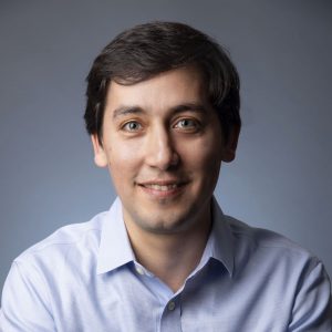 Hadi T. Nia合作论文数Department of Biomedical Engineering, College of Engineering, Boston University;Division of Materials Science and Engineering, Boston University5
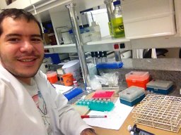 Igor L Gomes-Santos合作论文数Massachusetts General Hospital5
 Ivy Chen合作论文数5Shanmugarajan Krishnan合作论文数5Virginia Spanoudaki合作论文数4
 Kosuke Kawaguchi合作论文数4Nicole Henning合作论文数4Meghan J O'Melia合作论文数Emory Univ4
 Raphael Ferreira合作论文数Department of Genetics, Harvard Medical School4
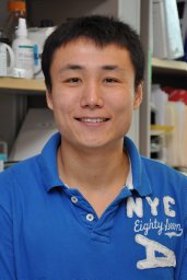 Hengbo Zhou合作论文数Department of Pharmacology, University of Colorado–Denver, Aurora, USA4Ahmed Ali合作论文数4Michelle J Wu合作论文数4Pereira Ethel R合作论文数Department of Genetics &Tumor Cell Biology, St. Jude Children's Research Hospital;Department of Genetics & Tumor Cell Biology, St. Jude Children's Research Hospital4Sivanand Sharanya合作论文数Department of Developmental Biology, University of Texas Southwestern Medical Center4
 Sanchez-Rivera Francisco J合作论文数4
 Kangsan Roh合作论文数Department of Genetic Engineering, Sungkyunkwan University4
 Gültekin Yetiş合作论文数Center for Life Sciences and Technology Research, Bogazici University4
 Do Brian T合作论文数4
 Dennis Hugh A Jones合作论文数Yale University, Yale Univ.4
 Somin Lee合作论文数4




