"The liver is the origin of the veins and the source of the blood."-De Usu Partium Galen.Galen's hepatocentric physiology foreshadowed the liver-brain axis, a network linking vagal signaling, cytokines, metabolites, and migratory immune cells. Central to the immune arm of this axis, the liver generates 25 to 50% of thoracic duct lymph, positioning hepatic lymphatic output as a significant, though not yet fully characterized, source of T cells and cytokines in systemic circulation, a contribution that increases with hepatic inflammation in chronic metabolic diseases. We propose that T lymphocytes are the key cellular effectors through which inflammation originating in the liver, propagating systemically, a process we term hepatosystemic inflammation, reaches the central nervous system (CNS). In metabolic dysfunction-associated steatotic liver disease, hepatosystemic inflammation generates peripheral "push signals" that may license T cells for CNS access, while neuroinflammatory "pull signals" at CNS barrier interfaces may establish permissive entry points. In conclusion, we propose that T cell licensing by hepatosystemic signals represents the immune arm of the liver-brain axis and a mechanistic bridge between metabolic liver disease and neuroimmune dysfunction and associated cognitive decline, and identifies candidate therapeutic targets along this cascade.
Integrin-targeted therapies are under investigation for HIV-associated neuroinflammation, yet their effect on CNS antiviral immunity remains undefined. We examined the role of alpha 4 integrin in T cell-mediated neuroimmune surveillance using SIV-infected macaques with alpha 4 blockade and T cell-specific alpha 4-deficient mice. In macaques, alpha 4 blockade preserved CD4* Th1 cell access to the brain parenchyma but impaired CD8 effector recruitment, disrupting antiviral control. Despite stable cerebrospinal fluid viral loads, hippocampal SIV RNA increased under blockade. Single-cell analyses revealed alpha 4 enrichment in CD8 effector memory (Tem) cells; blockade reduced inferred CD8* Tem-monocyte interactions and heightened innate immune activation in the hippocampus. Microscopy demonstrated persistent SIV-induced microglial simplification despite treatment. Th1 CD4 effectors correlated positively with gray matter viral RNA, whereas alpha 4 beta 7* CD8* T cells correlated inversely, implicating impaired CD8* Tem recruitment in elevated parenchymal viral burden. In mice, alpha 4 proved dispensable for CD4 trafficking to inflamed brain but essential for CD8 effector access across CNS compartments and for both subsets to reach skull marrow. These findings establish that alpha 4 integrin governs CD8-mediated neuroimmune surveillance through coordinated cellular positioning, with blockade enabling viral seeding while disrupting spatially organized antiviral defense.
The immunosuppressive T regulatory cells (Tregs) regulate immune responses and maintain immune homeostasis, yet their functions in metabolic dysfunction-associated steatotic liver disease (MASLD) remain controversial. Here we report increased accumulation of Tregs and effector T cells within the liver parenchyma of mice fed a Western diet (WD). This pattern was also observed in MASH patients, where an increase in intrahepatic Tregs was noted. In the absence of adaptive immune cells in Rag1 KO mice, WD promoted accumulation of intrahepatic neutrophils and macrophages and exacerbated hepatic inflammation and fibrosis. Similarly, targeted Treg depletion exacerbated WD-induced hepatic inflammation and fibrosis. In Treg-depleted mice, hepatic injury was associated with increased accumulation of neutrophils, macrophages, and activated T cells in the liver. Conversely, induction of Treg numbers using recombinant IL2/αIL2 mAb cocktail reduced hepatic steatosis, inflammation, and fibrosis in WD-fed mice. Analysis of intrahepatic Tregs from WD-fed mice revealed a phenotypic signature of impaired Treg function in MASLD. Ex vivo functional studies showed that glucose and palmitate, but not fructose, impaired the immunosuppressive ability of Treg cells. The findings indicate that the liver microenvironment in MASLD impairs the ability of Tregs to suppress effector immune cell activation, thus perpetuating chronic inflammation and driving MASLD progression.
Immunometabolism shapes vaccine immunogenicity, yet how HIV vaccine platforms affect metabolic programs and whether those programs predict immunogenicity remain poorly understood. We applied untargeted serum metabolomics to nine rhesus macaques receiving an HIV-1 DNA-prime/protein-boost regimen, sampling at weeks 0, 1, 2, and 8 after first protein boost. Vaccination induced acute and sustained metabolic changes. Lipid metabolism showed the broadest perturbation: free fatty acids were transiently depleted during the effector phase, while acylcholines, lysophospholipids, and endocannabinoid-like ethanolamides remained elevated through week 8. Glutamate, γ-glutamyl amino acids, and glycylvaline were elevated through week 8, with continued nitrogen recycling, glutathione turnover, and proteolytic activity. High antibody responders showed greater mitochondrial fatty acid oxidation and lower bile acid levels at baseline, and greater membrane lipid remodeling at the effector phase. These data define metabolic signatures of HIV vaccination and identify serum correlates of immunogenicity for prospective validation.
Integrin-targeted therapies are under investigation for HIV-associated neuroinflammation, yet their effect on CNS antiviral immunity remains undefined. We examined the role of α4 integrin in T cell-mediated neuroimmune surveillance using SIV-infected macaques with α4 blockade and T cell-specific α4-deficient mice. In macaques, α4 blockade preserved CD4+ Th1 cell access to the brain parenchyma but impaired CD8 effector recruitment, disrupting antiviral control. Despite stable cerebrospinal fluid viral loads, hippocampal SIV RNA increased under blockade. Single-cell analyses revealed α4 enrichment in CD8 effector memory (Tem) cells; blockade reduced inferred CD8+ Tem-monocyte interactions and heightened innate immune activation in the hippocampus. Microscopy demonstrated persistent SIV-induced microglial simplification despite treatment. Th1 CD4 effectors correlated positively with gray matter viral RNA, whereas α4β7+ CD8+ T cells correlated inversely, implicating impaired CD8+ Tem recruitment in elevated parenchymal viral burden. In mice, α4 proved dispensable for CD4 trafficking to inflamed brain but essential for CD8 effector access across CNS compartments and for both subsets to reach skull marrow. These findings establish that α4 integrin governs CD8-mediated neuroimmune surveillance through coordinated cellular positioning, with blockade enabling viral seeding while disrupting spatially organized antiviral defense.
First-line immune checkpoint inhibitor (ICI) combinations show responses in subsets of hepatocellular carcinoma (HCC) patients. Nearly half of HCCs are Wnt-active with mutations in CTNNB1 (encoding for β-catenin), AXIN1/2, or APC, and demonstrate heterogeneous and limited benefit to ICI due to an immune excluded tumor microenvironment. We show significant tumor responses in multiple β-catenin-mutated immunocompetent HCC models to a novel siRNA encapsulated in lipid nanoparticle targeting CTNNB1 (LNP-CTNNB1). Both single-cell and spatial transcriptomics reveal cellular and zonal reprogramming, along with activation of immune regulatory transcription factors IRF2 and POU2F1, re-engaged type I/II interferon signaling, and alterations in both innate and adaptive immunity upon β-catenin suppression with LNP-CTNNB1 at early- and advanced-stage disease. Moreover, ICI enhances response to LNP-CTNNB1 in advanced-stage disease by preventing T cell exhaustion and through formation of lymphoid aggregates (LA). In fact, expression of an LA-like gene signature prognosticates survival for patients receiving atezolizumab plus bevacizumab in the IMbrave150 phase III trial and inversely correlates with CTNNB1-mutatational status in this patient cohort. In conclusion, LNP-CTNNB1 is efficacious as monotherapy and in combination with ICI in CTNNB1-mutated HCCs through impacting tumor cell-intrinsic signaling and remodeling global immune surveillance, providing rationale for clinical investigations.
Integrin α4β7+ T cells perpetuate tissue injury in chronic inflammatory diseases, yet their role in hepatic fibrosis progression remains poorly understood. Here, we report increased accumulation of α4β7+ T cells in the liver of people with cirrhosis relative to disease controls. Similarly, hepatic fibrosis in the established mouse model of CCl4-induced liver fibrosis was associated with enrichment of intrahepatic α4β7+ CD4 and CD8 T cells. Monoclonal antibody (mAb)-mediated blockade of α4β7 or its ligand mucosal addressin cell adhesion molecule (MAdCAM)-1 attenuated hepatic inflammation and prevented fibrosis progression in CCl4-treated mice. Improvement in liver fibrosis was associated with a significant decrease in the infiltration of α4β7+ CD4 and CD8 T cells, suggesting that α4β7/MAdCAM-1 axis regulates both CD4 and CD8 T cell recruitment to the fibrotic liver, and α4β7+ T cells promote hepatic fibrosis progression. Analysis of hepatic α4β7+ and α4β7- CD4 T cells revealed that α4β7+ CD4 T cells were enriched for markers of activation and proliferation, demonstrating an effector phenotype. The findings suggest that α4β7+ T cells play a critical role in promoting hepatic fibrosis progression, and mAb-mediated blockade of α4β7 or MAdCAM-1 represents a promising therapeutic strategy for slowing hepatic fibrosis progression in chronic liver diseases.
CD4+ T cells survey and maintain immune homeostasis in the brain, yet their differentiation states and functional capabilities remain unclear. Our approach, combining single-cell transcriptomic analysis, ATAC-Seq, spatial transcriptomics, and flow cytometry, revealed a distinct subset of CCR7+ CD4+ T cells resembling lymph node central memory (TCM) cells. We observed chromatin accessibility at the CCR7, CD28, and BCL-6 loci, defining molecular features of TCM. Brain CCR7+ CD4+ T cells exhibited recall proliferation and interleukin-2 production ex vivo, showcasing their functional competence. We identified the skull bone marrow as a local niche for these cells alongside CNS border tissues. Sequestering TCM cells in lymph nodes using FTY720 led to reduced CCR7+ CD4+ T cell frequencies in the cerebrospinal fluid, accompanied by increased monocyte levels and soluble markers indicating immune activation. In macaques chronically infected with SIVCL757 and experiencing viral rebound due to cessation of antiretroviral therapy, a decrease in brain CCR7+ CD4+ T cells was observed, along with increased microglial activation and initiation of neurodegenerative pathways. Our findings highlight a role for CCR7+ CD4+ T cells in CNS immune surveillance, and their decline during chronic SIV highlights their responsiveness to neuroinflammation.
Background/Aim: Activin A is involved in the pathogenesis of human liver diseases, but its therapeutic targeting is not fully explored. Here, we tested the effect of novel, highly specific small-molecule-based activin A antagonists (NUCC-474/555) in improving liver regeneration following partial hepatectomy and halting fibrosis progression in models of chronic liver diseases (CLDs). Methods: Cell toxicity of antagonists was determined in rat hepatocytes and Huh-7 cells using the 3-(4,5-dimethylthiazol-2-yl)-2,5-diphenyl-2H-tetrazolium bromide assay. Hepatocytes and hepatic stellate cells (HSCs) were treated with activin A and NUCC-555 and analyzed by reverse transcription–polymerase chain reaction and immunohistochemistry. Partial hepatectomized Fisher (F)344 rats were treated with NUCC-555, and bromodeoxyuridine (BrdU) incorporation was determined at 18/24/36/120/240 h. NUCC-555 was administered into thioacetamide- or carbon tetrachloride-treated F344 rats or C57BL/6 mice, and the fibrosis progression was studied. Results: NUCC-474 showed higher cytotoxicity in cultured hepatic cells; therefore, NUCC-555 was used in subsequent studies. Activin A-stimulated overexpression of cell cycle-/senescence-related genes (e.g., p15INK4b, DEC1, Glb1) was near-completely reversed by NUCC-555 in hepatocytes. Activin A-mediated HSC activation was blocked by NUCC-555. In partial hepatectomized rats, antagonizing activin A signaling resulted in a 1.9-fold and 2.3-fold increase in BrdU+ cells at 18 and 24 h, respectively. Administration of NUCC-555 in rats and mice with progressing fibrosis significantly reduced collagen accumulation (7.9-fold), HSC activation indicated by reduced alpha smooth muscle actin+ and vimentin+ cells, and serum aminotransferase activity. Conclusions: Our studies demonstrate that activin A antagonist NUCC-555 promotes liver regeneration and halts fibrosis progression in CLD models, suggesting that blocking activin A signaling may represent a new approach to treating people with CLD.
Background and aims The immunosuppressive T regulatory cells (Tregs) regulate immune responses and maintain immune homeostasis, yet their functions in nonalcoholic fatty liver disease (NAFLD) pathogenesis remains controversial. Methods Mice were fed a normal diet (ND) or a western diet (WD) for 16 weeks to induce NAFLD. Diphtheria toxin injection to deplete Tregs in Foxp3DTR mice or Treg induction therapy in WT mice to augment Treg numbers was initiated at twelve and eight weeks, respectively. Liver tissues from mice and NASH human subjects were analyzed by histology, confocal imaging, and qRT-PCR. Results WD triggered accumulation of adaptive immune cells, including Tregs and effector T cells, within the liver parenchyma. This pattern was also observed in NASH patients, where an increase in intrahepatic Tregs was noted. In the absence of adaptive immune cells in Rag1 KO mice, WD promoted accumulation of intrahepatic neutrophils and macrophages and exacerbated hepatic inflammation and fibrosis. Similarly, targeted Treg depletion exacerbated WD-induced hepatic inflammation and fibrosis. In Treg-depleted mice, hepatic injury was associated with increased accumulation of neutrophils, macrophages, and activated T cells within the liver. Conversely, induction of Tregs using recombinant IL2/αIL2 mAb cocktail reduced hepatic steatosis, inflammation, and fibrosis in WD-fed mice. Analysis of intrahepatic Tregs from WD-fed mice revealed a phenotypic signature of impaired Treg function in NAFLD. Ex vivo functional studies showed that glucose and palmitate, but not fructose, impaired the immunosuppressive ability of Treg cells. Conclusions Our findings indicate that the liver microenvironment in NAFLD impairs ability of Tregs to suppress effector immune cell activation, thus perpetuating chronic inflammation and driving NAFLD progression. These data suggest that targeted approaches aimed at restoring Treg function may represent a potential therapeutic strategy for treating NAFLD. Lay summary In this study, we elucidate the mechanisms contributing to the perpetuation of chronic hepatic inflammation in nonalcoholic fatty liver disease (NAFLD). We show that dietary sugar and fatty acids promote chronic hepatic inflammation in NAFLD by impairing immunosuppressive function of regulatory T cells. Finally, our preclinical data suggest that targeted approaches aimed at restoring T regulatory cell function have the potential to treat NAFLD.
Background & AimsIntrahepatic cholangiocarcinoma (ICC) is a devastating liver cancer with extremely high intra- and inter-tumoral molecular heterogeneity, partly due to its diverse cellular origins. We investigated clinical relevance and the molecular mechanisms underlying hepatocyte (HC)-driven ICC development.MethodsExpression of ICC driver genes in human diseased livers at risk for ICC development were examined. The sleeping beauty and hydrodynamic tail vein injection based Akt-NICD/YAP1 ICC model was used to investigate pathogenetic roles of SRY-box transcription factor 9 (SOX9) and yes-associated protein 1 (YAP1) in HC-driven ICC. We identified DNA methyltransferase 1 (DNMT1) as a YAP1 target, which was validated by loss- and gain-of-function studies, and its mechanism addressed by chromatin immunoprecipitation sequencing.ResultsCo-expression of AKT and Notch intracellular domain (NICD)/YAP1 in HC yielded ICC that represents 13% to 29% of clinical ICC. NICD independently regulates SOX9 and YAP1 and deletion of either, significantly delays ICC development. Yap1 or TEAD inhibition, but not Sox9 deletion, impairs HC-to-biliary epithelial cell (BEC) reprogramming. DNMT1 was discovered as a novel downstream effector of YAP1-TEAD complex that directs HC-to-BEC/ICC fate switch through the repression of HC-specific genes regulated by master regulators for HC differentiation, including hepatocyte nuclear factor 4 alpha, hepatocyte nuclear factor 1 alpha, and CCAAT/enhancer-binding protein alpha/beta. DNMT1 loss prevented NOTCH/YAP1-dependent HC-driven cholangiocarcinogenesis, and DNMT1 re-expression restored ICC development following TEAD repression. Co-expression of DNMT1 with AKT was sufficient to induce tumor development including ICC. DNMT1 was detected in a subset of HCs and dysplastic BECs in cholestatic human livers prone to ICC development.ConclusionWe identified a novel NOTCH-YAP1/TEAD-DNMT1 axis essential for HC-to-BEC/ICC conversion, which may be relevant in cholestasis-to-ICC pathogenesis in the clinic.
Intrahepatic cholangiocarcinoma (iCCA) is a liver tumor of increasing incidence and devastating prognosis. The most critical barrier for developing a globally effective therapy for iCCA is molecular heterogeneity caused by diverse molecular drivers and cellular origins in the liver. Clinically, over 90% of iCCAs show alterations of YAP1 and/or SOX9. Therefore, we investigated the therapeutic potential of co‐repression of YAP1 and SOX9 as a wide range therapeutic option for advanced iCCA.
Hepatosteatosis, a hallmark of alcoholic and nonalcoholic fatty liver disease (NAFLD), is characterized by accumulation of lipid storage organelles called lipid droplets (LDs) within hepatocytes. LDs primarily function to maintain cellular metabolism and protect cells from lipotoxicity by storing excess free fatty acids and cholesterol esters during nutrient excess. Conversely, LDs serve as energy sources during periods of nutrient deprivation or tissue growth.1 During chronic or excess caloric intake when lipid availability exceeds the capacity of LDs to sequester excess free fatty acids, LDs accumulate in hepatocytes.
Nonalcoholic fatty liver disease (NAFLD) is a spectrum of conditions ranging from benign nonalcoholic fatty liver (NAFL) or bland steatosis to the more severe nonalcoholic steatohepatitis (NASH), characterized by excessive inflammation and varying degrees of fibrosis.1 An estimated 20%–25% of patients with NAFL progress to NASH, and up to 25% of patients with NASH progress to cirrhosis.1 Currently, NASH-related cirrhosis is the second leading indication for liver transplantation in the United States, and patients with NASH face an increased risk of developing hepatocellular carcinoma.
Nonalcoholic fatty liver disease (NAFLD) describes a spectrum of progressive liver diseases ranging from simple steatosis to steatohepatitis and fibrosis. Globally, NAFLD is the leading cause of morbidity and mortality associated with chronic liver disease, and NAFLD patients are at a higher risk of developing cirrhosis and hepatocellular carcinoma. While there is a consensus that inflammation plays a key role in promoting NAFLD progression, the underlying mechanisms are not well understood. Recent clinical and experimental evidence suggest that increased hepatic translocation of gut microbial antigens, secondary to diet-induced impairment of the intestinal barrier may be important in driving hepatic inflammation in NAFLD. Here, we briefly review various endogenous and exogenous factors influencing the intestinal barrier and present recent advances in our understanding of cellular and molecular mechanisms underlying intestinal barrier dysfunction in NAFLD.
word count: 146 14 Number of Figures: 8 15 Number of Supplementary Figures: 5 16 Number of Supplementary Tables: 2 17 Number of words in text: 4940 18 19 20 21
Expansion of biliary epithelial cells (BECs) during ductular reaction (DR) is observed in liver diseases including cystic fibrosis (CF), and associated with inflammation and fibrosis, albeit without complete understanding of underlying mechanism. Using two different genetic mouse knockouts of β-catenin, one with β-catenin loss is hepatocytes and BECs (KO1), and another with loss in only hepatocytes (KO2), we demonstrate disparate long-term repair after an initial injury by 2-week choline-deficient ethionine-supplemented diet. KO2 show gradual liver repopulation with BEC-derived β-catenin-positive hepatocytes and resolution of injury. KO1 showed persistent loss of β-catenin, NF-κB activation in BECs, progressive DR and fibrosis, reminiscent of CF histology. We identify interactions of β-catenin, NFκB, and CF transmembranous conductance regulator (CFTR) in BECs. Loss of CFTR or β-catenin led to NF-κB activation, DR, and inflammation. Thus, we report a novel β-catenin-NFκB-CFTR interactome in BECs, and its disruption may contribute to hepatic pathology of CF.
 Rai Ravi P合作论文数Department of Microbiology, Sanjay Gandhi Postgraduate Institute of Medical Sciences14
 Smita S. Iyer合作论文数Emory University, Emory University School of Medicine11
 Liu Silvia合作论文数Department of Biostatistics, University of Pittsburgh School of Medicine10
 Aaron Bell合作论文数10
 Minakshi Poddar合作论文数10Sucha Singh合作论文数10Satdarshan P. Monga合作论文数9Shikai Hu合作论文数9
 Aatur D. Singhi合作论文数Division of Anatomic Pathology, Department of Pathology, School of Medicine, University of Pittsburgh;Developmental Laboratory, University of Pittsburgh Medical Center9Sungjin Ko合作论文数Univ Pittsburgh, Pittsburgh, PA 15260 USA8Frank A. Anania合作论文数7
 Pradeep Kumar合作论文数7Yunshan Liu合作论文数7Junyan Tao合作论文数7Biki Gupta合作论文数6Nejak-Bowen Kari N合作论文数6
 Satdarshan Paul Singh Monga合作论文数Department of Pathology, School of Medicine, University of Pittsburgh6Michael Oertel合作论文数6
 Daniel Chopyk合作论文数4
 Laura Molina合作论文数Sch Med, Univ Pittsburgh4Smith Tekla合作论文数4Russell Jacquelyn合作论文数4Mohammed Hassan合作论文数4Anya Singh Varma合作论文数3
 Pabitra Bikash Pal合作论文数3Sudrishti Chaudhary合作论文数3Karis Kosar合作论文数3S. Ott合作论文数University of California3Zhong-Min Ma合作论文数Univ Calif Davis, Ctr Comparat Med, Davis, CA 95616 USA3Dhivyaa Rajasundaram合作论文数3
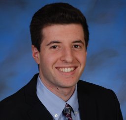 Brandon M Lehrich合作论文数Department of Otolaryngology–Head and Neck Surgery, University of California3
 Asma Nusrat合作论文数Department of Pathology, University of Michigan Medical School3
 Beckman Danielle合作论文数Institute of Medical Biochemistry Leopoldo de Meis, Federal University of Rio de Janeiro3
 John H. Morrison合作论文数California National Primate Research Center, University of California, Davis;Department of Neurology, Davis School of Medicine, University of California, Davis3
 Charles Parkos合作论文数Parkos & Nusrat Labs, Department of Pathology, Medical School, University of Michigan3Chirayu Desai合作论文数BRD School of Biosciences, Sardar Patel University3Catherine Cao合作论文数2Matthew Rolston合作论文数2Yekaterina Krutsenko合作论文数2
 Anil K. Verma合作论文数2Edward Hurley合作论文数2Shuchang Liu合作论文数2
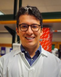 Sonny R Elizaldi合作论文数Univ Calif Davis, Ctr Immunol & Infect Dis, Davis, CA 95616 USA2
 Anil Verma合作论文数Univ Calif Davis, Ctr Immunol & Infect Dis, Davis, CA 95616 USA2
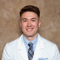 Tyler M Yasaka合作论文数Department of Otolaryngology–Head and Neck Surgery, University of California2
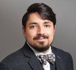 Giovanne Baroni Diniz合作论文数University of California2
 Ashok R Dinasarapu合作论文数Emory University2Durbin-Johnson Blythe合作论文数Dept Publ Hlth Sci, Univ Calif Davis2
 Alton Brad Farris合作论文数Dept Pathol & Lab Med, Emory Univ2




