We report on the fabrication and femtosecond-laser-damage characterization of ion-beam-sputtered TiO 2 /SiO 2 multilayer mirror coatings designed for operation at 1030 nm under high-repetition-rate irradiation. The 41-layer dielectric stack deposited on fused silica substrates demonstrates a non-damaging fluence threshold of 0.80J/cm 2 under 275 fs pulse irradiation at 1 MHz repetition rate. No measurable optical degradation or structural modification was observed following exposure exceeding 10 9 laser pulses at sub-threshold fluence. Damage probability exhibits a sharp transition between 0.80 and 1.00J/cm 2 , with catastrophic optical damage occurring at 1.00J/cm 2 . The coatings maintain transmission below 0.008% throughout testing, indicative of minimal absorption and high reflectivity stability. Despite the lower intrinsic bandgap of TiO 2 compared to conventionally preferred high-index oxides, the demonstrated performance approaches that of high-bandgap multilayer systems, indicating that optimized multilayer engineering and defect suppression can compensate for intrinsic material limitations. These results establish TiO 2 /SiO 2 multilayer coatings as viable candidates for high-power, MHz-regime femtosecond laser systems requiring both high damage resistance and long-term operational stability.
In order to maintain the heritage-listed Sydney Harbour Bridge, the steel surface must be regularly protected with a paint coating. Before applying a new coating, the steel needs to be cleaned of the previous paint layer and any contaminants, such as rust that may have formed. Laser ablation offers a more efficient method for cleaning steel compared with traditional techniques, provided that the underlying steel does not experience changes in microstructure or mechanical properties. This study investigated the effects of nanosecond laser cleaning on the steel components of this iconic bridge. To provide a comprehensive evaluation, the steel samples subjected to nanosecond laser ablation at 120 Hz were characterized by detailed residual stress analysis (including subsurface measurements), surface profiling, microstructural characterization, and hardness testing. The results show that increasing the number of laser passes promotes recrystallisation at the ablated surface and leads to an increase in surface hardness, accompanied by tensile residual stresses confined to the top surface layer. In addition, residual stress measurements indicate that subsurface and through-thickness regions beyond a depth of 0.5 mm are largely unaffected. These findings indicate that while nanosecond laser ablation does not influence residual stresses beyond a depth of 0.5 mm in the steel components, the substantial tensile residual stresses introduced at the steel surface may have implications for fatigue performance. This should be carefully considered in engineering assessments and is likely to limit the applicability of this technology for the conservation of steel structures. (c) 2026 The Authors. Published by Elsevier Masson SAS. This is an open access article under the CC BY-NC-ND license (http://creativecommons.org/licenses/by-nc-nd/4.0/)
This study explores the use of femtosecond laser pulses to remove iron ore slurry used by mining industries to stabilise blast-hole structures, aiming to preserve wall stability and the chemical composition of the underlying rock. In blast holes, minerals are often coated with deposits such as dust or slurry, which must be removed to analyse the rock beneath. The ablation depth per pulse and ablation efficiency of the slurry were assessed, followed by studies on the ablation thresholds and rates of economically significant Australian rocks, including banded iron, limonite, goethite, shale, and hematite. Surface damage was evaluated using optical microscopy, profilometry, colourimetry, VIS/NIR spectroscopy, and Fourier Transform Infrared spectroscopy (FTIR) spectroscopy. Changes in the Fe3+ electronic transition band at 0.60 mu m suggest alterations in iron's oxidation state, while shifts in OH bands at 3400 cm(-1) indicate water or molecular OH loss. Surface discoloration was observed, and safe laser fluence thresholds were established to minimise this effect. Despite these changes, the rocks remained identifiable, highlighting the potential of femtosecond laser cleaning for rock analysis with minimal alteration.
Fast quenching dynamics in confined laser-induced microexplosions have been shown to lead to localized shockwaves that can create nanometre-scale domains in novel high-pressure crystalline phases. In the case of silicon, new silicon polymorphs such as bt8-Si and st12-Si have been recently observed, which are predicted to have bandgaps desirable for photovoltaic applications. Identification of these phases has been previously achieved by analysis of selected-area electron diffraction (SAED) patterns taken from laser-shock-affected areas. However, this analysis was complicated by pattern overlap from the many crystallites in the selected area, and many spots were found to agree with multiple potential phases. To overcome this ambiguity and enable the identification of the phase of Bragg spots observed in SAED patterns from polymorphic nanomaterials, we developed a new algorithm that we termed poly. This method is based on maximizing the magnitude and angular correlation between observed diffraction spots and those values derived from a known potential phase. We present the performance of this algorithm on simulated electron diffraction patterns as well as experimental SAED patterns measured from laser-shock-affected silicon samples. We find that the most abundant phases in the affected areas are t32-Si and t32*-Si and report on their relaxation into other high-pressure silicon phases over the course of 90 days after the laser-induced confined microexplosion.
Nanosecond laser ablation generates steep thermal gradients and residual stresses that influence the performance of structural steel. This study develops a finite element (FEM) framework for multi-pulse, multi-scan laser ablation, extending beyond conventional single-spot models to more realistically capture industrial practice. Experiments were performed on Sydney Harbour Bridge steel using a 1000 W Q-switched Nd:YAG laser. Residual stresses were characterized by X-ray diffraction (XRD) and neutron diffraction (ND), with back-surface temperatures were also recorded for validation. XRD measurements revealed similar to 220 MPa tensile stress at the surface, in close agreement with the FEM prediction of similar to 215 MPa when averaged over the XRD sampling depth. Beyond 0.5 mm, ND measurements indicated negligible residual stresses within +/- 30 MPa, a trend also reproduced by the simulation. These results demonstrate that laser-induced residual stresses are confined to the top similar to 0.5 mm of the material. Furthermore, the simulated back-surface temperature after 20 laser passes (363 K) closely matched the experimental value (369 K), confirming the model's thermal accuracy. This combined experimental-numerical approach provides a validated framework for predicting residual stresses in laser-processed steels and supports the optimization of short-pulse laser processing for both research and industrial applications.
Laser cleaning has recently gained attention as a non-contact, precise, and environmentally friendly method for removing contaminants, oxides, or coatings from surfaces using controlled laser ablation. However, even at low fluence, where no visible damage is observed, thermal effects from nanosecond laser pulses can induce residual stress, potentially affecting the long-term reliability of the material. This study introduces a finite element method model to predict residual stress formation in single-pulse laser ablation of stainless steel, providing detailed insight into the localized thermomechanical response. The model captures individual pulse effects, including stress evolution, thermal expansion, and localized plastic deformation. Experimental validation via X-ray diffraction is also performed. Based on this model, a parametric study is conducted to investigate the influence of pulse duration and fluence on residual stress development. The results reveal how thermal input parameters affect stress field formation and redistribution, even in the absence of visible material removal. These findings enhance understanding of stress generation in laser-processed metals and offer a framework for predicting residual stress and identifying truly damage-free thresholds in metallic substrates.
Recent research and development into the formation of nanoscale channels as a central component of nanofluidic biochip systems revolutionized the biological and chemical fields. Exploration of new pathways to form nanochannels is increasingly necessary to provide a new generation of analytical tools with accurate control of liquid fluid flow, high selectivity and increased mass flow rate. Here, we demonstrate that a single 9-keV pulse from X-ray free-electron-laser can form a nanoscale mm-long cavity in LiF. The laser-generated shock pressure results in channel formation with >1,000 length-to-diameter aspect ratio. The development of void is analyzed via continuum and atomistic simulations revealing a sequence of processes leading to the final long cavity structure. This work presents the study of mm-long nanochannel formation by a single high-brilliance X-ray free-electron laser pulse. With MHz repetition rate X-ray free electron laser opens a new avenue for the development of lab-on-chip applications in any material, including those non-transparent to optical lasers.
Silicon polymorphs with exotic electronic and optical properties have recently attracted significant attention due to their wide range of useful band gap characteristics. They are typically formed by static high-pressure techniques, which limits the crystal structures that can be made. This constitutes a major obstacle to study these polymorphs and their incorporation into existing technology. Approaches have attempted to address this shortcoming through using dynamic conditions and chemical precursor materials. Here, we report on an approach to create unusual crystal structures deep in the bulk of a silicon crystal by irradiating it with a laser pulse at ultrarelativistic intensity of up to 7 . 5 x 10 19 W/cm 2 . Laser -generated electrons with MeV energy swiftly penetrate the target with speed close to the speed of light and deposit their energy into a large volume across the whole thickness of the sample. The relativistic electron current creates, via branching propagation and ionization, high -energy -density conditions for thermodynamically nonequilibrium phase transformation paths into new crystal polymorphs. X-ray microdiffraction and synchrotron x-ray diffraction analyses indicate, along with conventional dc -Si, the presence of exotic silicon structures in the bulk of the laser intact target volume. These structures are identified as body -centered bc8-Si, rhombohedral r8 -Si, hexagonal -diamond hd-Si, and the tetragonal Si -VIII, all phases of Si that have previously been made through static techniques. Additionally, simple -tetragonal st12-Si and body -centered tetragonal bt8-Si were observed along with signatures of not yet identified diffraction spots. Both st12-Si and bt8-Si have only been observed in ultrafast laser microexplosion conditions at much lower laser intensity similar to 10 14 W/cm 2 and within a micron -thin surface layer. The findings here are supported by direct observation of nanoparticles with high -resolution transmission electron microscopy and corresponding fast Fourier transform analysis of their interatomic distances. The presented analyses of absorbed laser energy, generation of the MeV electron current, and deposition of energy across the whole target thickness provide a solid basis for drawing the conclusion that the observed silicon polymorphs were produced because of laser -generated high-energy electrons fast -penetrating deeply into the bulk of silicon. In contrast to solid -solid transformations, the plasma -solid transitions offer a paradigm for the creation of exotic, high-energy density materials inside the bulk of the sample by using laser pulses at relativistic intensities.
Sub-picosecond optical laser processing of metals is actively utilized for modification of a heated surface layer. But for deeper modification of different materials a laser in the hard x-ray range is required. Here, we demonstrate that a single 9-keV x-ray pulse from a free-electron laser can form a um-diameter cylindrical cavity with length of ~1 mm in LiF surrounded by shock-transformed material. The plasma-generated shock wave with TPa-level pressure results in damage, melting and polymorphic transformations of any material, including transparent and non-transparent to conventional optical lasers. Moreover, cylindrical shocks can be utilized to obtain a considerable amount of exotic high-pressure polymorphs. Pressure wave propagation in LiF, radial material flow, formation of cracks and voids are analyzed via continuum and atomistic simulations revealing a sequence of processes leading to the final structure with the long cavity. Similar results can be produced with semiconductors and ceramics, which opens a new pathway for development of laser material processing with hard x-ray pulses.
Objectives: To develop and practically test high-precision femtosecond laser ablation models for dental hard tissue that are useful for detailed planning of automated laser dental restorative treatment. Methods: Analytical models are proposed, derived, and demonstrated for practical calculation of ablation rates, ablation efficiency and ablated morphology of human dental enamel and dentin using femtosecond lasers. The models assume an effective optical attenuation coefficient for the irradiated material. To achieve ablation, it is necessary for the local energy density of the attenuated pulse in the hard tissue to surpass a predefined threshold that signifies the minimum energy density required for material ionization. A 1029 nm, 40 W carbide 275 fs laser was used to ablate sliced adult human teeth and generate the data necessary for testing the models. The volume of material removed, and the shape of the ablated channel were measured using optical profilometry. Results: The models fit with the measured ablation efficiency curve against laser fluence for both enamel and dentin, correctly capturing the fluence for optimum ablation and the volume of ablated material per pulse. The detailed shapes of a 400-micrometer wide channel and a single-pulse width channel are accurately predicted using the superposition of the analytical result for a single pulse. Conclusions: The findings have value for planning automated dental restorative treatment using femtosecond lasers. The measurements and analysis give estimates of the optical properties of enamel and dentin irradiated with an infrared femtosecond laser at above-threshold fluence and the proposed models give insight into the physics of femtosecond laser processing of dental hard tissue.
We investigated the effect of femtosecond (fs) laser ablation of enamel and dentin for different pulse wavelengths: infrared (1030 nm), green (515 nm), and ultra-violet (343 nm) and for different pulse separations to determine the optimal irradiation conditions for the precise removal of dental hard tissues with the absence of structural and compositional damage. The ablation rates and efficiencies were established for all three laser wavelengths for both enamel and dentin at room temperature without using any irrigation or cooling system, and the surfaces were assessed with optical and scanning electron microscopy, optical profilometry, and Raman spectroscopy. We demonstrated that 515 nm fs irradiation provides the highest rate and efficiency for ablation, followed by infrared. Finally, we explored the temperature variations inside the dental pulp during the laser procedures for all three wavelengths and showed that the maximum increase at the optimum conditions for both infrared and green irradiations was 5.5 °C, within the acceptable limit of temperature increase during conventional dental treatments. Ultra-violet irradiation significantly increased the internal temperature of the teeth, well above the acceptable limit, and caused severe damage to tooth structures. Thus, ultra-violet is not a compatible laser wavelength for femtosecond teeth ablation.
Cleaning with laser light has become a popular technique for the removal of unwanted surface layers. It provides numerous benefits compared to conventional cleaning methods, such as avoiding the use of abrasives and chemicals and eliminating problems of corrosive residues and loss of surface detail. Conventional pulse lasers are the most widely used, and already commercially available, with portable units deployable on-site. However, those lasers rely on thermal mechanisms of ablation, which generate heat and shock waves that can result in undesirable side-effects such as melting, formation of cracks, exfoliation of flakes from the surface, and annealing/softening of thinner sections of the bulk material. Here we explore an alternative heat-free femtosecond laser cleaning technique based on powerful ultrashort pulse lasers. We discuss the capability of the technique, illustrating the significant advantages of femtosecond pulse lasers in removal of old paint and rust without alteration of underlying structure, and discuss the development of a portable femtosecond laser cleaning unit for the maintenance and preservation of large-scale assets around the world using Sydney Harbour Bridge as a real-world field test.
We explore femtosecond laser cleaning of materials used in the construction of historic monuments, such as stone and steel covered in typical contaminants caused by harsh environments that may be found in urban areas. We address the cleaning of these materials from a conservation perspective, taking as examples the preservation and cleaning of iconic structures such as the steel and the granite of the Sydney Harbour Bridge, Hawkesbury sandstone, a popular building material of a variety of monuments in Sydney (Australia), Makrana marble taken from the Soami Bagh Samadh temple of Agra in India, and also graffiti removal. We demonstrate that femtosecond laser pulses can clean a range of different contaminants such as biofilm, environmental soiling, rust, and spray paints, while preserving the integrity of the underlying substrates. Femtosecond laser cleaning is a fast and effective method and a safer alternative to lasers with longer pulse durations for the preservation of historic monuments.
This paper presents for the first time a micro-size MEMS mirror design that achieves large rotational mechanical angles of two axes using single electromagnetic field. The mirror features special packaging method that corrects the coupling effect between the axes, resulting in highly stable mechanical angle response. Additionally, the mirror includes honeycomb structures on the backside that distribute weight uniformly, improving stability. At resonant mode, The MEMS mirror reaches rotational mechanical angles of 10 degrees in the fast axis and 20 degrees in the slow axis with the current of mA order, which allows this mirror working for close-range scanning applications, such as dental ablation. The design, simulation, and fabrication processes are described, and the experimental results demonstrate this MEMS mirror has the largest mechanical angles which is currently not commercially available.
This study examines the use of heat-free femtosecond pulse laser technology for the cleaning of Makrana marble and semi-precious stones from the Soami Bagh Samadh temple in Agra, India. We determined the ablation thresholds of the semi-precious stones used in the inlay stonework with femtosecond laser pulses and demonstrated that laser ablation can effectively remove dust layers and environmental staining from the marble surfaces without damaging the original material. We demonstrated, by using optical microscopy, colorimetry, scanning electron microscopy and Raman spectroscopy, that femtosecond laser processing of surfaces reduced the risk of thermal damage due to minimal heat generation and allowed the preservation of the original surface structure. This research suggests that femtosecond pulse laser technology can be a sustainable and effective cleaning method for heritage places such as the Holy Samadh temple.
We investigated the removal of spray paint from heritage granite surfaces using a nanosecond pulse laser and compared the results, in terms of the effectiveness of treatment and preservation of the stone surface from potential damage, with results obtained using a femtosecond pulse laser. We show that, with the nanosecond laser, the ablation threshold of the stone was measured at 0.5 J·cm-2, but it was only possible to remove certain types of spray paints, such as blue and green paints, at a higher fluence of 0.9 J·cm-2. To remove other types of paints, such as red and yellow, it was necessary to increase even further the laser fluence to 1.5 J·cm-2, well above the ablation threshold of the stone. In this case, damage was induced in the minerals, such as melting of biotite, and a general roughening of the other minerals’ surfaces was observed. Despite nanosecond pulse lasers being more widespread for cleaning purposes, we demonstrate that femtosecond pulse lasers allowed better effectiveness in removing various colours of paint, without leaving residues, while keeping the laser energy below the damage threshold of the underlying stone, allowing complete preservation of the substrate, and avoiding the melting of the most sensitive mineral grains (particularly biotite). We determined the ablation efficiency of spray paint using the femtosecond pulse laser and found a maximum ablation regime centered around 6.2 J·cm-2, at 8.36 mm3·(min·W)-1. This study highlights the benefits of using a laser with femtosecond pulses instead of longer pulse durations (nanosecond range) for the preservation and maintenance of heritage stone.
We studied femtosecond pulse lasers for cleaning Makrana marble, used in the construction of Soami Bagh Samadh, Agra, India. We investigated laser irradiation at different wavelengths (1029 nm, 515 nm, and 343 nm) and found that ultraviolet radiation had the lowest ablation threshold at 0.23 J cm(-2), followed by green at 0.34 J center dot cm(-2), and infrared at 1.3 J center dot cm(-2). Green irradiation was the most efficient, ablating marble at 0.9 mm(3)center dot(min W)(-1) at 4 J center dot cm(-2) compared to 0.73 mm(3)center dot(min W)(-1) at 12 J center dot cm(-2) for 343 and 1029 nm. We evaluated the wavelength's effect on the stone. Infrared irradiation, having a higher ablation threshold, allowed for a broader range of laser fluence for the safe removal of contaminants, without risking inducing any chemical or morphological changes to the stone, and thus was selected for cleaning. We explored the cleaning effectiveness of infrared femtosecond laser pulses on a sculpted Makrana marble covered by environmental soiling. Optical and scanning electron microscopy, optical profilometry, Raman spectroscopy, and colorimetry demonstrated that satisfactory cleaning was achieved, with the removal of the unwanted layers without causing damage or chemical alteration to the marble structure. This study demonstrates the potential of femtosecond lasers for safe and effective heritage marble cleaning.
Controlling electronic and optical properties of semiconducting materials could substantially expand their functionality and enable new pathways for the formation of ‘smart’ materials for emerging innovative applications. An emerging new path for the synthesis of exotic silicon (Si) polymorphs is irradiation of bulk diamond-cubic Si by ultrashort laser pulses at relativistic intensity above 1019 W/cm2. Exotic polymorphs are formed due to interaction with MeV-energy electrons generated in laser-produced high-temperature plasma state. The unusual Si structures are found in the form of 5–10 nm nanoparticles confined deep in the bulk of 500-μm-thick Si samples. Raman spectroscopy, electron microscopy and X-ray diffraction studies provide an unequivocal evidence of exotic Si phase formation. A key advantage of such relativistic irradiation lies thereby in the fact that large quantities of such exotic Si is synthesised embedded within the bulk diamond-cubic Si and that further the formed Si polymorphs are stable at ambient temperature and pressure. They are thus available for further studies for electronic applications in selective band gap engineering.
High fluence focused femtosecond laser pulses were used to perform fast, high precision and minimally damaging cavity cutting of teeth at room temperature without using any irrigation or cooling system. The optimal ablation rates were established for both enamel and dentin, and the surfaces were assessed with optical and scanning electron microscopy, Raman spectroscopy and optical profilometry. No chemical change in the composition of enamel and dentin was observed. We explored temperature variations inside the dental pulp during the laser procedure and showed the maximum increase was 5.5°C, within the acceptable limit of temperature increase during conventional dental treatments.
 Andrei V Rode合作论文数School of Physical Science and Engineering, Research;Australian National University;School of Physical Science and Engineering, Australian National University30
 Anne Patricia Alloncle合作论文数28
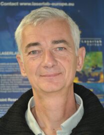 Philippe Delaporte合作论文数Centre National de la Recherche Scientifique27
 Francois Courvoisier合作论文数Optique Department, Femto-st Institute, Centre National de la Recherche Scientifique, Universite de Franche-Comte18
 Remo Giust合作论文数18
 Luca Furfaro合作论文数17
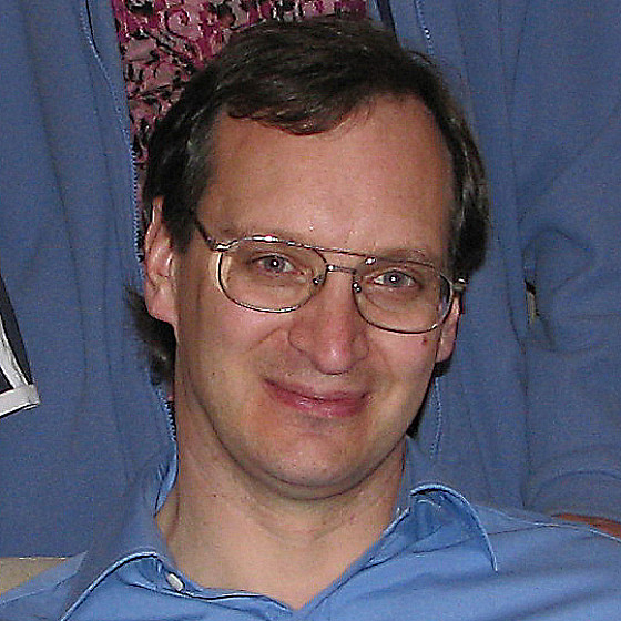 Stephen Madden合作论文数Department of Quantum Science & Technology, Australian National University16
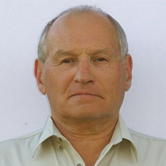 Eugene Gamaly合作论文数Department of Quantum Science & Technology, Research School of Physics, Australian National University15
 Saulius Juodkazis合作论文数School of Science, Computing and Engineering Technologies, Swinburne University of Technology14
 John M. Dudley合作论文数Optics Department, Franche-Comte Electronique Mecanique Thermique et Optique-Sciences et Technologies Institute, Universite de Franche-Comte14
 Christine Videlot-Ackermann合作论文数14
 Julia Brand合作论文数10
 Bianca Haberl合作论文数Neutron Scattering Division, Neutron Sciences Directorate, Oak Ridge National Laboratory10
 Ralph Meyer合作论文数Department of Oncology, Faculty of Health Sciences, McMaster University;Escarpment Cancer Research Institute, McMaster University10
 Alison Wain合作论文数The Australian War Memorial Collection Services Section10
 Sebastien Nenon合作论文数Centre Interdisciplinaire de Nanoscience de Marseille (CINaM),, Université de la Méditerranée9M. Frédéric Fages合作论文数Centre Interdisciplinaire de Nanoscience de Marseill9
 Jim Williams合作论文数College of Science, Australian National University;Acton Semiconductors (Aust) Pty Ltd;Research School of Physical Sciences and Engineering, Australian National University8
 Luc Froehly合作论文数FEMTO-ST8
 Catalin Constantinescu合作论文数Plasma and Radiation Physics PPAM-Lasers Department Bvd. Atomistilor, INFLPR-National Institute for Laser8
 Jodie Bradby合作论文数Physics Education Centre, Research School of Physics, College of Science and Medicine, The Australian National University;Department of Materials Physics, Research School of Physics, College of Science and Medicine, The Australian National University;Australian Institute of Physics8IO Idrissi合作论文数6
 Norimasa Ozaki合作论文数6
 Maxime Jacquot合作论文数CNRS, Univ Franche Comte6
 Abdou Karim Diallo合作论文数6A. V. Rode合作论文数5Rémi Meyer合作论文数5Timothy R Hirst合作论文数5Alaa Habeb合作论文数5Omar Zuaiter合作论文数5
 Heiko Spallek合作论文数School of Dentistry, Faculty of Medicine and Health, The University of Sydney5
 Cyril Billet合作论文数Institut FEMTO-ST, Université Bourgogne Franche Comté5
 Laurence Walsh合作论文数School of Dentistry, Faculty of Health and Behavioural Sciences, The University of Queensland5
 Christophe Cibert合作论文数Faculté des Sciences, UMR No 6638 CNRS5A. Rode合作论文数4Tatiana Pikuz合作论文数4Aki Tominaga合作论文数4
 Lachlan A. Smillie合作论文数Dept Elect Mat Engn, Australian Natl Univ4
 Gwenaelle Proust合作论文数School of Civil Engineering, Faculty of Engineering, The University of Sydney4Pierreambroise Lacourt合作论文数4Ailuno Julie合作论文数4
 Chen Xie合作论文数Tianjin University, Tianjin Univ.4
 Thomas Lippert合作论文数Paul Scherrer Institut, ETH Zurich4
 Chris Pickard合作论文数Materials Theory Group, Department of Materials Science & Metallurgy, University of Cambridge4
 Ryosuke Kodama (兒玉了祐)合作论文数Division of Electrical, Electronic and Information Engineering, Graduate School of Engineering, Osaka University4
 Ksenia Maximova合作论文数4Matthias Nagel合作论文数4Daisuke Sagae合作论文数3Keiichiro Mukai合作论文数3




