Cardiac mapping is a crucial procedure for diagnosing and treating cardiac arrhythmias. Still, current clinical techniques face limitations including insufficient electrode coverage, poor conformability to complex heart chamber geometries, and high costs. This study explores the design, testing, and validation of a 64-electrode soft robotic catheter that addresses these challenges in cardiac mapping. A dual-layer flexible printed circuit board (PCB) was designed and integrated with sensors into a soft robotic sensor array (SRSA) assembly. Design considerations included flex PCB layout, routing, integration, conformity to heart chambers, sensor placement, and catheter durability. Rigorous SRSA in vitro testing evaluated the burst/leakage pressure, block force for electrode contact, mechanical integrity, and environmental resilience. For in vivo validation, a porcine model was used to demonstrate the successful deployment, conformability, and acquisition of electrograms in both the ventricles and atria. This catheter-deployable SRSA represents a meaningful step towards translating the integration of soft robotic actuators and stretchable electronics for clinical use, showcasing the unique mechanical and electrical performance that these designs enable. The high-density electrode array enabled rapid 2 s data acquisition with detailed spatial and temporal resolution, as illustrated by the clear and consistent cardiac signals recorded across all electrodes. The future of this work will lie in enabling high-density, anatomically conformable devices for detailed cardiac mapping to guide ablation therapy and other interventions.
The authors present in this study the development of a novel method for creating stretchable electronics from dual-layer flex printed circuit boards (flex-PCBs) as a platform for soft robotic sensor arrays (SRSAs) for cardiac voltage mapping applications. There is a crucial need for devices that utilize multiple sensors and provide high performance signal acquisition for cardiac mapping. Previously, our group demonstrated how single-layer flex-PCB can be postprocessed to create a stretchable electronic sensing array. In this work, a detailed fabrication process for creating a dual-layer multielectrode flex-PCB SRSA is presented, along with relevant parameters to achieve optimal postprocessing with a laser cutter. The dual-layer flex-PCB SRSA's ability to acquire electrical signals is demonstrated both in vitro as well as in vivo on a Leporine cardiac surface. These SRSAs could be extended into full-chamber cardiac mapping catheter applications. Our results show a significant contribution towards the scalable use of dual-layer flex-PCB for stretchable electronics.
This paper describes the methodology for rapid prototyping of nitinol structures by heat setting. Nitinol is a shape memory alloy commonly used in implantable medical devices. The proposed technique, based on 3D printing, can be used to effectively iterate multiple nitinol designs for different types of medical devices. We describe a rapid and low-cost process of ceramic replica molding of standard 3D printed parts to create high-temperature resistant fixtures, suitable for heat setting of nitinol. The technique represents a low cost (<$20 materials per fixture) and rapid (as quickly as 16 h for a volume less than 1.25 × 105 mm3) method for shaping nitinol, a technique that typically is costly, labor intensive, and requires specialized equipment. Our method satisfies a need for cost-effective, rapid prototyping of nitinol for implantable medical devices, and we show an example set of shaped nitinol wires, clips, and stents. This method is straightforward and can be easily applied by researchers to rapidly iterate medical device designs.
While there is a strong need to assess the safety and efficacy of novel therapies prior to evaluation in human patients, the ability to accurately model the complexities of coronary disease in-vitro and/or animal models is somewhat limited. Animal models, even those with genetic predispositions toward coronary disease do not exhibit high grade stenoses, similar to those that require treatment in human patients. At the same time, most in-vitro models and simulations of fluid-structure interactions cannot simultaneously capture the complex geometries, hemodynamics, and biomechanical response of human coronary disease. Here, we introduce a robust workflow to replicate an in-vitro platform incorporating compliant, realistic diseased coronary arteries in a flow loop under physiological conditions. Through ex-vivo imaging of cadaveric specimens, coupled with co-registered measurements of coronary biomechanics, we developed protocols that allow for fabrication and testing of highly realistic 3D printed models. The anatomical and biomechanical features of the coronary arteries, including local variations associated with the observed disease burdens were extracted and 3D printed, in high spatial resolution, using commercially available printers, making this methodology conveniently reproducible. Subsequently, the models were incorporated in a testing platform made of flow loops and valve resistance to mimic circulation and microvascular resistance respectively. Under physiological boundary conditions, we successfully collected measurements along the length of the vessels that exhibit a range of biomechanical characteristics from low to high values of modulus and assessed the impact of biomechanics on gold standard diagnostic and prognostic measures such as Fractional Flow Reserve. We demonstrated that incorporating the effects of local biomechanics significantly improves the predicted hemodynamics metrics with respect to ex-vivo coronaries.
Objective To develop a novel in vitro method for evaluating coronary artery ischemia using a combination of non-invasive coronary CT angiograms (CCTA) and 3D printing (FFR3D). Methods Twenty eight patients with varying degrees of coronary artery disease who underwent non-invasive CCTA scans and invasive fractional flow reserve (FFR) of their epicardial coronary arteries were included in this study. Coronary arteries were segmented and reconstructed from CCTA scans using Mimics (Materialize). The segmented models were then 3D printed using a Carbon M1 3D printer with urethane methacrylate (UMA) family of rigid resins. Physiological coronary circulation was modeled in vitro as flow-dependent stenosis resistance in series with variable downstream resistance. A range of physiological flow rates (Q) were applied using a peristaltic steady flow pump and titrated with a flow sensor. The pressure drop (ΔP) and the pressure ratio (Pd/Pa) were assessed for patient-specific aortic pressure (Pa) and differing flow rates (Q) to evaluate FFR3D using the 3D printed model. Results There was a good positive correlation (r = 0.87, p < 0.0001) between FFR3D and invasive FFR. Bland-Altman analysis revealed a good concordance between the FFR3D and invasive FFR values with a mean bias of 0.02 (limits of agreement: −0.14 to 0.18; p = 0.2). Conclusions 3D printed patient-specific models can be used in a non-invasive in vitro environment to quantify coronary artery ischemia with good correlation and concordance to that of invasive FFR.
Flexible electronics have found a wide variety of applications, especially in biomedical engineering, where their intrinsic safety and conformability provide for configurations of sensors and actuators that can accommodate complex anatomies and movement. Current scalable techniques in the manufacture of flexible electronics are limited by the challenges of working with novel, intrinsically stretchable materials, or using cost‐intensive clean room fabrication techniques needed to create strain accommodating geometries. Recently, a technique that allows for scalable postprocessing of flexible printed circuit boards (flex PCBs) to convert them to stretchable configurations is described. Herein, a mechanism for understanding this process based on a thermal masking phenomenon is described. Thermal simulation provides an understanding of the phenomenon and various aspects of the process. Copper traces are modeled along with Kapton insulating layer on ANSYS workbench and a Gaussian waveform is introduced to simulate a laser beam. An inhouse laser cutter is used to calibrate the simulations and an accurate predictive model is used to determine working conditions, resolution, and other parameters that affect the standardized usage of this process for various thicknesses of copper and Kapton that can be produced by most mass manufacturers. This process provides a path for cost‐effective manufacture of stretchable electronics.
The quantitative assessment of structures from Cardiac Computed Tomography Angiography (CCTA) usually requires manual segmentation and adjustment which can be time-consuming. We used Deep Learning to segment the proximal ascending and descending aorta (PAA, DA), superior and inferior vena cavae (SVC
Background: Virtual functional assessment index (vFAI), an alternative approach for assessing hemodynamic significance of stenosis has been shown to enhance the diagnostic performance of coronary computed tomography angiography (CCTA) based on evaluating the area under pressure drop-flow curve for a stenosis. Previously, this was assessed via computational fluid dynamics. We investigated the evaluation of vFAI from CCTA images using 3D printing and an in vitro flow loop and its efficacy as compared to the invasively measured fractional flow reserve (FFR). Methods and Results: Eighteen patients with varying degrees of coronary artery disease who underwent non-invasive CCTA scans and invasive FFR of their left anterior descending coronary artery (LAD) were included. The LAD artery was segmented and reconstructed using Mimics (Materialise inc.,). The segmented models were then 3D printed using Carbon 3D printer (Carbon Inc.,) with rigid resins. An in vitro flow circulation system representative of invasive measurements in a cardiac catheterization laboratory was developed to experimentally evaluate the hemodynamic parameters of pressure and flow (Fig A). For each model, a range of physiological flow rates was applied by a peristaltic steady flow pump and titrated by a flow sensor. The pressure drop and the pressure ratio (Pd/Pa) were assessed for patient-specific aortic pressure and differing flow rates. vFAI was evaluated as the normalized area under the P d /P a vs Q curve from 0 to 240 mL/min. There was a strong correlation between vFAI and FFR, (R = 0.83, p < 0.001; Fig B) and a very good agreement between the two parameters by Bland-Altman analysis. The mean difference of measurements from the two methods was 0.06 (SD = 0.08, p=0.0063; Fig C), indicating a small systematic overestimation of the FFR by vFAI. Conclusions: vFAI can be effectively derived from 3D CTCA datasets using 3D-printed in vitro models, based on evaluation over a range of hemodynamic conditions.
Introduction: Dual-energy CT (DECT) can improve the accuracy of myocardial perfusion CT with projection-based monochromatic (DECT-MCE) and quantification of myocardial iodine in material decomposition (DECT-MD) reconstructions. However, evaluation of multiple reconstructions is laborious and the optimal re-construction to detect myocardial perfusion defects is unknown. Methods: Left ventricular (LV) phantoms with artificial perfusion defects were scanned using DECT and single energy cardiac computed tomography angiography (SECT). Reconstructions of DECT-MCE at 40, 70, 100 and 140 keV, DECT-MD pairs of water, iodine, iron and fat, and SECT were evaluated using a 17-segment myocardial model. The diagnostic performance of each reconstruction was calculated on a per-segment basis and compared across DECT reconstructions. Results: Over 34 phantoms with artificial perfusion defects were found in 64/578 (11%) of segments, the sensitivity of DECT-MCE at 40, 70, 100, and 140 keV was 100% (95% confidence interval (CI): 93-100), 100% (95% CI: 93-100), 71% (95% CI: 56-83), and 25% (95% CI: 14-40), respectively, with a significant decline between 70 keV and 100 keV (p < 0.001). The specificity of DECT-MCE was 100% at all energies (95% CI: 99-100). As a group, the DECT-MD iodine background reconstructions had significantly lower sensitivity than the remaining modes (2.1% [95% CI, 0.05-11.1], vs. 100% [95% CI, 92.6-100], p < 0.001). Specificity of all material pair modes remained 100%. Conclusions: Using LV phantom models, the approach with the best sensitivity and specificity to assess myocardial perfusion defects with DECT are reconstructions of DECT-MCE at 40 or 70 KeV and DECT-MD without iodine background.
OBJECTIVES:To develop, demonstrate and evaluate an automated deep learning method for multiple cardiovascular structure segmentation. BACKGROUND:Segmentation of cardiovascular images is resource-intensive. We design an automated deep learning method for the segmentation of multiple structures from Coronary Computed Tomography Angiography (CCTA) images. METHODS:Images from a multicenter registry of patients that underwent clinically-indicated CCTA were used. The proximal ascending and descending aorta (PAA, DA), superior and inferior vena cavae (SVC, IVC), pulmonary artery (PA), coronary sinus (CS), right ventricular wall (RVW) and left atrial wall (LAW) were annotated as ground truth. The U-net-derived deep learning model was trained, validated and tested in a 70:20:10 split. RESULTS:The dataset comprised 206 patients, with 5.130 billion pixels. Mean age was 59.9 ± 9.4 yrs., and was 42.7% female. An overall median Dice score of 0.820 (0.782, 0.843) was achieved. Median Dice scores for PAA, DA, SVC, IVC, PA, CS, RVW and LAW were 0.969 (0.979, 0.988), 0.953 (0.955, 0.983), 0.937 (0.934, 0.965), 0.903 (0.897, 0.948), 0.775 (0.724, 0.925), 0.720 (0.642, 0.809), 0.685 (0.631, 0.761) and 0.625 (0.596, 0.749) respectively. Apart from the CS, there were no significant differences in performance between sexes or age groups. CONCLUSIONS:An automated deep learning model demonstrated segmentation of multiple cardiovascular structures from CCTA images with reasonable overall accuracy when evaluated on a pixel level.
Devices that perform cardiac mapping and ablation to treat atrial fibrillation provide an effective means of treatment. Current devices, however, have limitations that either require tedious point-by-point mapping of a cardiac chamber or have limited ability to conform to the complex anatomy of a patient's cardiac chamber. In this work, a detailed, scalable, and manufacturable technique is reported for fabrication of a multielectrode, soft robotic sensor array. These devices exhibit high conformability (~85 to 90%) and are equipped with an array of stretchable electronic sensors for voltage mapping. The form factor of the device is intended to match that of the entire left atrium and has a hydraulically actuated soft robotic structure whose profile facilitates deployment from a 13.5-Fr catheter. We anticipate that the methods described in this paper will serve a new generation of conformable medical devices that leverage the unique characteristics of stretchable electronics and soft robotics.
A novel method for the rapid prototyping of personalized inflatable left atrial appendage occluders, suitable for minimally invasive delivery, is demonstrated. The method allows for the design, fabrication, and quantitative testing of implants based on an individual's computed tomography (CT) scan. The performance of a left atrial appendage occluder by characterizing its ability to anchor, prevent leaks, and not obstruct the left atrium is reported on. The design of the occluder by varying features such as size, inflation fluids, and surface patterns is optimized. The implant, and its delivery via a minimally invasive catheter, is demonstrated in a 3D‐printed left atrial model. Herein, the way for leveraging advanced manufacturing and imaging technologies for the development of personalized intracardiac implants for transcatheter delivery is described.
This paper presents the design, fabrication, and test results for a novel basket catheter that utilizes soft robotic technology, which can conform to complex patient anatomy. Two designs of basket-shaped balloons in three sizes are fabricated based on a CO2 laser cutting method from thin (<50 µm) thermoplastic polyurethane. The balloons are deployed in four soft-material 3D printed left atria, whose geometries are based on volume rendered segmentation of cardiac computed tomography (CT) scans. The coverage and conformability to the realistic patient anatomies is tracked with the small patches of pH paper that indicate, via a color change, contact with a basic solution that lined the 3D printed atriums. The conformability of these inflatable basket catheters is demonstrated as high as (85%) for the optimized design. To visualize the balloon's performance, microCT images of balloons deployed in 3D printed models are shown. These images show the ability of the balloons to adapt to complex patient anatomy and do not exhibit any spline bunching or other deleterious mechanical behavior. This platform has the potential to be coupled with electrical sensors for simultaneous multisensor mapping of atrial fibrillation and other cardiac arrhythmias.
Soft robotic technology has great potential for advancing the performance of transcatheter cardiac devices, due to its inherent properties for conformability and simple actuation. This work provides a proof-of-concept tricuspid valve that can be delivered via a catheter and made with soft balloons in order to avoid critical anatomical features in the tricuspid valve and to enhance anchoring. This soft valve is inflatable and thus can be repositioned during delivery to conform and anchor well to the native anatomy. Using a 3D printed model of the tricuspid valve, which is segmented from a patient CT scan, the anchoring force, functionality of the valve, and prevention of paravalvular leakage are demonstrated.
Patients treated by current ventricular assist devices (VADs) suffer from various post implantation complications including gastrointestinal bleeding and arteriovenous malformation. These issues are related to intrinsically mismatch of generated flow by VADs and the physiological flow. In addition, the common primary drawback of available VADs is excessive surgical dissection during implantation, which limits these devices to less morbid patients. We investigated an alternative soft VAD (SVAD) system that generates physiological flow, and designed to be implanted using minimally invasive surgery by leveraging soft materials. A soft VAD (which is an application of intraventricular balloon pump) is developed by utilizing a polyurethane balloon, which generates pulsatile flow by displacing volume within the left ventricle during its inflation and deflation phases. Our results show that the SVAD system generates an average ejection fraction of 50.18 ± 1.52% (n = 6 ± SD) in explanted porcine hearts. Since the SVAD is implanted via the apex of the heart, only a minithoracotomy should be required for implantation. Our results suggest that the SVAD system has the performance characteristics that could potentially make it useful for patients in acute and/or chronic heart failure, thus serving as a bridge-to-transplantation or bridge-to-recovery.
New technologies are emerging which allow us to manipulate and assemble 2-dimensional (2D) building blocks, such as graphene, into synthetic van der Waals (vdW) solids. Assembly of such vdW solids has enabled novel electronic devices and could lead to control over anisotropic thermal properties through tuning of inter-layer coupling and phonon scattering. Here we report the systematic control of heat flow in graphene-based vdW solids assembled in a layer-by-layer (LBL) fashion. In-plane thermal measurements (between 100 K and 400 K) reveal substrate and grain boundary scattering limit thermal transport in vdW solids composed of one to four transferred layers of graphene grown by chemical vapor deposition (CVD). Such films have room temperature in-plane thermal conductivity of ~400 Wm−1 K−1. Cross-plane thermal conductance approaches 15 MWm−2 K−1 for graphene-based vdW solids composed of seven layers of graphene films grown by CVD, likely limited by rotational mismatch between layers and trapped particulates remnant from graphene transfer processes. Our results provide fundamental insight into the in-plane and cross-plane heat carrying properties of substrate-supported synthetic vdW solids, with important implications for emerging devices made from artificially stacked 2D materials.
 Bobak Mosadegh合作论文数Department of Chemistry Chemical Biology, Harvard University32
 James K. Min合作论文数Cleerly;Weill Cornell Medical College23
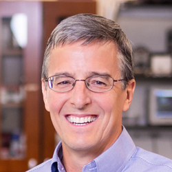 John Rogers合作论文数Department of Materials Science and Engineering, McCormick School of Engineering, Northwestern University22
 Alexandre Caprio合作论文数Dept Radiol, Weill Cornell Med16
 Alaie Seyedhamidreza合作论文数University of New Mexico, University of New Mexico13
 Xu Xie合作论文数12
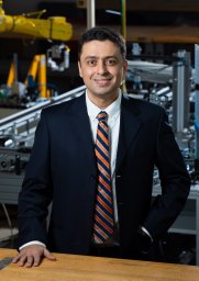 Amir-Ali Amiri Moghadam合作论文数12
 Yonggang Huang合作论文数Department of Mechanical Engineering, McCormick School of Engineering, Northwestern University;Departments of Civil and Environmental Engineering, McCormick School of Engineering, Northwestern University11
 Jizhou Song (宋吉舟)合作论文数School of Aeronautics and Astronautics, Zhejiang University;Department of Engineering Mechanics, Zhejiang University11Frank Du合作论文数10
 Subhi J. Al'Aref合作论文数University of Arkansas for Medical Sciences10
 Sanlin Robinson合作论文数9
 Yuhang Li (李宇航)合作论文数School of Aeronautic Science and Engineering, Beihang University8
 Sung Hun Jin合作论文数Department of Electronic Engineering, Incheon National University8
 Ahmad Ehteshamul Islam合作论文数Air Force Res Lab, Mat & Mfg Directorate, Wright Patterson AFB, OH 45433 USA8Lohendran Baskaran合作论文数7
 Sun-Joo Jang合作论文数Bridgeport Hosp, Yale New Haven Hlth7Fay Y. Lin合作论文数Department of Medicine, Weill Cornell Medical College—New York Presbyterian Hospital7Jordyn Auge合作论文数6
 Chaofeng Lv (吕朝锋)合作论文数College of Civil Engineering and Architecture, Zhejiang University;Ningbo University6
 Kranthi Kumar Kolli合作论文数School of Dynamic Systems, Mechanical Engineering Program6Tejas Doshi合作论文数5
 William P. King合作论文数Department of Biomedical and Translational Sciences, Carle Illinois College of Medicine, University of Illinois at Urbana-Champaign;Department of Mechanical Science and Engineering, Grainger College of Engineering, University of Illinois at Urbana-Champaign;Department of Electrical and Computer Engineering, University of Illinois at Urbana-Champaign;Department of Materials Science and Engineering, University of Illinois at Urbana-Champaign;SyBridge Technologies;EDM Intelligent Solutions5Chenxi Zhang (张晨曦)合作论文数4
 Eva Romito合作论文数Cardiovasc Translat Res Ctr, Univ South Carolina4
 Varun Kashyap合作论文数Weill Cornell Med, Dalio Inst Cardiovasc Imaging4
 Saleh Gharaie合作论文数Sch Engn, Deakin Univ4
 Robert F. Shepherd合作论文数Sibley School of Mechanical and Aerospace Engineering, College of Engineering, Cornell University4
 William L. Wilson合作论文数Center for Nanoscale Systems, Harvard University4
 Kyle L. Grosse合作论文数4
 Jun Liu (劉軍)合作论文数The University of Hong Kong4
 Elton Graugnard合作论文数School of Materials Science and Engineering;Georgia Institute of Technology;School of Materials Science and Engineering, Georgia Institute of Technology4Leslee Shaw合作论文数4
 Muhammad A. Alam合作论文数Elmore Family School of Electrical and Computer Engineering, College of Engineering, Purdue University;Laboratory for Classical and Emerging Electronic Devices, Purdue University3
 Gurpreet Singh合作论文数3
 Abdellatif Ait Lahcen合作论文数Sensors Lab, King Abdullah Univ Sci & Technol KAUST3
 Sahar Jalal合作论文数Department of Aerospace Engineering and Mechanics, University of Minnesota3
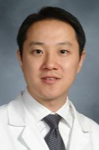 Christopher F Liu合作论文数Joan and Sanford I. Weill Department of Medicine, Weill Cornell Medicine, Cornell University3
 Mohamed Zgaren合作论文数3
 Kimberly Elmore合作论文数3Ha Uk Chung合作论文数3
 Hyuk-Jae Chang合作论文数Department of Internal Medicine, College of Medicine, Yonsei University;Division of Cardiology, Severance Cardiovascular Hospital, College of Medicine, Yonsei University;Division of Medical Information and Technology, Severance Hospital;Ontact Healthcare3
 Eric Pop合作论文数Materials Science & Engineering Department, School of Engineering, Stanford University;Precourt Institute for Energy, Stanford University3
 Habib Samady合作论文数Department of Medicine, Emory University School of Medicine;Covanos Inc.3Xinning Ho合作论文数3
 Md Abdul Wahab合作论文数3David Molony合作论文数2f du合作论文数2
 Suborna Deb Nath合作论文数2




