Aims \u0026 Objectives: Since it\u0027s origin in December 2019 Novel Corona virus infection has behaved in an unprecedented manner A number of studies had been done so far to find out peculiar characteristics about this disease However, few studies have shown varied hematological manifestations of this which have been found to have prognostic implications In this study we aim to assess the hematological parameters of COVID-19 positive cases and in their different clinical categories (mild, moderate and severe) Patients/Materials \u0026 Methods: It is an observational study done in Himalayan Institute of medical sciences, Dehradun (1) Number of cases included were 118 over 3 months duration (July, August, September-2020) (2) The relevant clinical details of all the COVID-19 positive patients such as name, age, UHID were recorded (3) EDTA sample was run on DXH800 (Beckmencoulter automated analyzer) and all parameters were recorded including the VCS Inclusion criterion: Patients diagnosed with COVID-19 on Rapid antigentestor RTPCR of all age groups Exclusioncriterion:Patients with missing hematological parameters Results: In our study, total number of cases included were 118, out of which 84 (71 18%) were males and 34 (28 81%) were females Patients above 60 years were 35 (29 6%) The patients were categorized into mild, moderate and severe on the basis of clinical findings and those findings were co-related with the VCS parameters The significant findings include decreased absolutely mphocytecount in 43 2% cases and absolute eosinopenia in 83 05% of cases, absolute neutrophil count was increased in 44 9% cases and platelet count was reduced in 30 5% of the cases Discussion \u0026 Conclusion: In major number of cases absolute lymphocyte count, absolute eosinophil count and neutrophil to lymphocyte ratio was found significant;so it is advised to categorize patients on the basis of clinical findings and related VCS parameters for early COVID-19 testing
Introduction: Analysis of clinical, molecular features and prognostic factors in aggressive histology MCL (AH-MCL; blastoid or pleomorphic) is not described. AH-MCL can be [de novo (dnMCL) or transformed from classic morphology (t-MCL)]. Methods: We reviewed 183 pts with AH-MCL (108 were dn-MCL and 75 were t-MCL). Overall, 152 were blastoid and 31 pleomorphic. Pt characteristics were collected at the time of diagnosis in dnMCL and at transformation. Overall survival (OS) - time of initial diagnosis of AH-MCL to death/last follow-up and failure free survival (FFS) - time of starting first-line treatment of AH-MCL to treatment discontinuation. Whole-exome sequencing (WES) with SureSelect Human All Exon V6 was performed from biopsy samples from 44 pts (dnMCL = 32, t-MCL = 12). Results: Median follow up after diagnosis of AH-MCL was 19.6 months (0.1-168). Clinical features of AH-MCL were - 20% B symptoms, 6% had ECOG PS (3-4), CNS involvement in 4%, 19% with GI involvement, 67% with marrow involvement and 27% had leukemic phase. The median Ki-67% was 70% (10-100), complex karyotype 13%, LDH > ULN in 45%, Sox-11 positive in 81%, median β2M of 2.9 mg/dL. Pts with t-MCL were distinct from dnMCL in having significantly higher median age, poor PS, lower WBC count, lower marrow involvement and lower rate of achieving CR with first line treatments. All pts with t-MCL had prior treatment for MCL before transformation with median lines of prior therapy 2 (range 1-8). The median OS after diagnosis of AH-MCL was 33 months (48 and 14 months for dnMCL and t-MCL respectively; p = 0.001; Figure-1A-B). Univariate and multivariate (MVA) analyses were performed. Recursive partitioning analysis revealed that Ki-67% ≥50% (Fig. 1C), LDH ≥1519, β2M ≥ 4, hemoglobin <14 and platelet count <63,000 had increased risk of death. In MVA, factors significantly associated with inferior OS in AH-MCL were age (>72 yrs), t-MCL category, Ki-67% ≥50%, ECOG-PS (1-4 compared to 0). Presence of t-MCL, high Ki-67%, CNS involvement were predictive of inferior FFS. Pts who received ibrutinib based or R-HCVAD based therapies had lower hazard ratio for FFS. Subset analysis of blastoid vs pleomorphic MCL did not reveal any significant difference in clinical features of survival but FFS was inferior in pleomorphic category. Frequently mutated genes in AH-MCL were ATM, CCND1, NOTCH1, KMT2D, KMT2C, TP53, SPEN, SMARCA4 and NSD2. There was no statistically significant difference in the mutation profile of dnMCL vs t-MCL. Significant differences in the mutation burden between pts with high Ki-67 (≥50%) and those with low Ki-67%, were noted - ATM (p = 0.001), CCND1 (p<0.001), NOTCH1(p = 0.001), TP53 (p = 0.006), SMARCA4 (p = 0.006), NSD2 (p = 0.02), NOTCH3 (p = 0.02) Figure-1E. Keywords: mantle cell lymphoma (MCL).
Background: Standard therapy for first relapsed Diffuse Large B-cell Lymphoma (DLBCL) is platinum-based chemotherapy (PBC) and autologous stem cell transplant (AutoSCT), however only half of patients respond. To date, there are no patient or tumor factors that are predictive for response. Early identification of lack of response could allow for alternative therapy selection, avoidance of toxic and futile therapy, and potentially impact clinical outcomes. Positron emission tomography–computed tomography (PET-CT) is commonly utilized to assess response after two cycles of PBC, but the utility at earlier time points remains unknown. We conducted an investigator initiated pilot study (NCT02405078) of PET-CT on day 4 and day 21 of therapy for patients with relapsed DLBCL in comparison with standard time points, along with paired collection of blood for circulating tumor (ct) DNA analysis. Methods: Adult patients with relapsed DLBCL who were eligible for PBC and AutoSCT were eligible. Therapy was selected by the treating physician and not restricted per protocol. The primary objectives were to evaluate the ability of early PET-CT to predict response to standard immunochemotherapy, in comparison with PET-CT at standard time points and with blood based detection of a ctDNA. Patients underwent standard 18F-fluorodeoxyglucose (FDG) PET-CT during screening and at the conclusion of 2 cycles of PBC per standard clinical practice, and research FDG PET-CT on day 4 and day 21 of cycle 1 of PBC. Treating physicians were not blinded to the research PET-CT results, but were encouraged to manage the patients per standard practice. Results: The protocol has accrued 28 patients from February 2016 to March 2019. The median age was 62 years (range: 25-82), and 36% were female. 61% had poor risk IPI, 71% had advanced stage. 24 patients had a PET-CT on day 4 (4 missing due to patient status factors and logistical issues). The response rates on day 4 were: complete response 21% (CR, n=5), partial response 33% (PR, n=8), stable disease 37.5% (SD, n=9), and progressive disease 8% (n=2). Among the 5 patients with CR on day 4, all had CR at the end of therapy and have not relapsed. Among the 8 patients with PR on day 4, 2 achieved a CR at the end of therapy and 6 had progressive disease. Among the 11 patients with SD or PD on day 4, 2 patients achieved a PR but both had eventual relapse and all others had PD at end of therapy. Additional data from day 21 PET-CT and ctDNA testing may be presented at the meeting. Conclusions: Our pilot study suggests that very early FDG PET-CT may be able to predict for end of therapy response in patients with relapsed DLBCL. Patients who achieve less than a complete response on day 4 of salvage chemotherapy have a high probability of therapeutic failure and could be considered for alternative therapeutic options. The limitations of our study include small patient numbers, heterogeneity of therapy, and availability of PET-CT at very early time points off study. Keywords: diffuse large B-cell lymphoma (DLBCL); positron emission tomography (PET); prognostic indices.
Introduction: We reported the efficacy of ibrutinib with rituximab (IR) combination in relapsed MCL. Here we present the first efficacy/safety analysis of single center, phase II clinical trial of IR combination in untreated elderly pts with MCL. Methods: Previously untreated elderly (>65 years) MCL pts (n=42) were enrolled in this study (NCT01880567). Pts with Ki-67% ≥ 50% and blastoid/pleomorphic histology were excluded. Pts received IR combination - ibrutinib 560 mg orally daily for 28 days (one cycle) continued until disease progression or discontinued for any reason. Rituximab was given on days 1, 8, 15 and 22 +/- 1 day by intravenous infusion (IV) at a fixed dose of 375 mg/m2 (cycle 1), followed by rituximab on day 1 of every cycle starting in cycles 3 – 8. Following cycle 8, rituximab was given on day 1 of every other cycle for up to 2 years. The primary objective was to assess the safety and the efficacy. Among evaluable samples, minimal residual disease (MRD) by flow cytometry at best response and other molecular studies for clonal evolution at various time points were performed. Results: The median age was 71 years (range 65-84), ECOG PS was (0/1) in 41 (98%) pts, 20% had high risk MIPI score, Ki-67% was low (<30%) in 30 (73%) and high (≥30-50%) in 11 (27%) pts, and 10% had complex karyotype. Median number of IR cycles was 19 (1-43). Best overall response (ORR) was 95% (69% CR, 26% PR, 5% stable disease). Median number of IR cycles to reach CR 4 (2-31). Thirty two patients had PET- based response assessment and all had CR (100%). MRD negative CR by flow cytometry was 65%. Overall, the median follow up was 24 months (6-42). Median PFS and OS were not reached. Among pts with low and high Ki-67%, median PFS was (not reached; p=0.28) and OS (not reached; p=0.02). Median duration on study was 19 months (range 1-39). Overall, 4 pts progressed (2 transformed to blastoid MCL) on study after taking IR for 4, 9, 13 and 32 months. Two pts died. At the time of last follow up, 24 pts remained on study and 18 (43%) discontinued therapy. Dose reduction was performed in 23 (55%) pts for various reasons. Most frequent grade 3-4 toxicities were 17% myalgias, 14% fatigue, 12% shortness of breath, 9% neutropenia and 5% new onset atrial fibrillation. Conclusions: IR combination in elderly pts with MCL as a frontline treatment was very effective and safe. This strategy provides an excellent frontline alternate to chemotherapy in elderly pts with MCL. Keywords: ibrutinib; mantle cell lymphoma (MCL). Disclosures: Wang, M: Consultant Advisory Role: Pharmacyclics and Janssen.
Introduction: Hepatitis C virus (HCV) seropositive patients who undergo autologous hematopoietic stem cell transplantation (HSCT) are at risk of HCV acute exacerbation (AE). Furthermore impact of high dose chemotherapy in patients who have hepatitis C is unknown. We sought to determine the risk factors for AE and the impact of HCV seropositivity on survival post-autologous HSCT. Methods: We retrospectively reviewed the medical records of all 64 HCV seropositive patients who had undergone autologous HSCT at our institution. COX regression model was used to evaluate the predictive risk factors for AE. Results: Twenty (32%) of the 64 patients had AE, defined as a 3-fold or greater increase in serum alanine aminotransferase level (ALT) from the pre-transplant value. Age above 60 years was found to be a risk factor for post-transplant AE (Figure 1). The occurrence of AE (analyzed as time-dependent variable) had no impact on the overall survival of patients who had undergone autologous HSCT. The overall and progression-free survival rates for chemosensitive lymphoma patients 2 years after autologous HSCT were 64% (range 41-80) and 47% (range 27-64), respectively. Median overall and progression-free survival durations in myeloma patients were 54 and 22 months, respectively.Figure 1: Cumulative incidence of acute exacerbation (AE) for all patients in the study and for the two age groups >60 years and ≤60 years.Conclusion: Our results suggest that the survival post autologous HSCT is not impacted by either the development of AE or the pre-transplant HCV seropositivity, as the survival of HCV seropositive patients post-transplant is similar to the survival of non-HCV seropositive historical controls. Hence, our study implies that HCV seropositivity is not a contraindication for autologous transplant.
We hypothesized that IV busulfan (Bu) dosing could be safely intensified through pharmacokinetic (PK-) dose guidance to minimize the inter-patient variability in systemic exposure (SE) associated with body-sized dosing, and that this should improve outcome of AML/MDS patients undergoing allogeneic stem cell transplantation. To test this hypothesis, we treated 218 patients (median age 50.7 years, male/female 50/50%) with fludarabine 40 mg/m2 once daily x4, each dose followed by IV Bu, randomized to 130 mg/m2 (N=107) or PK-guided to average daily SE, AUC of 6000 μM min (N=111), stratified for remission status and allo-grafting from HLA-matched donors. Toxicity and GvHD rates in the groups were similar; the risk of relapse or treatment-related mortality remained higher in the fixed-dose group throughout the 80-month observation period. Further, PK-guidance yielded safer disease control, leading to improved overall and PFS, most prominently in MDS patients and in AML patients not in remission at allogeneic stem cell transplantation. We conclude that AML/MDS patients receiving pretransplant conditioning treatment with our 4-day regimen may benefit significantly from PK-guided Bu dosing. This could be considered an alternative to fixed-dose delivery since it provides the benefit of precise dose delivery to a predetermined SE without increasing risk(s) of serious toxicity and/or GvHD.
Myeloproliferative neoplasms with myelofibrosis (MPN-MF) demonstrate constitutive activation of Janus kinase/signal transducer and activator of transcription (JAK/STAT) signaling that responds to treatment with the JAK1 and 2 kinase inhibitor (JAKi) ruxolitinib. However, MPN-MF often progresses (~20%) to secondary acute myeloid leukemia (sAML), where standard induction chemotherapy or ruxolitinib is relatively ineffective, necessitating the development of novel therapeutic approaches. In the present studies, we demonstrate that treatment with BET (bromodomain and extraterminal) protein inhibitor (BETi), for example, JQ1, inhibits growth and induces apoptosis of cultured and primary, patient-derived (PD), post-MPN sAML blast progenitor cells. Reverse-phase protein array, mass-cytometry and Western analyses revealed that BETi treatment attenuated the protein expressions of c-MYC, p-STAT5, Bcl-xL, CDK4/6, PIM1 and IL-7R, whereas it concomitantly induced the levels of HEXIM1, p21 and BIM in the sAML cells. Co-treatment with BETi and ruxolitinib synergistically induced apoptosis of cultured and PD sAML cells, as well as significantly improved survival of immune-depleted mice engrafted with human sAML cells. Although BETi or heat shock protein 90 inhibitor (HSP90i) alone exerted lethal activity, cotreatment with BETi and HSP90i was synergistically lethal against the ruxolitinib-persister or ruxolitinib-resistant sAML cells. Collectively, these findings further support in vivo testing of BETi-based combinations with JAKi and HSP90i against post-MPN sAML cells.
We are entering a very exciting era in umbilical cord blood transplantation (UCBT), where many of the associated formidable challenges may become treatable by ex vivo graft manipulation and/or adoptive immunotherapy utilizing specific cellular products. We envisage the use of double UCBT rather than single UCBT for most patients; this allows for greater ability to treat larger patients as well as to manipulate the graft. Ex vivo expansion and/or fucosylation of one cord will achieve more rapid engraftment, minimize the period of neutropenia and also give certainty that the other cord will provide long-term engraftment/immune reconstitution. The non-expanded (and future dominant) cord could be chosen for characteristics such as better HLA matching to minimize GvHD, or larger cell counts to enable part of the unit to be utilized for the development of specific cellular therapies such as the production of virus-specificT-cells or chimeric-antigen receptor T-cells which are reviewed in this study.
Solitary plasmacytoma (SP) is a malignancy in bone (BP) or extramedullary soft tissue (EP) at risk for progression to MM. We present preliminary characteristics, outcomes and predictors of progression to MM in a cohort of 61 patients (pt). We retrospectively searched our database for “plasmacytoma” from 1999 until 2014. 192 patients were identified. 131 patients were excluded due to incorrect diagnosis, lack of baseline data or pt duplication. Panel 1A shows baseline characteristics; median follow-up time was 5 years for censored observations. Definitive radiation was a component of therapy in 95% of patients. Median overall survival (OS) was 12.5 years for BP and not reached for EP (p=0.89). Univariate cox hazard model showed larger tumor size and serum paraprotein (SePa) of ≥ 1g/dL at diagnosis was associated with worse OS (HR 1.16, p=0.08 and HR 0.24 [for SePa < 1g/dL], p=0.09, respectively). At 5 years, only 17% of EP pts vs. 39% of BP pts had progressed to myeloma (TTM) (p=0.10) (Panel 1B). Univariate analysis of baseline characteristics for TTM identified higher serum calcium (HR 1.30) and location (head and neck versus long bones, HR 0.07) as risk factors (p<0.05). Lower SePa, higher hemoglobin and absence of Bence Jones (BJ) proteinuria, serum paraprotein or immunoparesis at diagnosis had a lower risk of progression (HR 0.42, p=0.09; HR 0.72, p<0.01; HR 0.4, p=0.054; HR 0.40, p=0.05; HR 0.23; p=0.01; respectively). Multivariate analysis showed immunoparesis and higher calcium levels at diagnosis predicted for TTM (HR 4.63, p<0.01; HR 1.34, p=0.03; respectively). Among 33 patients who had available data 1 year after diagnosis (range 0.6-1.4 years), higher SePa level and serum free light chain ratio (sFLCR), were associated with a higher risk for TTM (HR 5.8, p<0.01; HR 1.16, p=0.03, respectively). Absence of urine BJ proteinuria was associated with delayed TTM (HR 0.10, p<0.01). We confirm that SP patients have excellent survival outcomes in the era of novel imaging techniques and use of sFLCR. Baseline immunoparesis, calcium levels, SePa and BJ proteinuria predict for progression to myeloma. A landmark analysis for TTM at one year after diagnosis also identified sFLCR, SePa and urinary BJ as predictors for progression. In the future, molecular evaluation of SP might shed light into prognosis and treatment.
Autologous hematopoietic stem cell transplantation in light chain amyloidosis (AL) with renal involvement
We hypothesized that during conditioning chemotherapy for allogeneic stem cell transplant (allo-SCT), the disruption of stromal–leukemia interactions using G-CSF in combination with the CXCR4-specific inhibitor, plerixafor, may promote the release of leukemic cells from the niche and increase tumor elimination. In a phase 1/2 investigation, we treated 45 AML/myelodysplastic syndrome (MDS)/CML patients (34 AML, 7 MDS and 4 CML) with G-CSF (10 μg/kg daily for 6 days starting on day −9) plus plerixafor (doses of 0, 80, 160 or 240 μg/kg daily for 4 days starting on day −7) along with the busulfan–fludarabine (Bu–Flu) conditioning regimen. In the phase 1 part, we determined that G-CSF plus plerixafor is safe in this setting. We compared the clinical effects and outcomes of AML/MDS study patients (n=40) with 164 patients from a historical data set who received Bu–Flu alone before allo-SCT by stratifying on cytogenetics and disease status to correct for bias. Study patients had increased myeloid chimerism and lower rates of GvHD. There was no significant difference in relapse-free survival or overall survival. The G-CSF plus plerixafor combination increased circulating WBCs, CD34+ cells and CXCR4+ cells, and preferentially mobilized FISH+ leukemic cells.
Cardiac involvement in light-chain amyloidosis (AL) predicts poor prognosis and is associated with higher TRM and morbidity during high-dose therapy and auto-SCT (HDT–ASCT). We studied the outcomes of 30 patients with cardiac amyloidosis undergoing HDT–ASCT at our center between January 1998 and March 2012. The median age of the patients was 53 years (range, 36–74) with a median follow-up of 35 months (range, 0.4–97 months). Twenty-seven patients (90%) had more than one organ involved besides the heart with 37% with cardiac stage ⩾3. Melphalan-based conditioning regimen (140–200 mg/m2) was used for HDT–ASCT. One-year TRM is 10%. Three-year OS and EFS from HDT–ASCT was 83% and 56.8%, respectively. Cumulative incidence of relapse at 3 years was 38.5%. Negative factors affecting survival included age >60 years, lack of novel induction therapy and BM plasmacytosis >10%. We conclude that HDT–ASCT is well tolerated in patients with high-risk cardiac amyloidosis and can lead to improved overall outcomes.
Abstract Introduction While high dose chemotherapy and autologous hematopoietic stem cell transplantation (auto-HCT) is an accepted part of up front therapy for patients with multiple myeloma (MM), the role of this treatment modality for relapsed patients is still evolving. In light of data suggesting safety and synergy in combining novel therapeutics with traditional cytotoxic chemotherapy, we hypothesized that lenalidomide could be safely combined with high dose melphalan in the salvage auto-HCT setting and yield a meaningful duration of disease control. Methods We conducted a phase I/II study of lenalidomide and high dose melphalan + auto-HCT. MM patients with relapsed or progressive disease were treated with 7 days of oral lenalidomide (doses of 25, 50, 75 or 100 mg daily for the 7 days) on days (-8) to (-2). High dose melphalan (total of 200 mg/m2) was administered as 100 mg/m2 IV on days (-3) and (-2) followed by auto-HCT on day 0. The Eff-Tox method of Thall, Cook, and Estey was used for dose escalation with cohorts of 3 to maximize the trade-off between efficacy and toxicity, defined as CR at day 90 and regimen-related death, graft failure, or select grade 3+ events within 30 days after transplant, respectively. Kaplan-Meier method was used to estimate progression-free survival (PFS) and overall survival (OS) and the log-rank test was used to assess univariate differences between dose levels. Bayesian logistic regression and survival time models were used for multivariable analyses, with posterior probabilities greater than 0.95 or less than 0.05 considered significant. Initial results after 12.3 months of follow-up were published in 2015; we now present an update with 39.8 months of follow-up. Results 57 patients were enrolled, of which 18 (32%) had received a prior auto-HCT. A total of 3, 5, 24 and 25 patients received 25, 50, 75 and 100 mg of lenalidomide, respectively. Median age at auto-HCT was 60 (34-72) years. Median prior lines of treatment were 3 (1-11). Twenty-two patients (39%) were lenalidomide-refractory at study entry. Patient characteristics did not differ significantly between the lenalidomide dose levels. In total, only 2 dose-limiting toxicities were seen, both at dose level 75 mg. Two patients died of nonrelapse causes (viral infection 1, cardiac failure 1) for a treatment-related mortality of 4%. Median time to both neutrophil and platelet engraftment was 11 days. One patient developed a second primary malignancy (squamous cell cancer of the skin). 63% received maintenance therapy, (54% lenalidomide-based). By day +90, 8 patients (14%) had achieved a complete response (CR), 17 (30%) a very good partial response (VGPR), and 17 (30%) a partial response (PR), with no significant differences in response rates among the 4 lenalidomide dose levels. Best responses were PR: 26%, VGPR: 18%, near CR: 18%, CR: 7%, stringent CR: 23% for a ≥VGPR rate of 66. 23% achieved bone marrow minimal residual disease negativity by flow cytometry. Median time to achieve best response was 92 days (range: 16-732). One patient (2%) had progressive disease and 3 patients (5%) achieved only stable disease. Multivariable Bayesian logistic regression revealed that high-risk cytogenetics, (deletion 13q, t(4:14) or del 17p) by conventional cytogenetics or (t(4:14), t(14:16) or del17p by fluorescent in-situ hybridization), bone marrow disease burden and number of prior lines of treatment were each significantly associated with a lower probability of reaching CR by day 90. With a median follow up of 39.8 months (range: 0.5- 66.9), median PFS was 17.1 months (95% CI: 10.8 - 23.0, Figure 1) and median OS was 48.0 months (95% CI: 22.6 months, not estimated, Figure 2). There was no significant effect of dose level on PFS or OS. Multivariable Bayesian survival time models found high-risk cytogenetics to be significantly harmful to both OS and PFS. In addition, degree of plasma cell infiltration of bone marrow before auto-HCT was significantly harmful to PFS. Conclusion: Lenalidomide up to 100 mg PO daily x 7 can be safely combined with high dose melphalan and auto-HCT. Longer follow-up demonstrates PFS and OS as comparable to other salvage treatments for MM, suggesting that this regime can be applied as a part of the sequence of therapies for these patients. Figure 1 PFS of 17.1 months (95% CI: 10.8 - 23.0; N=57, Events=48) Figure 1. PFS of 17.1 months (95% CI: 10.8 - 23.0; N=57, Events=48) Figure 2 OS of 48.0 months (95% CI: 22.6 months, not estimated; N=57, Deaths=28) Figure 2. OS of 48.0 months (95% CI: 22.6 months, not estimated; N=57, Deaths=28) Disclosures Orlowski: Takeda Pharmaceuticals: Research Funding. Champlin:Intrexon: Equity Ownership, Patents & Royalties; Ziopharm Oncology: Equity Ownership, Patents & Royalties.
With improving expertise and supportive care, cord blood (CB) transplants are now associated with outcomes comparable to unrelated and sibling donor transplants.1 There is a need to increase the size and diversity of international CB inventories and to retain cryopreserved CB units for as long as safely possible. There are in vitro data supporting >90% recovery of hematopoietic progenitor cells from frozen CB cells stored for up to 12 years,2 and data showing that progenitor cell recoveries from short-term freeze samples ranging from 2 to 8 weeks are comparable to those cryopreserved for 10–15 years.3,4 Broxmeyer et al.5 reported adequate recovery (80–100%) of granulocyte–macrophage and multi-potential hematopoietic progenitors from functional CB units cryopreserved for up to 23.5 years as well as recovery of viable progenitor cells from CBs frozen for 15 years, which were able to generate CD45+ human cell engraftment when infused into sublethally irradiated NOD-SCID mice and were comparable to that reported with fresh CB.6 Despite this in vitro evidence, transplant physicians still remain concerned about the possible loss of integrity of frozen CB units associated with longer durations of storage.7 In one report, the incidence of bag breaks over a 6.5-year period was 3.5%, where 75% of the breaches occurred in units that had been cryopreserved for >2 years.7 Therefore, it is important to determine whether prolonged time of cryopreservation and storage may adversely affect clinical transplant outcomes, and with the new US Food and Drug Administration licensing regulations for CB units this question has become even more relevant: 'is there an expiration date for CB unit in storage?'. We reviewed 86 consecutive single CB transplant recipients in the period from March 1996 to June 2011, where 15 patients received CB units older than 5 years (CB age 12.2 years=1; >8 years=3; >7 years= 1; >6 years=2; >5 years=8). The vast majority of the CB units were obtained from NMDP (National Marrow Donor Program) or Netcord Banks (American Red Cross Portland=1; AUNCB=1; Australia=1; Barcelona=5; Belgium=5; Bergan Paramus, NJ, USA=1; Caitlin Raymond, UK=1; NC, USA=1; Colorado=9; Dusseldorf=10; France=1; JP McCarthy, USA=1; London=5; MD Anderson=2; Italy=9; NYCB=16; Puget Sound, OR, USA=1; Spain=2; St Louis=13; Tokyo=1). All CB units underwent washing and RBC depletion before freezing. The median length of storage of CB units was 2 years (range, 0.03–12 years). For the purpose of analysis, duration of CB unit storage was divided into four equal quartiles as well as an arbitrary cutoff of 5 years based on the common practice adopted by some public and private CB banks. Standard thawing procedures were followed at the MD Anderson Cell Therapy Laboratory.8 The pre-infusion viabilities were >90%. Patient and CB unit characteristics are described in Table 1. Seventy-six (89%) patients achieved engraftment (early death=4; graft failure=11). In univariate analysis, only the degree of HLA mismatch was significantly associated with engraftment (P=0.04; χ2-test). For the whole cohort, median time to neutrophil recovery was 22 days (range 13–68 days) and platelet recovery was 43 days (range 20–247 days). Median time to neutrophil and platelet recovery specifically for ‘old’ CB units was as follows: CB age >5 years: 24 and 40 days; age >6 years: 19 and 42 days; age >7 years: 28 and 85 days; age >8 years: 13 and 25 days; age >12 years: 56 and 109 days, respectively. No significant differences were observed in the time to neutrophil or platelet recovery when comparing ‘old’ vs ‘new’ CB unit recipients (Figures 1a and b), among CB quartiles (Figures 1c and d), CB storage duration (Figures 1e and f) or the year of CB storage (Figures 1g and h). Significant factors affecting the time to neutrophil recovery included recipient age <8 years (20 vs 23 days; P=0.05); weight <30 kg (19 vs 24 days; P=0.011); total nucleated count (TNC; x106/kg) count >47 (18 vs 25 days; P=0.034); and CD34+ (x106/kg) cell count >0.235 (18 vs 23 days; P=0.002). No differences were observed for duration of hospitalization and storage duration or storage year (Figures 1i and j). Significant factors influencing the duration of hospitalization to <45 days included: age <8 years (62% vs 39%; P=0.04), engraftment status (55% vs 10%; P=0.008) and HLA match (75% vs 45%; P=0.09). Median follow-up for survivors was 8 years. Day-100 and 1-year non-relapse mortality for the whole cohort was 19% and 35%, respectively. The Kaplan–Meir method was used to calculate survival from the date of transplant.9 The estimated 2-year, 5-year and 10-year survival for whole cohort were 43.5%, 40% and 35%, respectively. No survival differences were seen based on the CB storage age. The 3-, 6- and 12-month survival for ‘old’ vs ‘new’ CB units was 80%, 80% and 60% vs 79%, 62% and 50%, respectively. The 3-, 6- and 12-month survival breakdown for the four quartiles were 76%, 67% and 62% vs 73%, 59% and 41% vs 86%, 67% and 53% vs 81%, 67% and 52%, respectively. Significant factors affecting the survival in univariate analysis included myeloablative conditioning (P=0.007) and degree of HLA mismatch (P=0.05). This is the first clinical report of human single CB transplants evaluating the effect of duration of CB storage. Although a majority of the CB transplant recipients in our analysis were children, which may have accounted for a higher TNC and CD34+ cell dose as compared with adults, the overall age distribution was comparable in all different cohorts. Even though a lower number of ‘old’ CB patients received ablative conditioning, which was an independent predictor of survival, no differences were seen in the survival outcomes when comparing old vs new CB recipients. Major limitations of our analyses include its retrospective nature as well as small number of cases. We conclude that, in our restricted data set, the cryopreserved storage time of CB units did not affect engraftment, hematopoietic recovery, duration of hospitalization or survival. CB units within this age range should be considered a viable source of hematopoietic stem cells for allo-SCTs. Larger studies are needed to confirm our findings as well as to examine longer durations of cell storage. The authors declare no conflict of interest.
Exact mechanism of action of umbilical cord blood (CB)-derived regulatory T cells (Tregs) in the prevention of GVHD remains unclear. On the basis of selective overexpression of peptidase inhibitor 16 in CB Tregs, we explored the related p53 pathway, which has been shown to negatively regulate miR15a/16 expression. Significantly lower levels of miR15a/16 were observed in CB Tregs when compared with conventional CB T cells (Tcons). In a xenogeneic GVHD mouse model, lower levels of miR15a/16 were also found in Treg recipients, which correlated with a better GVHD score. Forced overexpression of miR15a/16 in CB Tregs led to inhibition of FOXP3 and CTLA4 expression and partial reversal of Treg-mediated suppression in an allogeneic mixed lymphocyte reaction that correlated with the reversal of FOXP3 demethylation in CB Tregs. On the other hand, miR15a/16 knockdown in CB Tcons led to expression of FOXP3 and CTLA4 and suppression of allogeneic lymphocyte proliferation. Using a luciferase-based mutagenesis assay, FOXP3 was determined to be a direct target of miR15a and miR16. We propose that miR15a/16 has an important role in mediating the suppressive function of CB Tregs and these microRNAs may have a 'toggle-switch' function in Treg/Tcon plasticity.
Optimal treatment approach continues to remain a challenge for systemic light chain amyloidosis (AL). So far, Auto-SCT is the only modality associated with long-term survival. However, failure to show survival benefit in randomized study raises questions regarding its efficacy. We present a comparative outcome analysis of Auto-SCT to conventional therapies (CTR) in AL patients treated over a 14-year period at our institution. Out of the 145 AL amyloidosis patients, Auto-SCT was performed in 80 patients with 1-year non-relapse mortality rate of 12.5%. Novel agents were used as part of induction therapy in 56% of transplant recipients vs 46% of CTR patients. Hematological and organ responses were seen in 74.6% and 39% in the Auto-SCT arm vs 53% and 12% in the CTR arm, respectively. The projected 5-year survival for Auto-SCT vs CTR was 63% vs 38%, respectively. Landmark analysis of patients alive at 1-year after diagnosis showed improved 5-year OS of 72% with Auto-SCT vs 65% in the CTR arm. In the multivariate analysis, age <60 years, induction therapy with novel agents, kidney only involvement and Auto-SCT were associated with improved survival. In conclusion, Auto-SCT is associated with long-term survival for patients with AL amyloidosis.
Abstract Abstract 4522 Introduction: POEMS syndrome (polyradiculoneuropathy, organomegaly, endocrinopathy, monoclonal plasma cell disorder, and skin changes) is a rare plasma cell disorder often associated with papilledema, extravascular volume overload, sclerotic bone lesions, Castleman disease, and high vascular endothelial growth factor (VEGF) levels. High-dose chemotherapy and autologous hematopoietic stem cell transplant (auto-HCT) can be used with good clinical response, but has significant post-transplant morbidity. Here we present our experience in 5 patients with POEMS syndrome who underwent auto-HCT at the MD Anderson Cancer Center (MDACC). Method: Between January 1999 and October 2010, 5 patients (4 males, 1 female) with POEMS syndrome received auto-HCT at MDACC. Diagnosis of POEMS was based on criteria of Dispenzieri et. al.1 All 5 patients had received systemic therapy prior to auto-HCT as follows: bortezomib+dexamethasone (dex) in 2, lenalidomide+dex in 1, cyclophosphamide+dex in 1, and pulse dex in 1. Two patients also received localized radiation for bone disease. Peripheral blood stem cells were collected with granulocyte colony-stimulating factor (G-CSF) in 4 patients and with cyclophosphamide+G-CSF in 1. The preparative regimen was melphalan 200 mg/m2 in 4 patients, while 1 patient received 180 mg/m2 due to renal insufficiency. We evaluated the response rate, toxicity, transplant-related mortality, progression-free survival (PFS) and overall survival (OS). Hematologic response was defined by the International Myeloma Working Group (IMWG) criteria2. Result: Median age at auto-HCT was 48 years (range: 39–58) and median time from diagnosis to auto-HCT was 16.6 months (6.4 – 89 months). All 5 patients had osteosclerotic bone lesions and monoclonal gammopathy: IgA lambda in 3, IgG lambda in 1, and IgG kappa in 1. Two patients had biopsy proven Castleman disease. Four patients had debilitating polyneuropathy. Other features were: skin involvement in 3, endocrinopathy in 2, ascites in 1, anasarca in 2, pulmonary hypertension in 1, and papilledema in 1. Median follow up after auto-HCT was 11.6 months (8.6 – 83.5 months). Median Karnofsky performance status was 80% (70–100) at the time of transplant and 90% (80–100) by the one year follow up visit. Two patients (40%) had complete response, 2 (40%) had a very good partial response, and 1 (20%) had a partial response by IMWG criteria. Engraftment syndrome was not seen in any of the 5 patients. Significant post auto-HCT complications were fungal pneumonia in 2 patients and pulmonary embolism in one. Patients fully recovered from these complications. One-year transplant-related mortality was 0%. All 5 patients had complete or significant resolution of their clinical symptoms after auto-HCT. Median PFS and OS have not yet been reached. With a median follow up of 11.6 months, 1-year PFS and OS were 100%. Four patients are alive and in remission, while one patient died 6 years after his auto-HCT secondary to gastrointestinal bleeding unrelated to his underlying disease. Conclusion: High-dose therapy followed by auto-HCT is safe and effective in selected patients with POEMS syndrome and is associated with durable responses and survival. Disclosures: No relevant conflicts of interest to declare.
 Elizabeth J. Shpall合作论文数Department of Stem Cell Transplantation and Cellular Therapy, Division of Cancer Medicine, The University of Texas MD Anderson Cancer Center19
 Richard E. Champlin合作论文数Department of Stem Cell Transplantation and Cellular Therapy, Division of Cancer Medicine, The University of Texas MD Anderson Cancer Center;Division of Internal Medicine, The University of Texas Health Science Center12
 Qaiser Bashir合作论文数12M de Lima合作论文数10Kebriaei Partow合作论文数10
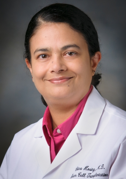 Chitra Hosing合作论文数Department of Stem Cell Transplantation and Cellular Therapy, Division of Cancer Medicine, The University of Texas MD Anderson Cancer Center10
 Muzaffar H. Qazilbash合作论文数Department of Stem Cell Transplantation, Division of Cancer Medicine, The University of Texas MD Anderson Cancer Center;James A. Baker III Institute for Public Policy, Rice University10Uday Popat合作论文数10G Rondon合作论文数9N Shah合作论文数9
 Leonidas C Platanias合作论文数Robert H. Lurie Comprehensive Cancer Center, Feinberg School of Medicine, Northwestern University;Department of Biochemistry and Molecular Genetics, Feinberg School of Medicine, Northwestern University9
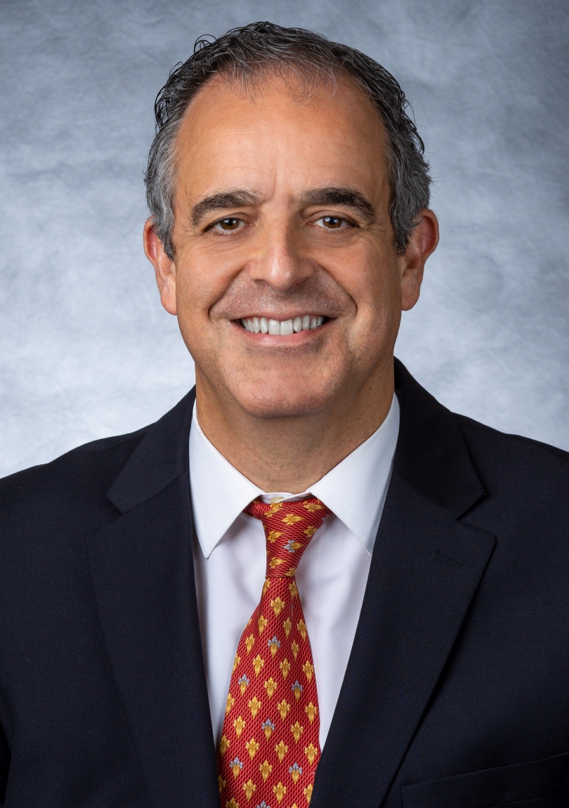 Yago L. Nieto合作论文数Department of Stem Cell Transplantation and Cellular Therapy, Division of Cancer Medicine, The University of Texas MD Anderson Cancer Center9
 Antonella Sassano合作论文数9
 Robert Z. Orlowski合作论文数Department of Lymphoma - Myeloma, Division of Cancer Medicine, The University of Texas MD Anderson Cancer Center;Department of Experimental Therapeutics, Division of Cancer Medicine, The University of Texas MD Anderson Cancer Center8
 Sergio A. Giralt合作论文数Division of Hematologic Malignancies, Memorial Sloan Kettering Cancer Center;Weill Cornell Medical College, Memorial Sloan Kettering Cancer Center7Martin S. Tallman合作论文数7Li Yong-Zhong (李永忠)合作论文数6Kambhampati Suman合作论文数6Hong Yang合作论文数6
 Yvonne Trang Dinh合作论文数6Richard E Champlin合作论文数6D Weber合作论文数5
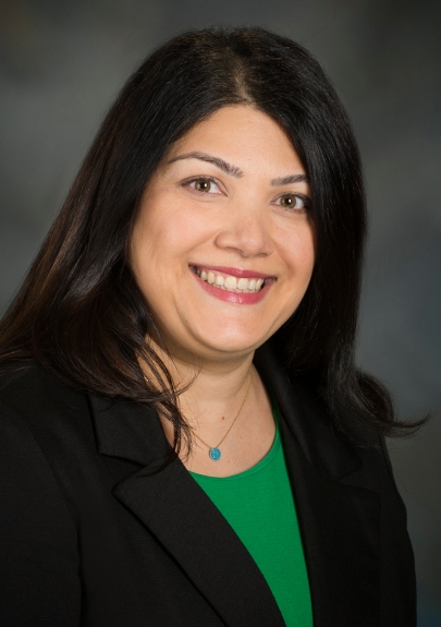 Sairah Ahmed合作论文数Department of Lymphoma/Myeloma, Division of Cancer Medicine, The University of Texas MD Anderson Cancer Center5
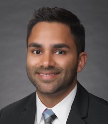 Patel Krish合作论文数5
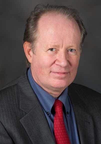 Borje S. Andersson合作论文数Department of Stem Cell Transplantation and Cellular Therapy, Division of Cancer Medicine, The University of Texas MD Anderson Cancer Center;College of Pharmacy, University of Houston5
 Shahab Uddin合作论文数Translational Research Institute, Hamad Medical Corporation;Laboratory of Animal Research Center, Qatar University5
 Nina Shah合作论文数School of Medicine, University of California, San Francisco5Beata Majchrzak合作论文数5Simon N Robinson合作论文数5John D Mcmannis合作论文数5
 Eleanor Fish合作论文数Department of Immunology, University of Toronto;Division of Cell & Molecular Biology, Toronto General Research Institute, University Health Network;Trinity College, University of Toronto;Ness Therapeutics5
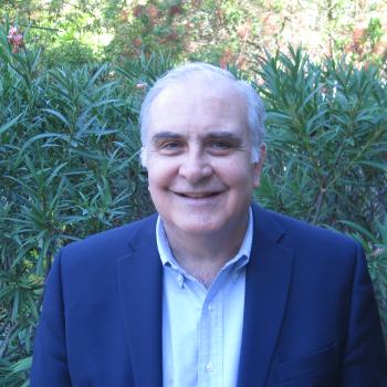 Marcelo Fernandez Vina合作论文数Department of Pathology, Histocompatibility, Immunogenetics and Disease Profiling Laboratory, School of Medicine, Stanford University5A. Alousi合作论文数5Rm Saliba合作论文数4J. Shah合作论文数4
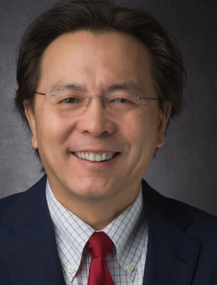 Luhua Wang(Michael)合作论文数The University of Texas MD Anderson Cancer4
 William K. Decker合作论文数Department of Molecular and Human Genetics, Baylor College of Medicine4Muzaffar H Qazilbash合作论文数4
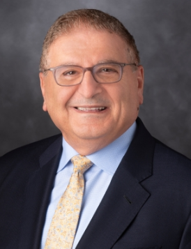 Issa F. Khouri合作论文数Department of Stem Cell Transplantation and Cellular Therapy, Division of Cancer Medicine, The University of Texas MD Anderson Cancer Center4Efstratios Katsoulidis合作论文数4S. Qureshi合作论文数3M. Munsell合作论文数3P Anderlini合作论文数3
 Cord Sturgeon合作论文数Department of Surgery, Feinberg School of Medicine, Northwestern University3
 Nadim Mahmud合作论文数Department of Biostatistics, Epidemiology and Informatics, Perelman School of Medicine, University of Pennsylvania3
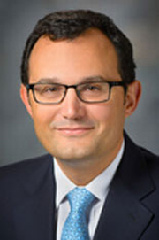 Guillermo Garcia-Manero合作论文数Section of Myelodysplastic Syndromes, Department of Leukemia, Division of Cancer Medicine, The University of Texas MD Anderson Cancer Center;UTHealth Houston Graduate School of Biomedical Sciences3Tara Sadeghi合作论文数3
 Loretta Nastoupil合作论文数The University of Texas MD Anderson Cancer Center3




