BACKGROUND: Transesophageal echocardiography (TEE) is an essential skill in perioperative and critical care practice. Traditional lecture-based instruction remains the standard for foundational training, but virtual reality (VR)–based platforms may enhance engagement and provide scalable, self-directed learning. This exploratory study evaluated the feasibility, educational impact, and learner satisfaction of a low-cost VR TEE training system for anesthesiology residents. MATERIALS AND METHODS: In this randomized, single-center study, 19 anesthesiology residents (Post-Graduate Years 1–3) without prior TEE experience were assigned to lecture instruction or VR-based training. Participants completed baseline, immediate post-training, 1-month, and 3-month knowledge assessments evaluating TEE principles and clinical correlation. A post-training survey assessed satisfaction and perceived educational value. RESULTS: Both groups showed improvement in knowledge scores with retention at 1 and 3 months. There were no significant differences in mean scores between groups (control vs. VR: baseline 14.4 ± 3.6 vs. 14.9 ± 4.2, P = 0.779; postintervention 15.4 ± 6.1 vs. 16.5 ± 4.2, P = 0.670; 1-month 16.0 ± 6.5 vs. 16.8 ± 5.1, P = 0.771; 3-month 17.9 ± 6.7 vs. 17.6 ± 3.8, P = 0.911). However, VR participants reported significantly higher satisfaction, citing greater engagement, ease of use, and perceived clinical relevance. The VR system was deployed without need for dedicated simulation staff or technical support, demonstrating practical feasibility. CONCLUSIONS: A low-cost VR platform for TEE education is feasible and well received by trainees. Although knowledge outcomes were similar to lecture-based instruction, higher learner satisfaction supports VR as a valuable supplement to traditional methods, particularly in programs with limited access to high-fidelity simulators.
The training of Adult Cardiothoracic Anesthesiology (ACTA) fellows in transesophageal echocardiography (TEE) is essential for their development, but assessing technical proficiency remains challenging. While hands-on experience is the cornerstone of learning, traditional evaluation methods primarily focus on image interpretation, neglecting the assessment of image acquisition techniques and visual attention. We describe our experience using wearable eye-tracking technology to objectively assess the TEE skills of ACTA fellows. Since 2023, our ACTA fellows have been equipped with Tobii Pro Glasses 3 (Tobii Pro, Danderyd, Sweden) during weekly preoperative TEE examinations. These glasses record video and track gaze patterns in real-time, allowing for the analysis of fellows' focus and performance over time. The gaze data is processed using Tobii Pro Lab Analyzer, creating heat maps and measuring the time spent on specific TEE views. One attending physician, considered an expert, also participates to provide a benchmark for comparison. Our initial experience suggests that eye-tracking technology can provide valuable insights into the fellows' gaze patterns, enabling objective measurement of their focus during TEE procedures. While limitations such as scheduling conflicts, case type, and variable imaging windows were noted, our experience demonstrates the feasibility of this technology in evaluating technical skills. This approach offers a promising method for refining TEE education and feedback in cardiac anesthesia fellowship. Further research is needed to explore additional metrics such as gaze efficiency and time to image acquisition to enhance TEE training for fellows.
Much of anesthesiology takes place outside moments of crisis, in the steady work of watching, anticipating, and shaping the environment of care. What may appear as quiet detachment is in fact an active form of attentiveness. From the vantage point of standing back, anesthesiologists witness the full rhythm of the operating room: gestures of trust, moments of strain, and the subtle dynamics that influence both safety and teamwork. This skill, often unnamed in their training, aligns with what social scientists describe as an ethnographic sensibility, a way of noticing meaning in context. It matters for patients, for colleagues, and for anesthesiologists themselves. By recognizing and cultivating this capacity, anesthesiology can be seen not only as a field of technical expertise and physiologic insight but also as one that contributes to the culture in which care takes place.
Background The right ventricle (RV) plays a central role in the maintenance of effective cardiac pump function. Despite overwhelming evidence that perioperative RV dysfunction (RVD) and failure (RVF) are associated with poor clinical outcomes, there are very few published recommendations or guidelines for comprehensive, evidence-based RV assessment on the risk of developing either during the perioperative period. Main text To address this gap, the Perioperative Quality Initiative-IX (POQI-IX) investigators group, comprised of clinical experts in anesthesiology, cardiovascular surgery, internal medicine, critical care medicine, and advanced practice nursing, has developed a consensus statement based on current literature, published society recommendations, and the clinical expertise of the group. Herein, the group provides recommendations and evidence-based tools related to perioperative RV assessment, functional screening, staging, and the clinical implications of each. These assessment tools are based on comprehensive patient evaluation consisting of physical examination, biomarker data, imaging, and hemodynamic assessment. Conclusion This review presents a comprehensive tool for assessing perioperative RV function. We hope that this simple, intuitive tool can be applied to all phases of perioperative care and thereby improve patient outcomes.
The Society of Cardiovascular Anesthesiologists (SCA) is committed to improving the quality, safety, and value that cardiothoracic anesthesiologists bring to patient care. To fulfill this mission, the SCA supports the creation of peer-reviewed manuscripts that establish standards, produce guidelines, critically analyze the literature, interpret preexisting guidelines, and allow experts to engage in consensus opinion. The aim of this report, commissioned by the SCA President, is to summarize the distinctions among these publications and describe a novel SCA-supported framework that provides guidance to SCA members for the creation of these publications. The ultimate goal is that through a standardized and transparent process, the SCA will facilitate up-to-date education and implementation of best practices by cardiovascular and thoracic anesthesiologists to improve patient safety, quality of care, and outcomes.
University of Minnesota, Minneapolis, Minnesota, USA Correspondence to Tjorvi E. Perry, MD, MMSc, University of Minnesota, 420 Delaware St SE, MMC 294, Mayo Memorial Building, 8294A, Minneapolis, MN, USA. E-mail: [email protected]
ObjectiveRight ventricular (RV) dysfunction in cardiac surgery can lead to RV failure which is associated with increased morbidity and mortality. Abnormal RV function can be identified using RV pressure monitoring. The primary objective of the study is to determine the proportion of patients with abnormal RV early to end-diastole diastolic pressure gradient (RVDPG) and abnormal RV end-diastolic pressure (RVEDP) before initiation and after cardiopulmonary bypass (CPB) separation. Secondary objective is to evaluate if RVDPG before CPB initiation is associated with difficult and complex separation from CPB, RV dysfunction and failure at the end of cardiac surgery.DesignProspective study.Setting patientsTertiary care cardiac institute.ParticipantsCardiac surgical patients.InterventionCardiac surgery.Measurements and Main ResultsAutomated electronic quantification of RVDPG and RVEDP were obtained. Hemodynamic measurements were correlated with cardiac and extracardiac parameters from TEE and post-operative complications. Abnormal RVDPG were present in 80% of the patients (n=105) at baseline, with a mean RVEDP of 14.2±3.9 mmHg. Patients experienced a RVDPG >4 mmHg for a median duration of 50.2% of the intra-operative period before CPB initiation and 60.6% after CPB separation. A total of 46 (43.8%) patients had difficult/complex separation from CPB, 18 (38.3%) patients with RV dysfunction and eight (17%) with RV failure. Abnormal RVDPG before CPB was not associated with post-operative outcome.ConclusionElevated RVDPG and RVEDP are common in cardiac surgery. RVDPG and RVEDP before CPB initiation are not associated with RV dysfunction and failure but can be used for their diagnosis.
ObjectiveTo demonstrate the value of a viscoelastic-based intraoperative transfusion algorithm to reduce non-RBC product administration in adult cardiac surgical patients.DesignA prospective observational study.SettingAt a quaternary academic teaching hospital.ParticipantsCardiac surgical patients.InterventionsViscoelastic-based intraoperative transfusion algorithm.Measurements and Main ResultsThe study authors compared intraoperative blood product transfusion rates in 184 cardiac surgical patients to 236 historic controls after implementing a viscoelastic-based algorithm. The authors found a non-significant reduction in transfusion of 23.8% for fresh frozen plasma (FFP) units (0.84 ± 1.4 v 0.64 ± 1.38; p = ns), 33.4% for platelet units (0.90 ± 1.39 v 0.60 ± 131; p = ns), and 15.8% for cryoprecipitate units (0.19 ± 0.54 v 0.16 ± 0.50; p = ns). They found a 43.9% reduction in red blood cell (RBC) units transfused (1.98 ± 2.24 v 0.55 ± 1.36; p = 0.008). There were no statistically significant differences in time to extubation (8.0 hours (4.0-21.0) v 8.0 (4.0-22.3), reoperation for bleeding (15 [12.3%] v 10 [10.6%]), intensive care unit length of stay (ICU LOS) (51.0 hours [28.0-100.5] v 53.5 [33.3-99.0]) or hospital LOS (9.0 days [6.0-15.0] v 10.0 [7.0-17.0]). Deviation from algorithm adherence was 32.7% (48/147). Packed RBC, FFP, platelets, cryoprecipitate, and cell saver were significantly reduced in the Algorithm Compliant Cohort compared with historic controls, whereas times to extubation, ICU LOS, and hospital LOS did not reach significance.ConclusionsAfter the implementation of a viscoelastic-based algorithm, patients received fewer packed RBC, FFP, platelets, cryoprecipitate, and cell saver. Algorithm-compliant patients received fewer transfusions; however, reductions in times to extubation, ICU LOS, and hospital LOS were not statistically significant compared with historic controls.
OBJECTIVE:The aim of this study was to use wearable video-recording technology to measure precisely the timing of discrete events during perioperative central venous catheter (CVC) placements. DESIGN:A single-center, observational, exploratory study on the use of wearable video-recording technology during intraoperative CVC placement. SETTING:The study was conducted at a University Hospital. PARTICIPANTS:Clinical anesthesia residents, cardiothoracic anesthesia fellows, and attending anesthesiologists participated in this study. INTERVENTIONS:Participants were asked to use eye-tracking glasses prior to the placement of a CVC in the cardiac operating rooms. No other instruction was given to the participants. MEASUREMENTS AND MAIN RESULTS:The authors measured the total time to complete the CVC placement, phase-specific time, and specific times of interest. They compared these times across 3 training levels and tested differences with analysis of variance. The authors' findings indicated significant differences in total CVC placement time when the procedure included a pulmonary artery catheter insertion (1,170 ± 364, 923 ± 272, and 596 ± 226 seconds; F2,63 = 12.71, p < 0.0001). Additionally, they found differences in interval times and times of interest. The authors observed a reduction of variability with increasing experience during the CVC placement phase. CONCLUSIONS:In this observational study, the study authors describe their experience using first-person wearable video-recording technology to precisely measure the timing of discrete events during CVC placement by anesthesia residents and anesthesiologists. Future work will leverage the eye-tracking capabilities of the existing hardware to identify areas of inefficiency to develop actionable targets for interventions that could improve trainee performance and patient safety.
Developing an accurate and detailed 3D mental model of cardiac anatomy is critical for electrophysiology (EP) trainees. This can be challenging in certain forms of palliated congenital heart disease. Surgical repair of d-TGA via a Mustard or Senning procedure requires a complex baffling technique that is particularly difficult to visualize from a 3D perspective and creates a propensity for arrhythmias. Traditional teaching methods may be sub-optimal for visualizing the interaction between the conduction system and this unusual anatomic substrate.
The clinical uses of perioperative transesophageal echocardiography have grown exponentially in recent years for both cardiac and noncardiac surgical patients. Yet, echocardiography is a complex skill that also requires an advanced understanding of human cardiac anatomy. Although simulation has changed the way echocardiography is taught, most available systems are still limited by investment costs, accessibility, and qualities of the input cardiac 3-dimensional models. In this report, the authors discuss the development of an online simulator using a high-resolution human heart scan that accurately represents real cardiac anatomies, and that should be accessible to a wide range of learners without space or time limitations.
Developing an accurate and detailed 3-dimensional (3D) mental model of cardiac anatomy is critical for electrophysiology (EP) trainees. Due to its immersive nature, virtual reality (VR) may provide a better learning environment than traditional teaching methods for assimilating 3D cardiac anatomy. The purpose of this pilot study was to evaluate the technical feasibility of an interactive, remote VR-based method for teaching cardiac anatomy to novice EP trainees. We created a shared, remote VR environment that allows the shared viewing of high-resolution 3D cardiac models. Eighteen trainees accepted for pediatric and adult EP fellowships were recruited. We performed a cohort study comparing the traditional teaching methods with the VR learning environment. Participants completed a demographic questionnaire and a satisfaction survey. The adult EP trainees were given a multiple-choice pre- and post-test exam to assess their anatomical knowledge. Both the adult and pediatric EP trainee cohorts rated the VR experience positively and preferred the VR environment to the more traditional teaching method. All the participants expressed interest in incorporating the VR learning environment into the EP fellowship curriculum. The usability of the system was relatively low, with approximately one-third of participants rating the system as hard to use. The impact of the VR session on exam performance was mixed among the adult cohort. We demonstrated the feasibility of gathering geographically dispersed EP fellows in training with a shared VR-based environment to teach cardiac anatomy. Although we were not able to demonstrate a learning benefit over the traditional lecture format in the adult cohort, the training environment was favorably received by all the participants.
Perioperative echocardiography requires an advanced understanding of the complex human cardiac anatomy. Cur-rently, conventional training simulators rely on handcrafted heart models that lack accuracy and details and undermine the complexities of the cardiac anatomy, both actual and relative. These simulators are expensive and difficult to transport, creat-ing barriers to widespread implementation. In this report, the authors describe a realistic, virtual reality simulator using high-resolution human heart scans that accurately represent the healthy and pathologic cardiac anatomies in ways that can be standardized and made accessible to a wide range of learn-ers at the cost of a virtual reality headset. Herein, the authors present a description, including the design of the transesopha-geal echocardiography and transthoracic echocardiography simulator, and their initial experiences during cardiac fellow-ship training.
 Simon C. Body合作论文数Brigham and Women's Hospital, Harvard Medical School23
 Stanton K. Shernan合作论文数Brigham and Women's Hospital, Harvard Medical School23
 Danny Muehlschlegel合作论文数Department of Anesthesiology and Critical Care Medicine, The Johns Hopkins University School of Medicine23Charles D. Collard合作论文数21Amanda A. Fox合作论文数Brigham and Women's Hospital, Harvard Medical School21Kuang-Yu Liu合作论文数17Susana Arango合作论文数14
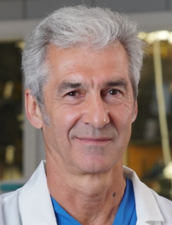 Paul Iaizzo合作论文数Departments of Surgery, School of Medical, University of Minnesota10
 John Edmund Mayer Jr.合作论文数Center for Bioethics, Harvard Medical School;Boston Children's Hospital10Benjamin Gorbaty合作论文数9
 Sary F. Aranki合作论文数Division of Cardiac Surgery, Brigham and Women's Hospital, Harvard Medical School9David Buyck合作论文数8Fraser W. H. Sutherland合作论文数8
 Sunjay Kaushal合作论文数Department of Surgery, Feinberg School of Medicine, Northwestern University6
 Christine E. Seidman合作论文数Cardiovascular Genetics Center, Brigham and Women's Hospital;Department of Genetics, Harvard Medical School;Howard Hughes Medical Institute6
 Joyce E. Bischoff合作论文数Boston Children's Hospital;Harvard Medical School5
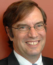 Jonathan G. Seidman合作论文数Blavatnik Institute, Harvard Medical School;Department of Genetics, Harvard Medical School5Andrew Shaffer合作论文数5Megan Lanigan合作论文数4Daniel S Herman合作论文数Pathology and Laboratory Medicine, University of Pennsylvania4
 Alexander Kaizer合作论文数Dept Biostat & Informat, Univ Colorado4Brian S Donahue合作论文数4
 Mias Pretorius合作论文数4
 Andrew L Wilkey合作论文数4
 Thomas Meitinger合作论文数Institut für Humangenetik, Technische Universität München4
 David C. Martin合作论文数Department of Materials Science and Engineering, College of Engineering, University of Delaware;Department of Biomedical Engineering, College of Engineering, University of Delaware4Dawood Darbar合作论文数4
 Joyce A Wahr合作论文数Department of Anesthesiology Ig323-0048, University of Michigan Medical School4
 Douglas Shook合作论文数Department of Anesthesiology and Perioperative and Pain Medicine, Harvard Medical School4Lynelle Schneider合作论文数3D. Siers合作论文数3Ryan Knoper合作论文数3Kevin Moses合作论文数3Marcus Burns合作论文数3Kristine Guleserian合作论文数3Jun Wang合作论文数3Darrell Randle合作论文数3P Lichtner合作论文数3
 Cristhian Potes合作论文数Edwards Lifesciences3
 Samantha T. Porter合作论文数Coll Liberal Arts, Liberal Arts Technol & Innovat Serv3
 Marylyn D Ritchie合作论文数Department of Genetics, Perelman School of Medicine, University of Pennsylvania;Center for Translational Bioinformatics, Institute for Biomedical Informatics, Perelman School of Medicine, University of Pennsylvania;Biomedical and Translational Informatics Laboratory, Perelman School of Medicine, University of Pennsylvania3
 Etienne Couture合作论文数Department of Anesthesiology and Critical Care, Université Laval3Sorajja Paul合作论文数3
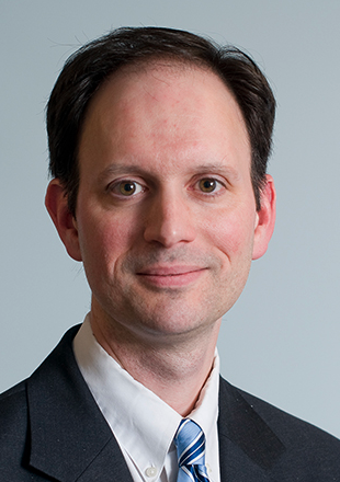 Patrick Ellinor合作论文数Cardiovascular Research Center, Massachusetts General Hospital;Harvard Medical School3
 Nancy J. Brown合作论文数Vanderbilt University3
 André Denault合作论文数Département d’Anesthésiologie et de Médecine de la Douleur, Faculté de Médecine, Université de Montréal;Institut de Cardiologie de Montréal, Université de Montréal3
 Joseph Philip Vacanti合作论文数Department of Pediatric Surgery, Massachusetts General Hospital;Harvard Medical School3
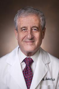 Dan Mark Rode合作论文数Division of Clinical Pharmacology, Department of Medicin, Vanderbilt University Medical Center3
 Mario Gössl合作论文数Minneapolis Heart Institute Foundation3




