OBJECTIVE:To implement parallel transmit (pTx) 7T magnetic resonance imaging (MRI) in the pre-surgical evaluation of 3T-negative patients with drug-resistant focal epilepsy, and to compare quality to conventional single transmit (specifically, circularly polarized [CP]) 7T MRI. METHODS:We implemented a comparative protocol comprising both pTx and CP 7T MRI in consecutive adult candidates for epilepsy surgery who had negative or equivocal 3T MRI imaging. Here we report the outcomes from the first 31 patients. We acquired pTx and CP T1, T2, fluid-attenuated inversion recovery (FLAIR) and edge-enhancing gradient echo (EDGE) images, all in the same three-dimensional (3D) 0.8 mm isotropic space. Two-dimensional (2D) high-resolution T2 and T2*-weighted sequences were acquired only in CP mode due to current technological limitations. Two neuroradiologists, a neurologist, and a neurosurgeon made independent, blinded quality and preference ratings of pTx vs CP images. Quantitative methods were used to assess signal dropout. RESULTS:7T revealed previously-unseen structural lesions in nine patients (29%), confirmed 3T-equivocal lesions in four patients (13%), and disproved 3T-equivocal lesions in four patients (13%). Lesions were better visualized on pTx than CP in 57% of cases, and never better visualized on CP. Clinical management was altered by 7T in 18 cases (58%). Nine cases were offered surgical resection and one laser interstitial thermal therapy (LITT). Three cases were removed from the surgical pathway because of bilateral or extensive lesions. Five cases were offered stereo-electroencephalography (sEEG) with better targeting (in three because the 7T lesion was deemed equivocal by the multi-disciplinary team (MDT), and in two because the lesion was extensive). Blinded comparison confirmed significantly better overall quality of pTx FLAIR images (F(2, 184) = 13.7, p = 2.88 × 10⁻⁶), whereas pTx MP2RAGE images were subjectively non-inferior and had improved temporal lobe coverage with quantitatively less signal drop-out. SIGNIFICANCE:pTx-7T is implementable in a clinical pathway, changed management in 58% of patients where 3T + FDG-PET had not enabled resection, and is superior to single transmit 7T MRI.
AbstractObjectiveTo implement parallel transmit (pTx) 7T MRI in the pre-surgical evaluation of patients with drug resistant focal epilepsy, and to compare quality and diagnostic yield to conventional single transmit (specifically, circularly polarised, CP) 7T MRI.MethodsWe implemented a comparative protocol comprising both pTx and CP 7T MRI in consecutive adult candidates for epilepsy surgery who had negative or equivocal 3T MRI imaging. Here we report the outcomes from the first 31 patients.We acquired pTx and CP T1, T2, FLAIR and EDGE images, all in the same 3D 0.8mm isotropic space. 2D high-resolution T2and T2*-weighted sequences were acquired only in CP mode due to current technological limitations.Two neuroradiologists, a neurologist and a neurosurgeon made independent, blinded quality and preference ratings of pTx vs CP images. Quantitative methods were used to assess signal dropout.ResultsBlinded comparison confirmed significantly better overall quality of pTx FLAIR images (F(2,184)=13.7, p=2.88×10⁻⁶), while pTx MP2RAGE images were subjectively non-inferior and had improved temporal lobe coverage with quantitatively less signal drop-out.7T-pTx revealed previously-unseen structural lesions in 9 patients (29%), confirmed 3T-equivocal lesions in 4 patients (13%), and disproved 3T-equivocal lesions in 4 patients (13%).Lesions were better visualised on pTx than CP in 57% of cases, and never better visualised on CP.Clinical management was altered by pTx-7T in 18 cases (58%). 9 cases were offered surgical resection and 1 LITT. 3 cases were removed from the surgical pathway because of bilateral or extensive lesions. 5 cases were offered sEEG with better targeting (in 3 because the 7T lesion was deemed equivocal by the MDT, and in 2 because the lesion was extensive).SignificanceParallel transmit 7T MRI is implementable in a clinical pathway, is superior to single transmit 7T MRI, and changed management in 58% of patients scanned.Key pointsWe scanned 31 patients with parallel transmit and conventional 7T MRI, finding previously-unreported structural lesions in 9 patients (29% of cases).In 13% of cases pTx 7T MRI showed that an equivocal lesion at 3T MRI was likely significant.In 13% of cases pTx 7T MRI showed that an equivocal lesion at 3T MRI could be disregarded.Both qualitative and quantitative quality assessments indicate superiority of pTx images over CP.Future clinical implementations of 7T MRI for epilepsy should utilise parallel transmit where possible.
Cerebral microdialysis (CMD) catheters allow continuous monitoring of patients' cerebral metabolism in severe traumatic brain injury (TBI). The catheters consist of a terminal semi-permeable membrane that is inserted into the brain's interstitium to allow perfusion fluid to equalize with the surrounding cerebral extracellular environment before being recovered through a central non-porous channel. However, it is unclear how far recovered fluid and suspended metabolites have diffused from within the brain, and therefore what volume or region of brain tissue the analyses of metabolism represent. We assessed diffusion of the small magnetic resonance (MR)-detectible molecule gadobutrol from microdialysis catheters in six subjects (complete data five subjects, incomplete data one subject) who had sustained a severe TBI. Diffusion pattern and distance in cerebral white matter were assessed using T1 (time for MR spin-lattice relaxation) maps at 1 mm isotropic resolution in a 3 Tesla MR scanner. Gadobutrol at 10 mmol/L diffused from cerebral microdialysis catheters in a uniform spheroidal (ellipsoid of revolution) pattern around the catheters' semipermeable membranes, and across gray matter-white matter boundaries. Evidence of gadobutrol diffusion was found up to a mean of 13.4 ± 0.5 mm (mean ± standard deviation [SD]) from catheters, but with a steep concentration drop off so that ≤50% of maximum concentration was achieved at ∼4 mm, and ≤10% of maximum was found beyond ∼7 mm from the catheters. There was little variation between subjects. The relaxivity of gadobutrol in human cerebral white matter was estimated to be 1.61 ± 0.38 L.mmol-1sec-1 (mean ± SD); assuming gadobutrol remained extracellular thereby occupying 20% of total tissue volume (interstitium), and concentration equilibrium with perfusion fluid was achieved immediately adjacent to catheters after 24 h of perfusion. No statistically significant change was found in the concentration of the extracellular metabolites glucose, lactate, pyruvate, nor the lactate/pyruvate ratio during gadobutrol perfusion when compared with period of baseline microdialysis perfusion. Cerebral microdialysis allows continuous monitoring of regional cerebral metabolism-the volume of which is now clearer from this study. It also has the potential to deliver small molecule therapies to focal pathologies of the human brain. This study provides a platform for future development of new catheters optimally designed to treat such conditions.
Post-mortem studies have shown that patients dying from severe acute respiratory syndrome coronavirus (SARS-CoV-2) infection frequently have pathological changes in their CNS, particularly in the brainstem. Many of these changes are proposed to result from para-infectious and/or post-infection immune responses. Clinical symptoms such as fatigue, breathlessness, and chest pain are frequently reported in post-hospitalized coronavirus disease 2019 (COVID-19) patients. We propose that these symptoms are in part due to damage to key neuromodulatory brainstem nuclei. While brainstem involvement has been demonstrated in the acute phase of the illness, the evidence of long-term brainstem change on MRI is inconclusive. We therefore used ultra-high field (7 T) quantitative susceptibility mapping (QSM) to test the hypothesis that brainstem abnormalities persist in post-COVID patients and that these are associated with persistence of key symptoms. We used 7 T QSM data from 30 patients, scanned 93-548 days after hospital admission for COVID-19 and compared them to 51 age-matched controls without prior history of COVID-19 infection. We correlated the patients' QSM signals with disease severity (duration of hospital admission and COVID-19 severity scale), inflammatory response during the acute illness (C-reactive protein, D-dimer and platelet levels), functional recovery (modified Rankin scale), depression (Patient Health Questionnaire-9) and anxiety (Generalized Anxiety Disorder-7). In COVID-19 survivors, the MR susceptibility increased in the medulla, pons and midbrain regions of the brainstem. Specifically, there was increased susceptibility in the inferior medullary reticular formation and the raphe pallidus and obscurus. In these regions, patients with higher tissue susceptibility had worse acute disease severity, higher acute inflammatory markers, and significantly worse functional recovery. This study contributes to understanding the long-term effects of COVID-19 and recovery. Using non-invasive ultra-high field 7 T MRI, we show evidence of brainstem pathophysiological changes associated with inflammatory processes in post-hospitalized COVID-19 survivors.
BACKGROUND:Patients with brain injury who are unresponsive to commands may perform cognitive tasks that are detected on functional magnetic resonance imaging (fMRI) and electroencephalography (EEG). This phenomenon, known as cognitive motor dissociation, has not been systematically studied in a large cohort of persons with disorders of consciousness. METHODS:In this prospective cohort study conducted at six international centers, we collected clinical, behavioral, and task-based fMRI and EEG data from a convenience sample of 353 adults with disorders of consciousness. We assessed the response to commands on task-based fMRI or EEG in participants without an observable response to verbal commands (i.e., those with a behavioral diagnosis of coma, vegetative state, or minimally conscious state-minus) and in participants with an observable response to verbal commands. The presence or absence of an observable response to commands was assessed with the use of the Coma Recovery Scale-Revised (CRS-R). RESULTS:Data from fMRI only or EEG only were available for 65% of the participants, and data from both fMRI and EEG were available for 35%. The median age of the participants was 37.9 years, the median time between brain injury and assessment with the CRS-R was 7.9 months (25% of the participants were assessed with the CRS-R within 28 days after injury), and brain trauma was an etiologic factor in 50%. We detected cognitive motor dissociation in 60 of the 241 participants (25%) without an observable response to commands, of whom 11 had been assessed with the use of fMRI only, 13 with the use of EEG only, and 36 with the use of both techniques. Cognitive motor dissociation was associated with younger age, longer time since injury, and brain trauma as an etiologic factor. In contrast, responses on task-based fMRI or EEG occurred in 43 of 112 participants (38%) with an observable response to verbal commands. CONCLUSIONS:Approximately one in four participants without an observable response to commands performed a cognitive task on fMRI or EEG as compared with one in three participants with an observable response to commands. (Funded by the James S. McDonnell Foundation and others.).
Post mortem studies have shown that patients dying from severe SARS-CoV-2 infection frequently have pathological changes in their central nervous system, particularly in the brainstem. Many of these changes are proposed to result from para-infectious and/or post-infection immune responses. Clinical symptoms such as fatigue, breathlessness, and chest pain are frequently reported in post-hospitalized COVID-19 patients. We propose that these symptoms are in part due to damage to key neuromodulatory brainstem nuclei. While brainstem involvement has been demonstrated in the acute phase of the illness, the evidence of long-term brainstem change on magnetic resonance imaging (MRI) is inconclusive. We therefore used ultra-high field (7T) quantitative susceptibility mapping (QSM) to test the hypothesis that brainstem abnormalities persist in post-COVID patients and that these are associated with persistence of key symptoms. We used 7T QSM data from 30 patients, scanned 93-548 days after hospital admission for COVID-19 and compared them to 51 age-matched controls without prior history of COVID-19 infection. We correlated the patients' QSM signals with disease severity (duration of hospital admission and COVID-19 severity scale), inflammatory response during the acute illness (C-reactive protein, D-Dimer and platelet levels), functional recovery (modified Rankin scale; mRS), depression (PHQ-9) and anxiety (GAD-7). In COVID-19 survivors the MR susceptibility increased in the medulla, pons and midbrain regions of the brainstem. Specifically, there was increased susceptibility in the inferior medullary reticular formation and the raphe pallidus and obscurus. In these regions, patients with higher tissue susceptibility had worse acute disease severity, higher acute inflammatory markers, and significantly worse functional recovery. Using non-invasive ultra-high field 7T MRI, we show evidence of brainstem pathophysiological changes associated with inflammatory processes in post-hospitalized COVID-19 survivors. This study contributes to understanding the mechanisms of long-term effects of COVID-19 and recovery. ### Competing Interest Statement Dr. Pattinson is named as co-inventor on a provisional U.K. patent application titled "Use of cerebral nitric oxide donors in the assessment of the extent of brain dysfunction following injury". Dr Pattinson is named as co-inventors on a provisional U.K. patent titled "Discordant sensory stimulus in VR based exercise" UK Patent office application: 2204698.1 filing date 31/3/2022. ### Funding Statement This work was supported by the NIHR Cambridge Biomedical Research Centre (BRC-1215-20014). The views expressed are those of the authors and not necessarily those of the NIHR or the Department of Health and Social Care. JBR was supported by the Wellcome Trust (103838; 220258) and Medical Research Council (M C\_UU\_00030/14). VFJN was supported by an Academy of Medical Sciences / The Health Foundation Clinician Scientist Fellowship. ETB was supported by an NIHR Senior Investigator award. BR, MC, SN are supported by NIHR Oxford BRC and BHF Oxford CRE. WTC was supported by funding from Wellcome Trust [225924/Z/22/Z]. CTR was supported by funding from Wellcome Trust [098436/Z/12/B]. For the purpose of open access, the authors have applied a CC-BY public copyright licence to any Author Accepted Manuscript version arising from this submission. ### Author Declarations I confirm all relevant ethical guidelines have been followed, and any necessary IRB and/or ethics committee approvals have been obtained. Yes The details of the IRB/oversight body that provided approval or exemption for the research described are given below: The study was approved by the following ethics committees: Cambridgeshire Research Ethics Committee HBREC.2016.13.am3, East of England Research Ethics Committee 17/EE/0025 , Norfolk Research Ethics Committee EE/0395, and North West Preston Research Ethics Committee 20/NW/0235. All participants provided informed consent in accordance with the Declaration of Helsinki. I confirm that all necessary patient/participant consent has been obtained and the appropriate institutional forms have been archived, and that any patient/participant/sample identifiers included were not known to anyone (e.g., hospital staff, patients or participants themselves) outside the research group so cannot be used to identify individuals. Yes I understand that all clinical trials and any other prospective interventional studies must be registered with an ICMJE-approved registry, such as ClinicalTrials.gov. I confirm that any such study reported in the manuscript has been registered and the trial registration ID is provided (note: if posting a prospective study registered retrospectively, please provide a statement in the trial ID field explaining why the study was not registered in advance). Yes I have followed all appropriate research reporting guidelines, such as any relevant EQUATOR Network research reporting checklist(s) and other pertinent material, if applicable. Yes We can provide average QSM χ extracted values from the brainstem and subregions upon reasonable request.
Human coronavirus disease 2019 (COVID-19) due to severe acute respiratory syndrome coronavirus-2 (SARS-CoV-2) has multiple neurological consequences, but its long-term effect on brain health is still uncertain. The cerebrovascular consequences of COVID-19 may also affect brain health. We studied the chronic effect of COVID-19 on cerebrovascular health, in relation to acute severity, adverse clinical outcomes and in contrast to control group data. Here we assess cerebrovascular health in 45 patients six months after hospitalisation for acute COVID-19 using the resting state fluctuation amplitudes (RSFA) from functional magnetic resonance imaging, in relation to disease severity and in contrast with 42 controls. Acute COVID-19 severity was indexed by COVID-19 WHO Progression Scale, inflammatory and coagulatory biomarkers. Chronic widespread changes in frontoparietal RSFA were related to the severity of the acute COVID-19 episode. This relationship was not explained by chronic cardiorespiratory dysfunction, age, or sex. The level of cerebrovascular dysfunction was associated with cognitive, mental, and physical health at follow-up. The principal findings were consistent across univariate and multivariate approaches. The results indicate chronic cerebrovascular impairment following severe acute COVID-19, with the potential for long-term consequences on cognitive function and mental wellbeing.
Purpose: To explore the diffusion-tensor (DT) imaging-defined invasive phenotypes of both isocitrate dehydrogenase (IDH-1)-mutated and IDH-1 wild-type glioblastomas.Materials and Methods: Seventy patients with glioblastoma were prospectively recruited and imaged preoperatively. All patients provided signed consent, and the local research ethics committee approved the study. Patients underwent surgical resection, and tumor samples underwent immunohistochemistry for IDH-1 R132H mutations. DT imaging data were coregistered to the anatomic magnetic resonance study and reconstructed to provide the anisotropic and isotropic components of the DT. The invasive phenotype was determined by using previously published criteria and correlated with IDH-1 mutation status by using the Freeman-Halton extension of the Fisher exact probability test.Results: Nine patients had an IDH-1 mutation and 61 had IDH-1 wild type. All of the patients with IDH-1 mutation had a minimally invasive DT imaging phenotype. Among the IDH-1 wild-type tumors, 42 of 61 (69%) were diffusively invasive glioblastomas, 14 of 61 (23%) were locally invasive, and five of 61 (8%) were minimally invasive (P < .001).Conclusion: IDH-mutated glioblastomas have a less invasive phenotype compared with IDH wild type. This finding may have implications for individualizing the extent of surgical resection and radiation therapy volumes.
We have previously shown that normobaric hyperoxia may benefit peri-lesional brain and white matter following traumatic brain injury (TBI). This study examined the impact of brief exposure to hyperoxia using diffusion tensor imaging (DTI) to identify axonal injury distant from contusions. Fourteen patients with acute moderate/severe TBI underwent baseline DTI and following one hour of 80% oxygen. Thirty-two controls underwent DTI, with 6 undergoing imaging following graded exposure to oxygen. Visible lesions were excluded and data compared with controls. We used the 99% prediction interval (PI) for zero change from historical control reproducibility measurements to demonstrate significant change following hyperoxia. Following hyperoxia DTI was unchanged in controls. In patients following hyperoxia, mean diffusivity (MD) was unchanged despite baseline values lower than controls (p < 0.05), and fractional anisotropy (FA) was lower within the left uncinate fasciculus, right caudate and occipital regions (p < 0.05). 16% of white and 14% of mixed cortical and grey matter patient regions showed FA decreases greater than the 99% PI for zero change. The mechanistic basis for some findings are unclear, but suggest that a short period of normobaric hyperoxia is not beneficial in this context. Confirmation following a longer period of hyperoxia is required.
IMPORTANCE Combined oxygen 15-labeled positron emission tomography (15O PET) and brain tissue oximetry have demonstrated increased oxygen diffusion gradients in hypoxic regions after traumatic brain injury (TBI). These data are consistent with microvascular ischemia and are supported by pathologic studies showing widespread microvascular collapse, perivascular edema, and microthrombosis associated with selective neuronal loss. Fluorine 18-labeled fluoromisonidazole ([18F]FMISO), a PET tracer that undergoes irreversible selective bioreduction within hypoxic cells, could confirm these findings. OBJECTIVE To combine [18F]FMISO and 15O PET to demonstrate the relative burden, distribution, and physiologic signatures of conventional macrovascular and microvascular ischemia in early TBI. DESIGN, SETTING, AND PARTICIPANTS This case-control study included 10 patients who underwent [18F]FMISO and 15O PET within 1 to 8 days of severe or moderate TBI. Two cohorts of 10 healthy volunteers underwent [18F]FMISO or 15O PET. The study was performed at the Wolfson Brain Imaging Centre of Addenbrooke's Hospital. Cerebral blood flow, cerebral blood volume, cerebral oxygen metabolism (CMRO2), oxygen extraction fraction, and brain tissue oximetry were measured in patients during [18F]FMISO and 15O PET imaging. Similar data were obtained from control cohorts. Data were collected from November 23, 2007, to May 22, 2012, and analyzed from December 3, 2012, to January 6, 2016. MAIN OUTCOMES AND MEASURES Estimated ischemic brain volume (IBV) and hypoxic brain volume (HBV) and a comparison of their spatial distribution and physiologic signatures. RESULTS The 10 patients with TBI (9 men and 1 woman) had a median age of 59 (range, 30-68) years; the 2 control cohorts (8 men and 2 women each) had median ages of 53 (range, 41-76) and 45 (range, 29-59) years. Compared with controls, patients with TBI had a higher median IBV (56 [range, 9-281] vs 1 [range, 0-11] mL; P < .001) and a higher median HBV (29 [range, 0-106] vs 9 [range, 1-24] mL; P = .02). Although both pathophysiologic tissue classes were present within injured and normal appearing brains, their spatial distributions were poorly matched. When compared with tissue within the IBV compartment, the HBV compartment showed similar median cerebral blood flow (17 [range, 11-40] vs 14 [range, 6-22] mL/100 mL/min), cerebral blood volume (2.4 [range, 1.6- 4.2] vs 3.9 [range, 3.4-4.8] mL/100 mL), and CMRO2 (44 [range, 27-67] vs 71 [range, 34-88] μmol/100 mL/min) but a lower oxygen extraction fraction (38% [range, 29%-50%] vs 89% [range, 75%-100%]; P < .001), and more frequently showed CMRO2 values consistent with irreversible injury. Comparison with brain tissue oximetry monitoring suggested that the threshold for increased [18F]FMISO trapping is probably 15 mm Hg or lower. CONCLUSIONS AND RELEVANCE Tissue hypoxia after TBI is not confined to regions with structural abnormality and can occur in the absence of conventional macrovascular ischemia. This physiologic signature is consistent with microvascular ischemia and is a target for novel neuroprotective strategies.
The growing recognition of diseases associated with dysfunction of mitochondria poses an urgent need for simple measures of mitochondrial function. Assessment of the kinetics of replenishment of the phosphocreatine pool after exercise using 31 P magnetic resonance spectroscopy can provide an in vivo measure of mitochondrial function; however, the wider application of this technique appears limited by complex or expensive MR-compatible exercise equipment and protocols not easily tolerated by frail participants or those with reduced mental capacity. Here we describe a novel in-scanner exercise method which is patient-focused, inexpensive, remarkably simple and highly portable. The device exploits an MR-compatible high-density material (BaSO 4 ) to form a weight which is attached directly to the ankle and a one-minute dynamic knee extension protocol produced highly reproducible measurements of post-exercise PCr recovery kinetics in both healthy subjects and patients. As sophisticated exercise equipment is unnecessary for this measurement, our extremely simple design provides an effective and easy-to-implement apparatus that is readily translatable across sites. Its design, being tailored to the needs of the patient, makes it particularly well suited to clinical applications and we argue the potential of this method for investigating in vivo mitochondrial function in new cohorts of growing clinical interest.
Purpose To use perfusion and magnetic resonance (MR) spectroscopy to compare the diffusion tensor imaging (DTI)‐defined invasive and noninvasive regions. Invasion of normal brain is a cardinal feature of glioblastomas (GBM) and a major cause of treatment failure. DTI can identify invasive regions. Materials and Methods In all, 50 GBM patients were imaged preoperatively at 3T with anatomic sequences, DTI, dynamic susceptibility perfusion MR (DSCI), and multivoxel spectroscopy. The DTI and DSCI data were coregistered to the spectroscopy data and regions of interest (ROIs) were made in the invasive (determined by DTI), noninvasive regions, and normal brain. Values of relative cerebral blood volume (rCBV), N‐acetyl aspartate (NAA), myoinositol (mI), total choline (Cho), and glutamate + glutamine (Glx) normalized to creatine (Cr) and Cho/NAA were measured at each ROI. Results Invasive regions showed significant increases in rCBV, suggesting angiogenesis (invasive rCBV 1.64 [95% confidence interval, CI: 1.5–1.76] vs. noninvasive 1.14 [1.09–1.18]; P < 0.001), Cho/Cr (invasive 0.42 [0.38–0.46] vs. noninvasive 0.35 [0.31–0.38]; P = 0.02) and Cho/NAA (invasive 0.54 [0.41–0.68] vs. noninvasive 0.37 [0.29–0.45]; P = < 0.03), suggesting proliferation, and Glx/Cr (invasive 1.54 [1.27–1.82] vs. noninvasive 1.3 [1.13–1.47]; P = 0.028), suggesting glutamate release; and a significantly reduced NAA/Cr (invasive 0.95 [0.85–1.05] vs. noninvasive 1.19 [1.06–1.31]; P = 0.008). The mI/Cr was not different between the three ROIs (invasive 1.2 [0.99–1.41] vs. noninvasive 1.3 [1.14–1.46]; P = 0.68). In the noninvasive regions, the values were not different from normal brain. Conclusion Combining DTI to identify the invasive region with perfusion and spectroscopy, we can identify changes in invasive regions not seen in noninvasive regions. J. Magn. Reson. Imaging 2016;43:487–494.
INTRODUCTION: Seizures are common in patients with a Glioblastoma. Although the epileptogenic mechanism remains unclear, it is suggested to be dependent upon peritumoural changes. Histological analysis of human intratumoural and peritumoural glioma tissue demonstrated that glutamate concentrations in tissue from both sites were increased in patients with seizures when compared to those without. It would be advantageous to have a non-invasive method for predicting which patients are most likely to be affected by seizures. METHOD: 80 patients with a Glioblastoma were imaged pre-operatively at 3T with sequences including anatomical and multivoxel spectroscopy. Regions of interest (ROIs) were selected in a circumferential consecutive pattern from the border of the T1-weighted enhancing region to normal appearing brain. Glutamate parameters (Glu + Gln/Cr) were calculated per voxel and expressed as a ratio to normal brain. Clinical variables were analysed in relation to tumour progression and survival. RESULTS: The mean Glu + Gln/Cr concentration remained more than 1.5 times higher compared to normal brain at a 3 cm radial distance from the T1-weighted enhancing margin in the seizure group. At this distance, the mean Glu + Gln/Cr concentration ratio had returned to 1 in the non-seizure group (Seizures 1.56 ± 0.1 vs. non-seizures 0.92 ± 0.06, p < 0.001). The presence of seizures was associated with a shorter time to progression on multivariate analysis (HR = 7.55; p = 0.01). CONCLUSION: We demonstrate that the presence of seizures is associated with elevated tumour and peritumoural glutamate concentrations and a shortened time to tumour progression in Glioblastomas. Glutamate may play a role in the pathogenesis of seizures and represent a potential biomarker for tumour progression.
INTRODUCTION: Identification of the invasive margin is a key limitation to treating Glioblastomas. Conventional T1-weighted and T2-weighted imaging methods are still used to assess the extent of tumour for resection. However, it is shown in histological specimens that malignant cells extend for several centimetres beyond the contrast enhancing regions. The transition between tumour edge, peritumoural oedema and normal brain remains ambiguous. Multi-modal MR (diffusion tensor, spectroscopy and perfusion) is a promising new imaging technique to study tumour invasion. An improvement in the understanding of the tumour margin would allow individualized tailoring of treatment. METHOD: 80 patients with glioblastomas were imaged pre-operatively at 3T with anatomical sequences, diffusion-tensor, dynamic susceptibility contrast and spectroscopy. Data was co-registered to the same imaging space. Regions of interest (ROIs) were selected in a circumferential consecutive pattern from the edge of the T1-weighted enhancing region to normal appearing brain. The parameters were calculated per voxel and expressed as a ratio to normal brain. RESULTS: The isotropic component of diffusion-tensor remained raised at a 30mm distance beyond the edge of the enhancing region (1.18 ± 0.02, p < 0.000 at 30mm). Perfusion was persistently elevated within each consecutive expanding circumference (1.56 ± 0.06 at 30mm). Choline remained raised in comparison to the contralateral hemisphere at 20mm (1.14 ± 0.04) beyond the edge of the T1-enhancing region. CONCLUSION: MRI biomarkers of tumour cell presence extend up to 3 cm beyond the conventionally determined tumour margin. Incorporating multimodal MRI data into treatment planning will allow more precise surgical resection and planning of radiotherapy.
The aim of these studies was to provide reference data on intersubject variability and reproducibility of metabolite ratios for Choline/Creatine (Cho/Cr), N-acetyl aspartate/Choline (NAA/Cho) and N-acetyl aspartate/Creatine (NAA/Cr), and individual signal-intensity normalised metabolite concentrations of NAA, Cho and Cr. Healthy volunteers underwent imaging on two occasions using the same 3T Siemens Verio magnetic resonance scanner. At each session two identical Metabolic Imaging and Data Acquisition Software (MIDAS) sequences were obtained along with standard structural imaging. Metabolite maps were created and regions of interest applied in normalised space. The baseline data from all 32 volunteers were used to calculate the intersubject variability, while within session and between session reproducibility were calculated from all the available data. The reproducibility of measurements were used to calculate the overall and within session 95% prediction interval for zero change. The within and between session reproducibility data were lower than the values for intersubject variability, and were variable across the different brain regions. The within and between session reproducibility measurements were similar for Cho/Cr, NAA/Choline, Cho and Cr (11.8%, 11.4%, 14.3 and 10.6% vs. 11.9%, 11.4%, 13.5% and 10.5% respectively), but for NAA/Creatine and NAA between session reproducibility was lower (9.3% and 9.1% vs. 10.1% and 9.9%; p <0.05). This study provides additional reference data that can be utilised in interventional studies to quantify change within a single imaging session, or to assess the significance of change in longitudinal studies of brain injury and disease.
INTRODUCTION: Glioblastoma multiforme (GBM) is the most aggressive primary brain tumour. Intra-tumour heterogeneity of GBM is a contributing factor to treatment failure and non-invasive methods are needed for analysis. This study aimed to identify imaging signatures of aggressive tumour belonging to different regions of GBM using multimodal imaging methods, including diffusion-weighted imaging and MR spectroscopy. METHOD: 32 Patients (mean age 57.2 years) with confirmed GBM were imaged at 3T on a Siemens Trio. Maps of apparent diffusion coefficient (ADC) were coregistered to T2-weighted images. Regions of interest (ROI) were selected: FLAIR enhancement with T1-weighted post-contrast enhancement (CE); and FLAIR enhancement without CE (FLAIR). Within these regions, an ADC cut-off value was developed using MATLAB. Metabolic maps of Cho/tNAA ratios belonging to these ADC ROIs were calculated using LCModel. Ratios from CE and FLAIR were compared to each other and to ratios of ROIs above the ADC threshold within tumour (high ADC) and normal appearing white matter (NAWM). RESULTS: Ratio values of CE and FLAIR were significantly higher than those of NAWM (0.26 ± 0.09) (0.60 ± 0.21, p < 0.001; 0.45 ± 0.22; p < 0.001 for CE and FLAIR, respectively). ROIs of high ADC had a mean ratio value (0.39± 0.16) that was significantly lower than that of CE (p < 0.01). CONCLUSION: Findings suggest that regional heterogeneity of GBM can be detected and characterised using advanced imaging. Low ADC ROIs within CE regions appear to have metabolic profiles representing aggressive tumour with higher Cho/tNAA ratios. Combining imaging modalities could potentially differentiate regions that will respond/fail to respond to treatment.
 David Menon合作论文数Division of Anaesthesia, Department of Medicine, Queens' College, University of Cambridge12
 Stephen Price合作论文数Queens' College, University of Cambridge11
 Timothy J Larkin合作论文数10
 Joanne Outtrim合作论文数Wolfson Brain Imaging Centre, University of Cambridge9
 Virginia Newcombe合作论文数Division of Anaesthesia, University of Cambridge;National Institute for Health and Care Research;Royal College of Emergency Medicine9Guy B. Williams合作论文数Addenbrooke's Hospital, Cambridge University8
 Ed Bullmore合作论文数Behavioural and Clinical Neuroscience Institute, University of Cambridge;Department of Psychiatry, University of Cambridge7
 Natalie R Boonzaier合作论文数6
 Jonathan Coles合作论文数Department of Medicine, University of Cambridge6
 Tonny Veenith合作论文数Institute of Inflammation and Ageing, College of Medical and Dental Sciences, University of Birmingham6Eleanor Carter合作论文数5Grossac Julia合作论文数5
 Marius Mada合作论文数MRC Cognition and Brain Sciences Unit, University of Cambridge5Natalie R. Boonzaier合作论文数4
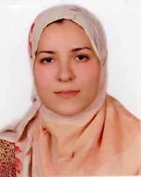 Mohsen Laila合作论文数Department of Radiology, Minia University4
 Belinda R. Lennox合作论文数Department of Psychiatry, University of Oxford4
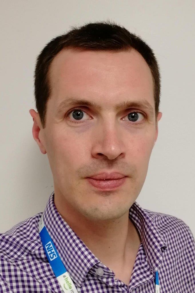 Chris Rodgers合作论文数Department of Clinical Neurosciences, University of Cambridge4
 Emmanuel Stamatakis合作论文数School of Health Sciences, Faculty of Medicine and Health, The University of Sydney;Charles Perkins Centre, Faculty of Medicine and Health, The University of Sydney3Manktelow Anne合作论文数3
 James B. Rowe合作论文数Cambridge Centre for Frontotemporal Dementia and Related Disorders, Department of Clinical Neurosciences, University of Cambridge;Dementias Platform UK;Alzheimer's Research UK;Medical Research Council Cognition and Brain Sciences Unit, University of Cambridge3
 Adam M H Young合作论文数Harvard Medical School, Massachusetts General Hospital3Doris A. Chatfield合作论文数3A. T. C. Carpenter合作论文数3
 Andrew J Calder合作论文数MRC Cognition and Brain Sciences Unit, University of Cambridge3
 Aurelien Massire合作论文数2Sarah A. Leir合作论文数2Chisomo Zimphango合作论文数2
 Matt Stovell合作论文数2Pascal P R Ruetten合作论文数2Owen Thomas合作论文数2Mark Philip Cassar合作论文数Royal Berkshire Hosp, Reading, Berks, England2Vincent Gras合作论文数2
 Mauconduit Franck合作论文数2Arturo Cardenas-Blanco合作论文数2
 Catarina Rua合作论文数2
 Betty Raman合作论文数Queen Elizabeth Hospital, University of Adelaide2Boulant Nicolas合作论文数CEA, UNIRS2Nallapareddy Sridhar合作论文数2
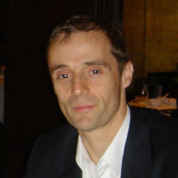 Stephen Sawcer合作论文数Department of Clinical Neurosciences, University of Cambridge;Cambridge University Hospitals NHS Foundation Trust2
 Peter J A Hutchinson合作论文数Department of Clinical Neurosciences, Robinson College, University of Cambridge2
 Martyn Ezra合作论文数Nuffield Department of Clinical Neurosciences, University of Oxford2Bisbrown-Chippendale R合作论文数2William L. Clarke合作论文数University of Virginia Medical Center2
 Stefan Neubauer合作论文数Radcliffe Department of Medicine, Medical Sciences Division, University of Oxford2R Jacob合作论文数2
 John Suckling合作论文数Department of Psychiatry, University of Cambridge2
 Mark Wilson合作论文数Department of Surgery & Cancer, Faculty of Medicine, Imperial College London2
 Marta Correia合作论文数MRC Cognition and Brain Sciences Unit, University of Cambridge2Tim Fryer合作论文数Wolfson Brain Imaging Centre, Department of Clinical Neurosciences, University of Cambridge2




