Biofilm-associated infections present a critical therapeutic challenge due to antibiotic resistance and impaired tissue healing. Here, we present a microrobotic system (MZ-8) that integrates real-time human-steered navigation with autonomous, microenvironment-responsive therapy to actively eradicate biofilms and promote tissue regeneration. This microrobotic system features a spine-inspired structure for mechanical biofilm disruption, a pH-responsive ZIF-8 coating for immunomodulatory Zn2+ release, and closed-loop actuation under second near-infrared fluorescence guidance. In a rat model of periprosthetic joint infection, MZ-8 achieved effective biofilm removal, induced a pro-regenerative immune response by polarizing macrophages toward the M2 phenotype, and significantly enhanced tissue regeneration. Transcriptomic analysis further revealed the activation of immunomodulatory pathways and upregulation of M2-associated genes, confirming the system's sequential shift from eradication to repair. Moreover, validation in a rabbit model and human knee joint confirmed its operational feasibility under clinical imaging guidance and excellent biosafety. This work establishes that integrating physical eradication, biochemical immunomodulation, and interactive control within a single system is essential for advancing from infection clearance to functional tissue restoration. Thus, it provides a therapeutic paradigm for biofilm-associated diseases and lays a foundation for future intelligent, clinically adaptive anti-infective systems.
Cell migration serves as a crucial factor in cell therapy, which has been extensively studied in vitro. However, the impact of in vivo migratory behavior of cells on therapeutic efficacy remains an uncharted territory, due to the complexity of biological processes within living organisms. Here, tendon stem/progenitor cells (TSPCs) with or without mechanical confinement simulating sedentary and migratory cell behaviors were sophisticatedly designed and transplanted to patellar tendon defect mouse model, with short-wave infrared (SWIR) fluorescence imaging adopted to dynamically monitor in vivo cell migration process. As a result, migratory TSPCs exhibited enhanced morphological plasticity with substantial changes in area (51.47% vs. 17.06%, p < 0.05) and width (41.11% vs. 8.69%, p < 0.05) of cell population. Besides, directional in vivo cell migration pattern from injection site to the proximal and medial region of patellar tendon defect was depicted, which further led to superior therapeutic efficacy in tendon regeneration based on histological scoring. In contrast, sedentary TSPCs demonstrated prolonged in vivo cell retention in large cell numbers (21 d vs. 14 d), and resulted in inferior therapeutic efficacy. Furthermore, transcriptomic analysis revealed activations of cell chemotaxis and migration pathways in mice with migratory TSPCs, while morbid pathways of excessive cell proliferation and ossification in sedentary TSPCs. In summary, in vivo cell migratory behavior was identified as a major driver of superior therapeutic efficacy in tendon regeneration by morphological plasticity and directional migration. This study highlights the pivotal role of in vivo migration competence in cell therapy optimization and provides a mechanistic framework for clinical translation strategies.
Angiogenesis is a critical process in the early stages of tendon healing but is always limited by its avascular structure, thus impairing effective healing. Nanopeptides targeting endogenous vascular endothelial growth factor (VEGF) presented a promising strategy for promoting early angiogenesis in tendon healing. In this study, we introduced a nanoscale electro-assembly technique to kinetically control the collagen assembling to form VEGF-binding protein (VEGF-BP) and PR1P-loaded oriented collagen matrix (BP/P Col) for tendon healing. The specific binding of VEGF-BP and PR1P to endogenous VEGF mediated the proangiogenic effects during the early stages of repair. In vitro cell experiments showed that BP/P Col could promote human umbilical vein endothelial cell migration and tube formation as well as new vessel formation in chick chorioallantois. In a rabbit model of tendon defect, it increased vascular area and new vessel formation within the first 2 weeks. Additionally, multiomics analysis further revealed the potential mechanism of BP/P Col in promoting early angiogenesis was to modulate the macrophage-regulated VEGF expression, providing positive feedback for early angiogenesis. This process induced the organized collagen structure along the oriented collagen matrix, which ultimately facilitated rapid tendon healing during the early stage. Overall, this study demonstrated that BP/P Col, through the efficient loading of VEGF-BP and PR1P, exerted a synergistic enhancement effect on endogenous VEGF. It could not only accelerate early angiogenesis but also promote subsequent rapid and functional tendon healing, offering a promising treatment strategy for tendon injuries.
Collagen-based biomaterials are gaining prominence in tissue engineering, attributed to their remarkable biocompatibility, inherent biodegradability, and unparalleled capacity to facilitate tissue repair and regeneration. However, the ability to dynamically visualize and quantitatively assess collagen degradation in vivo remains a critical challenge, hindering the development of optimized biomaterials for clinical applications. To address this, a novel approach was developed to monitor the injury microenvironment by conjugating second near-infrared quantum dots with solid collagen. This live imaging system offered high-resolution, real-time tracking of collagen degradation both in vitro and in vivo, enabling a deeper understanding of the degradation behavior under various conditions. This system was applied to mouse models with different cartilage defects, including critical-sized defect (CSD), minor defect (Minor) and sham surgery (Sham) groups for a 28-day in vivo monitoring. Among them, the CSD group exhibited the fastest and most stable collagen degradation, indicating that the degradation rate was closely linked to the severity of the injury. Transcriptomic analysis further identified key signaling pathways that might drive rapid collagen degradation by promoting collagenase activity and tissue remodeling in cartilage defect conditions. In summary, our study provided valuable insights into the mechanisms of collagen degradation under different injury conditions, contributing to innovative strategies for designing collagen-related biomaterials in the future.
Vascularization as a spatiotemporally interlaced process involving angiogenesis and vascular remodeling, has seldom been investigated comprehensively regarding the interrelationship of the two intertwining but sequential processes. Here, a shortwave infrared (SWIR) fluorescence imaging strategy based on quantum dots (QDs) was designed to dynamically visualize vascularization in vivo and in situ in a perforator transplantation mouse model. The vascularization process could be directly perceived from the established flap model with an optimal observation window at 10 min post-injection. Anchored in SWIR technology and image processing, it was revealed that temporally, angiogenesis lasted throughout 21 days after surgery while vascular remodeling took a dominant role after 14 days both in vivo and in situ. Moreover, four perforasomes of the flap in situ displayed spatially that Zone IV shortened the vascularization process with sufficient blood supply from the LDCIA, while Zone II recovered slowly from ischemia with a lack of blood supply. This study serves as a pioneer in adding novel cognition to spatiotemporal pattern of vascularization through visualizing angiogenesis and vascular remodeling simultaneously and dynamically, thus facilitating further investigation into the mechanisms behind.
BACKGROUND:Augmented repair is an alternative strategy for the treatment of acute ligament and tendon injuries that imparts time-zero biomechanical strength to allow early loading, thereby protecting the repaired structures during the early healing process. PURPOSE:To investigate the biomechanical properties and biological healing process after suture repair of acute anterior cruciate ligament (ACL) tears with polyethylene terephthalate (PET) augmentation and compare the findings with those obtained without PET augmentation. STUDY DESIGN:Controlled laboratory study. METHODS:A total of 48 rabbits were assigned to 3 groups: a PET-augmented group, a nonaugmented suture repair group, and a natural (control) group. All 3 groups were evaluated at 4, 12, and 16 weeks after surgery. Biomechanical performance was assessed using tensile strength testing, and ACL healing and maturation were assessed using histological assessments. RESULTS:The PET-augmented group showed less anterior knee laxity at 30° of knee flexion and superior structural continuity compared with the suture group. ACL repair with PET augmentation yielded recovery of the maximum tensile load as early as 4 weeks compared with that of the natural group (110.5 ± 6.5 vs 129.0 ± 8.6 N, respectively; P = .29) and a gradual improvement in linear stiffness from 4 weeks (58.4 ± 3.9 N/mm) to 16 weeks (83.1 ± 5.1 N/mm; P = .04), approaching that of the natural group (106.7 ± 5.8 N/mm). Furthermore, histological analyses revealed that in the PET-augmented group, the ACL healed back to the proximal insertion as early as 4 weeks with angiogenesis and collagen regeneration, and the increased ligament maturity score indicated a gradual healing process from 4 to 16 weeks. CONCLUSION:Compared with nonaugmented repair, repair augmented with a PET band enhanced early ACL stability and supported healing of ACL tears in a rabbit model. CLINICAL RELEVANCE:The biomechanical and histological findings support subsequent clinical investigations using PET augmentation in patients with acute ACL tears.
Anterior cruciate ligament (ACL) injury is one of the most common and severe sports-related knee injuries. Anterior cruciate ligament reconstruction (ACLR) is currently the primary clinical treatment. Compared with other grafts, artificial ligaments offer significant advantages in enabling early return to sports after surgery and, therefore, have been widely studied and applied in clinical practice. This review summarizes recent literature on the application of artificial ligaments in ACLR, outlining the classification of ligament materials, historical development, and advancements in biomaterial research. Although early artificial ligaments showed limited clinical efficacy, the continuous progress in materials science and tissue engineering has facilitated the use of various natural, synthetic, and composite materials in ACL artificial ligament construction, significantly improving their biocompatibility and mechanical properties. In addition, researchers have employed strategies such as surface modification, bioactive factor loading, and cell delivery to endow artificial ligaments with enhanced functionality, yielding promising results in animal studies. This review provides a comprehensive overview of the progress in artificial ligaments biomaterials, systematically summarizes the current changes and innovations in artificial ligament scaffold materials, surface modification technologies, and new auxiliary strategies and highlights auxiliary strategies used with artificial ligaments in ACLR to enhance tendon bone healing, with particular emphasis on the role of tissue engineering and biomaterials. The clinical translation prospects of these emerging strategies are also discussed.
Near-infrared fluorescence imaging in the second window has emerged as a valuable tool for the non-invasive and real-time assessment of vascular information in skin flaps. Enhancing flap images to provide more accurate flap vascularization information is critical for predicting flap viability. To address the limitations of existing methods in enhancing vessel images, we propose a novel and adaptive technique for enhancing flap microvessel images. Multiple strategies can be employed to effectively enhance the visualization of small-scale vessels. Firstly, the proposed method leverages the multiscale rolling guided filter to acquire the base layer and detail layers at different scales. Furthermore, correlation coefficients are utilized to weigh and fuse the detail layers effectively. To suppress noise amplification while enhancing vascular structures, an improved adaptive gamma correction method based on local visual saliency is introduced. Meanwhile, the bilateral gamma correction is used to enhance the base layer. Finally, the enhanced base layer and detail layer are fused using the weighted fusion strategy. We conducted experiments on skin flap vessel images, retinal fundus images, finger vein images, and low-light images. Our method achieved excellent results in metrics such as NIQE, AMBE, and WPSNR, demonstrating significant advantages in preserving the structural integrity and brightness consistency of the images. The obtained results validate the potential of this method in enhancing vascular images, indicating promising prospects in the field of medicine.
Determining the precise course of bacterial infection requires abundant in vivo real-time data. Synchronous monitoring of the bacterial load, temperature, and immune response can satisfy the shortage of real-time in vivo data. Here, we conducted a study in the joint-infected mouse model to synchronously monitor the bacterial load, temperature, and immune response using the second near-infrared (NIR-II) fluorescence imaging, infrared thermography, and immune response analysis for 2 weeks. Staphylococcus aureus (S. aureus) was proved successfully labeled with glucose-conjugated quantum dots in vitro and in subcutaneous-infected model. The bacterial load indicated by NIR-II fluorescence imaging underwent a sharp drop at 1 day postinfection. At the same time, the temperature gap detected through infrared thermography synchronously brought by infection reached lowest value. Meanwhile, the flow cytometry analysis demonstrated that immune response including macrophage, neutrophil, B lymphocyte, and T lymphocyte increased to the peak at 1 day postinfection. Moreover, both M1 macrophage and M2 macrophage in the blood have an obvious change at ~ 1 day postinfection, and the change was opposite. In summary, this study not only obtained real-time and long-time in vivo data on the bacterial load, temperature gap, and immune response in the mice model of S. aureus infection, but also found that 1 day postinfection was the key time point during immune response against S. aureus infection. Our study will contribute to synchronously and precisely studying the complicated complex dynamic relationship after bacterial infection at the animal level.
Purpose:To use a finite element method to construct a patch-bridge repair model for massive rotator cuff tears (MRCTs) and investigate the effects of different suture methods and knot numbers on postoperative biomechanics. Methods:A finite element model based on intact glenohumeral joint data was used for a biomechanical study. A full-thickness defect and retraction model of the supraspinatus tendon simulated MRCTs. Patch, suture, and anchor models were constructed, and the Marlow method was used to assign the material properties. Three suturing models were established: 1-knot simple, 1-knot mattress, and 2-knot mattress. The ultimate failure load, failure mode, stress distribution of each structure, and other biomechanical results of the different models were calculated and compared. Results:The ultimate failure load of the 1-knot mattress suture (71.3 N) was 5.6 % greater than that of the 1-knot simple suture (67.5 N), while that (81.5 N) of the 2-knot mattress was 14.3 % greater than that of the 1-knot mattress. The stress distribution on the patch and supraspinatus tendon was concentrated on suture perforation. Failure of the bridging reconstruction mainly occurred at the suture perforation of the patch, and the damage forms included cutting-through and isthmus pull-out. Conclusion:A finite element model for the patch-bridging reconstruction of MRCTs was established, and patch-bridging restored the mechanical integrity of the rotator cuff. The 2-knot mattress suture was optimal for patch-bridging reconstruction of MRCTs.
Background . There is still no consensus on the treatment of frozen shoulder (FS). Clinical studies on extracorporeal shockwave therapy (ESWT) combined with arthroscopic capsular release (ACR) for FS are still lacking. Objectives . To investigate the effect of ESWT on short-term functional recovery after arthroscopic capsular release (ACR). Methods . A randomized clinical trial including 49 patients who under ACR was included. The patients were assigned to the ESWT group ( n = 24) or non-ESWT group ( n = 25) 2 weeks after surgery. Routine advanced rehabilitation program are given to all participants, while the ESWT group additionally received 5 times shockwave therapy. Visual analog scale (VAS) pain score, Constant score, University of California Los Angeles score (UCLA), UCLA-satisfaction, and shoulder range of motion (ROM) in four directions of flexion, abduction, internal, and external rotation were analyzed at the baseline (2 weeks postsurgery), 2 weeks, 8 weeks, and 12 weeks of follow-up. Repeated measure ANOVA is used to analyze the above outcomes. Results . A total of 46 participants completed all assessments. Pain and function improved in both groups. Comparing between the groups, we found that lower VAS pain scores and higher internal rotation were found in the ESWT group at each follow-up stage (all P < 0.05, 12 weeks VAS-pain 95% CI: [ESWT: 1.55–2.19, non-ESWT: 2.11–2.76], and 12 weeks internal rotation 95% CI: [ESWT: 5.42–7.27, non-ESWT: 7.20–9.06]). Regarding functional scores, the UCLA and UCLA-satisfaction of the ESWT group was significantly higher than that of the control group at 8 weeks and 12 weeks of follow-up (all P < 0.05, 12 weeks UCLA 95% CI: [ESWT: 31.94-33.45, non-ESWT: 27.94-29.45], 12 weeks UCLA-satisfaction 95% CI: [ESWT: 4.32–5.00, non-ESWT: 2.50–4.32]), but there was no significant difference in Constant ( P > 0.05, 12 weeks Constant 95% CI: [ESWT: 82.98-92.94, non-ESWT: 78.24–88.20]). Conclusion . Extracorporeal shockwave therapy has a good analgesic effect and improves internal rotation after arthroscopic capsular release through short-term follow-up.
Background: Extracorporeal shock wave therapy (ESWT) promotes tissue healing by modulating inflammation, which has implications for meniscal tear healing in the avascular zone. Purpose: To evaluate the effects of a single dose of radial ESWT on the healing process and inflammation of the meniscus and knee joints after meniscal tears in the avascular zone. Study Design: Controlled laboratory study. Methods: Avascular tears were induced in the medial meniscus (MM) of 72 Sprague-Dawley rats. One week postoperatively, the rats received a single session of radial ESWT with a Power+ handpiece (ESWT group; n = 36) or with a fake handpiece (sham-ESWT group; n = 36). The rats were then euthanized at 2, 4, or 8 weeks postoperatively. The MMs were harvested for analysis of healing (hematoxylin-eosin, safranin O–Fast Green, and collagen type 2 staining) and inflammation (interleukin [IL]-1β and IL-6 staining). Lateral menisci and synovia were obtained to evaluate knee joint inflammation (enzyme-linked immunosorbent assay of IL-1β and IL-6). Cartilage degeneration was assessed in the femurs and tibial plateaus using safranin O–Fast Green staining. Results: The ESWT group showed significantly better meniscal healing scores than the sham-ESWT group at 4 ( P = .0066) and 8 ( P = .0050) weeks postoperatively. The IL-1β level was significantly higher in the sham-ESWT group than in the ESWT group at 2 (MM: P = .0009; knee joint: P = .0160) and 8 (MM: P = .0399; knee joint: P = .0001) weeks. The IL-6 level was significantly lower in the sham-ESWT group than in the ESWT group at 2 (knee joint: P = .0184) and 4 (knee joint: P = .0247) weeks but higher at 8 weeks (MM: P = .0169; knee joint: P = .0038). The sham group had significantly higher osteoarthritis scores than the ESWT group at 4 (tibial plateau: P = .0157) and 8 (femur: P = .0048; tibial plateau: P = .0359) weeks. Conclusion: A single dose of radial ESWT promoted meniscal tear healing in the avascular zone, modulated inflammatory factors in the menisci and knee joints in rats, and alleviated cartilage degeneration. Clinical Relevance: Radial ESWT can be considered a potential option for improving meniscal tear healing in the avascular zone because of its ability to modulate inflammation.
The administration time is a critical but long-neglected point in cell therapy based on macrophages because the incorrect time of macrophage administration could result in diverse outcomes regarding the same macrophage therapy. In this work, the second near-infrared (NIR-II) fluorescence imaging in vivo tracking of M2 macrophages during a pro-healing therapy in the mice model of rotator cuff injury revealed that the behavior of administrated macrophages was influenced by the timing of their administration. The delayed cell therapy (DCT) group had a longer retention time of injected M2 macrophages in the repairing tissue than that in the immediate cell therapy (ICT) group. Both Keller–Segel model and histological analysis further demonstrated that DCT altered the chemotaxis of M2 macrophages and improved the healing outcome of the repaired structure in comparison with ICT. Our results offer a possible explanation of previous conflicting results on reparative cell therapy and provoke reconsideration of the timing of these therapies.
Skeletal muscle has a robust regeneration ability that is impaired by severe injury, disease, and aging, resulting in a decline in skeletal muscle function. Therefore, improving skeletal muscle regeneration is a key challenge in treating skeletal muscle-related disorders. Owing to their significant role in tissue regeneration, implantation of M2 macrophages (M2Mø) has great potential for improving skeletal muscle regeneration. Here, we present a short-wave infrared (SWIR) fluorescence imaging technique to obtain more in vivo information for an in-depth evaluation of the skeletal muscle regeneration effect after M2Mø transplantation. SWIR fluorescence imaging was employed to track implanted M2Mø in the injured skeletal muscle of mouse models. It is found that the implanted M2Mø accumulated at the injury site for two weeks. Then, SWIR fluorescence imaging of blood vessels showed that M2Mø implantation could improve the relative perfusion ratio on day 5 (1.09 ± 0.09 vs 0.85 ± 0.05; p = 0.01) and day 9 (1.38 ± 0.16 vs 0.95 ± 0.03; p = 0.01) post-injury, as well as augment the degree of skeletal muscle regeneration on day 13 post-injury. Finally, multiple linear regression analyses determined that post-injury time and relative perfusion ratio could be used as predictive indicators to evaluate skeletal muscle regeneration. These results provide more in vivo details about M2Mø in skeletal muscle regeneration and confirm that M2Mø could promote angiogenesis and improve the degree of skeletal muscle repair, which will guide the research and development of M2Mø implantation to improve skeletal muscle regeneration.
BACKGROUND:Although meniscal injury is common after anterior cruciate ligament (ACL) injury, the underlying process in different meniscal regions remains unclear.PURPOSE:To investigate macroscopic and histological alterations in different meniscal regions in an ACL transection (ACLT) rabbit model.STUDY DESIGN:Controlled laboratory study.METHODS:ACLT was performed on New Zealand White rabbits. Both the medial meniscus (MM) and the lateral meniscus (LM) of the ACLT knees were obtained at 8 (n = 6) and 26 (n = 6) weeks postoperatively. MM and LM collected from nonoperated knees were considered 0 weeks (n = 6) postoperatively. Menisci were then divided into posterior, central, and anterior regions for macroscopic (width) and histological (hematoxylin and eosin, safranin O/fast green, collagen type 2 [COL2]) analysis.RESULTS:The macroscopic widths of MM and LM increased and then decreased over 26 weeks postoperatively, with all 3 MM widths at 8 weeks significantly wider than at 0 weeks (posterior: P < .01; central: P < .05; anterior: P < .05). In the MM, chondrocyte-like cell density increased and then decreased postoperatively, whereas in the LM, it decreased and then remained almost unchanged. Cell density was significantly higher in the central MM region at 8 weeks than at 0 weeks (P < .05). Glycosaminoglycan (GAG) and COL2 percentages of MM and LM decreased from 0 to 8 weeks and then returned to nearly normal levels at 26 weeks postoperatively. In the MM, the GAG percentage in the posterior (P < .05) and central (P < .01) regions and the COL2 percentage in the posterior region (P < .05) was significantly lower at 8 weeks than at 0 weeks.CONCLUSION:After ACLT in rabbit meniscus, the extracellular matrix (ECM) initially decreased and then increased to almost normal. Additionally, there were significant differences in the ECM percentage in the posterior and central regions of the MM in comparison with other meniscal regions between 0 and 8 weeks postoperatively.CLINICAL RELEVANCE:The results indicate that the time for meniscal injury after ACL injury is important, and attention should be paid to the posterior and central regions of the MM after ACLT.
Tendon and ligament injuries, prevalent requiring surgical intervention, significantly impact joint stability and function. Owing to excellent mechanical properties and biochemical stability, Nondegradable synthetic materials, including polyethylene terephthalate (PET) and polytetrafluoroethylene (PTFE), have demonstrated significant potential in the treatment of tendon and ligament injuries. These above materials offer substantial mechanical support, joint mobility, and tissue healing promotion of the shoulder, knee, and ankle joint. This review conclude the latest development and application of nondegradable materials such as artificial patches and ligaments in tendon and ligament injuries including rotator cuff tears (RCTs), anterior cruciate ligament (ACL) injuries, and Achilles tendon ruptures.
OBJECTIVE:Blood supply to the meniscus determines its recovery and is a reference for treatment planning. This study aimed to apply tissue clearing and three-dimensional (3D) imaging in exploring the quantitative distribution of blood vessels in the mouse meniscus.MATERIALS AND METHODS:In this experimental study, tissue clearing was performed to treat the bilateral knee joints of transgenic mice with fluorescent vascular endothelial cells. Images were acquired using a light sheet microscope and the vascular endothelial cells in the meniscus was analysed using 3D imaging. Quantitative methods were employed to further analyse the blood vessel distribution in the mouse meniscus.RESULTS:The traditional three-equal-width division of the meniscus is as follows: the outer one-third is the red-red zone (RR), the inner one-third is the white-white zone (WW), and the transition area is the red-white zone (RW). The division revealed significant signal differences between the RW and WW (P<0.05) zones, but no significant differences between the RR and RW zones, which indicated that the division might not accurately reflect the blood supply of the meniscus. According to the modified division (4:2:1) in which significant differences were ensured between the adjacent zones, we observed that the width ratio of each zone was 38 ± 1% (RR), 24 ± 1% (RW), and 38 ± 2% (WW). Furthermore, the blood supply to each region was verified. The anterior region had the most abundant blood supply. The fluorescence count in the anterior region was significantly higher than in the central and posterior regions (P<0.05). The blood supply of the medial meniscus was superior to the lateral meniscus (P<0.05).CONCLUSION:Analysis of the blood supply to the mouse meniscus under tissue clearing and 3D imaging reflect quantitative blood vessel distribution, which would facilitate future evaluations of the human meniscus and provide more anatomical references for clinicians.
CONTEXT Chronic ankle instability (CAI) has been considered a neurophysiological disease, having dysfunction in somatosensory and motor system excitability. However, few studies have so far explored the changes in cortical activation during balance tasks of CAI patients. OBJECTIVES To compare the cortical activity during single-leg stance among CAI, copers and uninjured controls and to compare dynamic balance across groups. DESIGN Cross-sectional study. SETTING Biomechanics laboratory. PATIENTS AND OTHER PARTICIPANTS Twenty-one uninjured controls (age = 25.0(10.5) years, height = 170.0(11.0) cm, mass = 64.0(16.5) kg), 17 copers (age = 27.0(14.0) years, height = 170.0(9.5) cm, mass = 66.5(16.5) kg) and 22 participants with CAI (age = 34.5(11.0) years, height = 170.0(15.8) cm, mass = 67.0(16.2) kg) participated in this study. MAIN OUTCOME MEASURES Participants performed single-leg stance while testing cortical activation with functional near-infrared spectroscopy. The peak response of oxyhemoglobin of the activated cortex was calculated and compared across groups. Y-balance test outcomes and patient-reported outcomes were assessed and compared across groups. RESULTS The CAI group had worse patient-reported outcomes and Y-balance test outcomes than copers and uninjured controls. Differences in the peak response of oxyhemoglobin were observed for the primary somatosensory cortex (S1, F(2,57)=4.347, p=0.017, η2p=0.132) and superior temporal gyrus (STG, F(2,57)=4.548, p=0.015, η2p=0.138). Specifically, copers demonstrated a greater activation in S1 and STG than CAI (d=0.73, p=0.034; d=0.69, p=0.043, respectively) and uninjured controls (d=0.77, p=0.036; d=0.88, p=0.022, respectively). No significant differences were found in the cortical activation between CAI participants and uninjured controls. CONCLUSIONS Copers displayed significantly greater cortical activation in S1 and STG when compared with CAI participants and uninjured controls. Greater activation in S1 and STG suggested a better ability to perceive somatosensory stimuli and may represent a compensatory mechanism of the copers to maintain good functional ability after the initial severe ankle sprain.
Background:Graft maturation is an important prognostic factor for hamstring autograft anterior cruciate ligament reconstruction (ACLR). It remains unclear whether extracorporeal shock wave therapy (ESWT) can promote graft healing after ACLR. Purpose:To evaluate the therapeutic and graft maturation effects of ESWT in hamstring autograft ACLR. Study Design:Randomized controlled trial; Level of evidence, 1. Methods:Between May 18, 2019, and September 20, 2019, we randomly assigned 30 patients who met study inclusion criteria to 2 groups. Patients in the control group followed a 5-week advanced rehabilitation training program (30 minutes/session, 5 times/week) starting at 3 months postoperatively. In the ESWT group, together with the 5-week advanced rehabilitation training, radial ESWT was applied once a week for 5 weeks. Functional scores (Lysholm, International Knee Documentation Committee, and Tegner scores), KT-1000 arthrometer knee laxity measurement, and magnetic resonance imaging scans were assessed at 3 months (baseline), 6 months, and 24 months postoperatively. To evaluate graft maturation, we assessed the graft signal-to-noise quotients (SNQs) of the tibial, intra-articular, and femoral sides on magnetic resonance imaging scans. Data were compared between the ESWT and control groups. Results:In total, 26 patients (13 with ESWT, 13 controls) were assessed. There were no significant between-group differences on any assessment at baseline, and no significant within-group or between-group differences were found in knee laxity at any point. At 24-month follow-up, the ESWT group had significantly higher Lysholm and Tegner scores compared with the controls (P = .012 and .017, respectively). Regarding graft maturation, at 6-month follow-up, the SNQ of the tibial intraosseous graft was significantly lower in the ESWT group versus controls (P = .006), but no differences were detected at the femoral intraosseous graft (P = .321) or the intra-articular graft (P = .314). At 24-month follow-up, the SNQs of the femoral intraosseous graft and intra-articular graft were significantly lower in the ESWT group versus controls (P = .020 and .044, respectively) but no difference was found at the tibial intraosseous graft (P = .579). Conclusion:Both enhanced graft maturation and improved functional scores at 24-month follow-up were seen in patients who received radial ESWT during rehabilitation after hamstring autograft ACLR. Registration:ChiCTR1900022853 (Chinese Clinical Trial Registry).
Advancements in lymphography technology are essential for comprehensive investigation of the lymphatic system and its function. Here, a shortwave infrared (SWIR) luminescence imaging of lymphatic vessels is proposed in both normal and lymphatic dysfunction in rat models with PbS quantum dots (PbS Qdots). The lymphography with PbS Qdots can clearly and rapidly demonstrate the normal lymphatic morphology in both the tail and hind limb. More importantly, compared to ICG, SWIR luminescence imaging with PbS Qdots can easily identify the dominant lymphatic vessel and node with higher luminescence signal in rats. Moreover, lymphatic pump is identified as segment contracting sections with a size of ≈1 cm in rat by in vivo SWIR lymphograhy, which propose a direct feature for precise evaluation of lymphatic function. Notably, in vivo SWIR luminescence imaging with PbS Qdots also clearly deciphers the in vivo pattern of morphological and function recovery from lymphatic system in rat model. In summary, SWIR luminescence imaging with PbS Qdots can improve the lymphography and thus deepen the understanding of the morphology and structure of the lymphatic system as well as lymphatic function such as lymphatic pump, which will facilitate the diagnosis of lymphatic dysfunction in the future.
 Jun Chen (陈钧)合作论文数School of Pharmaceutical Sciences, Fudan University32
 Sijia Feng合作论文数22
 Jian Zhang (张键)合作论文数复旦大学附属中山医院骨科13YueFeng Hao (郝跃峰)合作论文数11
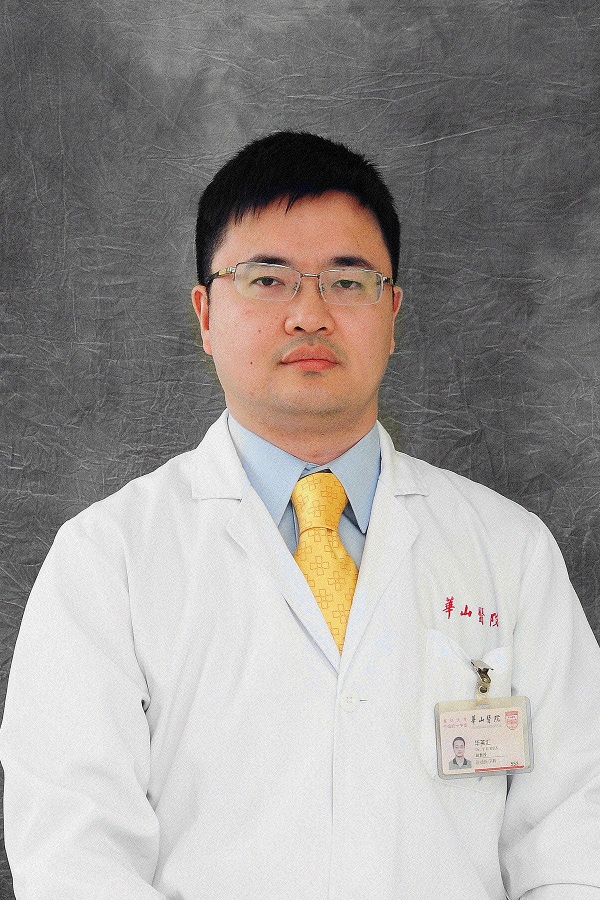 Yinghui Hua (华英汇)合作论文数Huashan Hospital, Fudan University11
 Yan Wo (沃雁)合作论文数College of Basic Medical Sciences, Shanghai Jiao Tong University11
 Chengxuan Yu合作论文数9
 Shiyi Chen (陈诗一)合作论文数School of Economics, Fudan University;Fudan International University School of Finance9Mo Chen合作论文数9
 Yuzhou Chen合作论文数8JiWu Chen (陈疾忤)合作论文数8Huaixuan Sheng合作论文数7Yueming Wang合作论文数7Yang Xing合作论文数6Yimeng Yang合作论文数5Fuchun Chen (陈福春)合作论文数5
 Dandan Sheng合作论文数4
 Luyi Sun合作论文数4Mingru Huang合作论文数4Liman Sai合作论文数4
 Ziying Wu (吴紫莺)合作论文数4
 Shurong Zhang合作论文数4Feng Shaoqing合作论文数4Shixian Dong合作论文数3Yu Dong合作论文数3Lu Fang合作论文数3Wei Yao合作论文数3Xing Yang合作论文数3Hongyun Li合作论文数3Zhu Junfeng合作论文数3
 Hong Li (力弘)合作论文数School of Pharmacy, Fudan University3
 Yixin Zhang (章一新)合作论文数Department of Plastic and Reconstructive Surgery, Shanghai Ninth People's Hospital, School of Medicine, Shanghai Jiao Tong University3
 Xiaogang Xu (徐晓刚)合作论文数3Tengjia Ma合作论文数2Zhai Weitao合作论文数2Xuanfeng Qin合作论文数2Han Gao合作论文数2MENG Xinxian (孟心娴)合作论文数2Zhengbiao Jin合作论文数2Xiliang Shang合作论文数2Wei Wang合作论文数2Tianwu Chen合作论文数2Yiran Wang合作论文数2
 Li Qianru合作论文数2Zifan LIN (林资范)合作论文数2
 Peng Zhang合作论文数Huashan Hospital2
 Xu Heng (许恒)合作论文数2Hong Shao (邵虹)合作论文数2




