Nerve regeneration after spinal cord injury (SCI) is severely hindered by a hostile microenvironment, where excessive reactive oxygen species (ROS) and uncontrolled inflammation form a vicious cycle, triggering secondary injury cascades. However, most current treatments are single-target strategies, obtaining marginal benefits for the intricate pathological mechanisms after SCI. Herein, we developed a nanozyme-switched efferocytosis initiation platform, termed CM-ApoV, by integrating mesenchymal stem cell-derived apoptotic vesicles (ApoVs) with cerium-melatonin nanozymes (Ce-MT). As a distinct subtype of extracellular vesicles, ApoVs are enriched with functional proteins that mediate immunomodulation. Besides, phosphatidylserine (PtdSer) exposed on the surface of ApoVs serves as a critical "eat me" signal that enables targeted recognition and efferocytosis by microglia, thereby promoting microglial repolarization and modulating their functions. Ce-MT nanozymes were anchored onto ApoVs to enhance their ROS scavenging capacity. In the meanwhile, the reversible attachment and detachment of Ce-MT mask PtdSer during systemic circulation and enable re-exposure of PtdSer in an oxidative microenvironment at the injured site. Consequently, the CM-ApoV system comprehensively remodels the pathological network and establishes a favorable microenvironment for neuronal repair. In a rodent model of SCI, CM-ApoV promoted neuronal survival, modulated microglial function, and reduced glial scar formation, ultimately leading to a significant improvement in motor function. Overall, this system highlights the synergistic therapeutic potential of the nanozyme-ApoV hybrid platform and provides a feasible strategy for multidimensional treatment of SCI.
Apoptotic cells release a highly heterogeneous population of extracellular vesicles, which can be categorized into apoptotic bodies, apoptotic microvesicles, and apoptotic exosomes. These apoptotic vesicles (apoVs) not only inherit materials from their parental cells but also encapsulate apoptosis-related factors, thereby playing multifaceted roles in signal transduction, homeostatic regulation, and the tumor microenvironment. This review highlights recent advances in the cell biology of apoptotic extracellular vesicles, with a focus on the unique characteristics of tumor-derived ApoVs and their mechanisms of interaction in immune evasion, angiogenesis, and the modulation of the tumor microenvironment. Additionally, the potential of ApoEVs as biomarkers for tumor diagnostics is explored. In the realm of tumor therapy, engineered and modified ApoEVs have emerged as promising drug delivery vehicles and vaccine carriers, enabling targeted delivery of therapeutic agents and showing significant potential in antitumor immunization strategies. Summarizes the role of apoptotic extracellular vesicles in the tumor microenvironment and their clinical applications. Evaluates the potential of these vesicles for use in tumor diagnosis and emerging therapies. Outlines future research directions and discusses challenges for clinical translation.
Ischemic stroke, a critical neurological disorder resulting from abrupt cerebral blood flow interruption, remains a leading cause of global mortality and chronic disability. Despite advancements in reperfusion therapies, many survivors still suffer significant neurological deficits, primarily attributed to ischemia-reperfusion injury-induced oxidative stress and neuroinflammation. To address these challenges, we designed cerium-curcumin hybrid nanoparticles (Ce-Cur NPs) by leveraging the coordination interaction between redox-active cerium ions and curcumin's beta-diketone moiety. These Ce-Cur NPs were engineered to alleviate ROS-mediated secondary injuries and promote neural repair by leveraging their remarkable ROS scavenging capabilities, which mimic enzymatic activities to effectively neutralize superoxide and hydroxyl radicals. Furthermore, M1 polarized macrophage-derived membranes were employed to enhance the targeted delivery of Ce-Cur NPs to ischemic regions, while transferrin receptor (TfR)-activated peptides enabled efficient receptor-mediated transcytosis across the blood-brain barrier. In vivo studies using a middle cerebral artery occlusion (MCAO) model demonstrated that TfR-M1-Ce-Cur NPs successfully localized to the ischemic brain, significantly reduced infarct volume, and preserved neuronal integrity. Enhanced neurogenesis and improved functional recovery were observed, underscoring the therapeutic potential of Ce-Cur NPs as a versatile platform for ischemic stroke treatment. This approach provides a robust and evidence-based solution to enhance clinical outcomes for patients with ischemic stroke.
Central nervous system (CNS) injury is a leading cause of death and long-term disability worldwide. Neurological deficits reflect disruption of central neural circuits. A major barrier to circuit repair is the intrinsically low regenerative potential of adult CNS neurons-linked in part to failure of injury-induced nuclear export of class IIa histone deacetylases (notably HDAC5)-together with a hostile post-injury microenvironment. Here we present a multifunctional nanosystem, encapsulating the class IIa HDAC4/5-selective inhibitor LMK-235 and featuring an electroactive polyaniline coating with asymmetrically distributed 5-hydroxytryptamine moieties. Upon reaching the lesion, our nanosystem assembles into large-pore scaffolds that (i) inhibit the activity of nuclear-retained class IIa HDACs in neurons and thereby reactivate intrinsic regenerative programs, (ii) regulate microglial activation to mitigate neuroinflammation, and (iii) provide an electroactive interface promoting activity-dependent synaptic reconnection. This multi-pronged approach illustrates an integrated platform with translational potential for CNS disorders in which circuit disconnection constrains recovery.
Biofilm-associated infections present a critical therapeutic challenge due to antibiotic resistance and impaired tissue healing. Here, we present a microrobotic system (MZ-8) that integrates real-time human-steered navigation with autonomous, microenvironment-responsive therapy to actively eradicate biofilms and promote tissue regeneration. This microrobotic system features a spine-inspired structure for mechanical biofilm disruption, a pH-responsive ZIF-8 coating for immunomodulatory Zn2+ release, and closed-loop actuation under second near-infrared fluorescence guidance. In a rat model of periprosthetic joint infection, MZ-8 achieved effective biofilm removal, induced a pro-regenerative immune response by polarizing macrophages toward the M2 phenotype, and significantly enhanced tissue regeneration. Transcriptomic analysis further revealed the activation of immunomodulatory pathways and upregulation of M2-associated genes, confirming the system's sequential shift from eradication to repair. Moreover, validation in a rabbit model and human knee joint confirmed its operational feasibility under clinical imaging guidance and excellent biosafety. This work establishes that integrating physical eradication, biochemical immunomodulation, and interactive control within a single system is essential for advancing from infection clearance to functional tissue restoration. Thus, it provides a therapeutic paradigm for biofilm-associated diseases and lays a foundation for future intelligent, clinically adaptive anti-infective systems.
Glioblastoma (GBM), the most aggressive primary brain tumor, remains a formidable therapeutic challenge, with a median survival under 15 months. Despite the current standard of care-comprising maximal safe surgical resection, radiotherapy, and temozolomide chemotherapy-patient outcomes have seen minimal improvement over the past two decades. A key barrier to effective treatment is GBM’s robust and multifaceted immunosuppressive network, which critically undermines antitumor immunity. While much of the research has focused on the local immunosuppressive tumor microenvironment, systemic immunosuppression represents an equally important yet often underappreciated obstacle, significantly impairing host immune competence. Effective immunotherapy relies on an intact and functional immune system capable of mounting durable T cell-mediated responses. However, GBM induces profound systemic immune dysfunction, manifested by severe lymphopenia and depletion of effector immune cells, which further limits immune-mediated tumor control. Therefore, a comprehensive understanding of both systemic and local immunosuppressive mechanisms is essential for the rational design of effective immunotherapeutic strategies. In this review, we examine the unique physiological features of the brain, dissect the immunosuppressive landscape of GBM at both local and systemic levels, and highlight recent insights into the underlying mechanisms. We also discuss current immunotherapeutic modalities, and emerging drug delivery strategies aimed at overcoming immunosuppression to improve therapeutic efficacy.
Stimuli-responsive hydrogels demonstrate an intelligent ability to respond to environmental triggers, such as pH, temperature, etc. Such an ability has been applied to the control release drug delivery system to specific sites with controllable kinetics. Natural macromolecules, especially pectin, have a high potential to develop into stimuli-responsive hydrogels for biomedical applications due to their versatile gelling properties, high biocompatibility, biodegradability, and nontoxicity. Furthermore, the chemical structure of pectin also offers modifications through various chemical reactions to enhance the ability of responding to external triggers under physiological conditions. While pectin has been widely studied in drug delivery, its potential as a smart hydrogel for site-specific, stimuli-responsive drug release remains underexplored. This review highlights recent advancements in pectin modifications to improve its responsiveness to pH and temperature variations, which are crucial for biomedical applications. Additionally, the underlying mechanisms governing these behaviors in pectin-based hydrogels are discussed.
The tissues spanning from tendon to bone exhibit a highly specialized extracellular matrix (ECM) architecture, characterized by hierarchical collagen alignment and a gradient mineral composition, which together enable efficient force transfer and guide spatially organized cellular phenotypes. However, recapitulating such complex multi-scale organization and compositional gradients to achieve integrated soft-hard tissue remains challenging. Here, we report the de novo construction of biomimetic collagen-mineral matrices that mimic both the hierarchical organization and mineral gradient distribution of the native tendon-to-bone ECM. Through synergistic electro-assembly and post-treatment, collagen molecules self-organized into aligned fibrillar matrices with multi-scale architecture, replicating tendon-side morphology while providing robust tensile mechanics. At the opposing end, intrafibrillar and interfibrillar minerals were spatially patterned to emulate the mineral gradient from tendon to bone. This structural and compositional continuum enables smooth mechanical transition across the soft-hard tissue interface and promotes region-specific regeneration of aligned tendon-like tissue, fibrocartilage and bone. In vivo studies in rabbit models confirm that these de novo constructed matrices support histological reconstruction of multiple tissues from tendon to bone at the rotator cuff, and significantly improve functional recovery. This work presents a bottom-up biomimetic strategy for engineering multiscale collagen-based scaffolds and demonstrates the therapeutic potential of de novo constructed matrices for multiple tissue regeneration.
The activation of cyclic guanosine monophosphate-adenosine monophosphate (cGAS)/stimulator of interferon genes (STING) pathway has emerged as a promising cancer immunotherapy strategy. However, the clinical efficacy of STING agonists is hindered by poor pharmacological properties, asynchronous tumor antigen delivery, and unwanted side effects. Inspired by the observation that tumor cells excrete extracellular vesicles (EVs) enriched with tumor antigens and various proteins for intercellular communication, we leveraged a biological self-assembling pathway to construct a tumor vaccine for cancer immunotherapy through cGAS/STING activation, overcoming these limitations. The vaccine enriches double-stranded DNA (dsDNA), which serves as a natural adjuvant, enhancing antigen presentation in EVs and promoting T-cell activation via the cGAS/STING pathway. Specifically, we incubated metformin-loaded positively charged poly(d, l-lactide-co-glycolide) nanoparticles (Met-PC-NPs) with tumor cells to elevate intracellular reactive oxygen species (ROS) levels and subsequently harvested "waste" EVs containing both dsDNA entrapped by Met-PC-NPs and tumor antigens (Met-PC-EVs). Upon subcutaneous injection, Met-PC-EVs efficiently migrated to lymph nodes and activated antigen-presenting cells, enabling the cross-presentation of tumor antigens to CD8+ T-cells. This process led to robust tumor eradication in both prophylactic and therapeutic models, and it established long-term immune memory. Furthermore, Met-PC-EVs demonstrated a significant synergistic effect when coadministered with immune checkpoint inhibitors. Our approach successfully transformed "waste" Met-PC-EVs into valuable vaccines, leveraging cGAS/STING activation to bypass the current limitation of STING agonists, and offered a clinically translatable method for developing EV-based vaccines.
Current immunotherapies primarily focus on intratumoral immune suppression, with limited consideration of peripheral immune exhaustion, resulting in suboptimal clinical outcomes. Tumor-derived exosomes (TEXs) play a crucial role in both intratumoral immune suppression and peripheral immune exhaustion, making them an attractive target for cancer immunotherapy. In this study, we developed an optimized TEXs modulation strategy to enhance cancer immunotherapy by reprogramming the immune-promoting phenotype of TEXs and boosting their secretion. We designed a TEXs-tuning nanoassembly (TEXT) coloaded with biguanides and photosensitizers. The nanoassembly leverages hyaluronic acid for targeted accumulation at tumor sites. Biguanides inhibit the AMPK/Yap pathway in tumor cells, thereby reprogramming the tumor metabolic landscape and remodeling TEXs to an immune-promoting phenotype. Photodynamic therapy (PDT) is employed to boost the release of immune-promoting TEXs. Through the reprogramming of TEXs' phenotype and enhancement of their release, TEXT effectively alleviates immune exhaustion in peripheral tissues and reverses intratumoral immune suppression. TEXT effectively overcame both local and peripheral immunosuppression and demonstrated robust inhibition of primary and metastatic tumors as a monotherapy. Our findings highlight TEXT as an effective TEXs modulator, showcasing exosome regulation as a highly efficient and low-toxicity approach to combat cancer immune evasion. Overall, this strategy achieves a functional shift of TEXs from immunosuppressive factors to therapeutic synergists, establishing a paradigm of proactive immune modulation in cancer immunotherapy.
Hepatic ischemia-reperfusion injury (IRI) is an important factor affecting the prognosis of patients undergoing surgery. Exosomes derived from mesenchymal stem cells (MSC-EXOs) are widely used and play a therapeutic role in hepatic IRI. However, natural exosomes lack liver-targeting ability and have low bioavailability. In this study, MSC-EXOs were simply modified with OPDEA-PCL or liver-targeting DSPE-PEG2000-Galactose, forming OPDEA-PCL-modified MSC-EXOs (OP-EXOs) or DSPE-PEG2000-Galactose-modified MSC-EXOs (GPEG-EXOs). In mouse hepatic IRI model, OP-EXOs and GPEG-EXOs both significantly reduced alanine aminotransferase (ALT), aspartate aminotransferase (AST), and lactate dehydrogenase (LDH) levels in serum after hepatic IRI, alleviating liver injury. Transcriptomic and proteomic analyses showed that OP-EXOs and GPEG-EXOs reduced hepatic IRI by downregulating the expression of S100A8, S100A9, SELP, and ANXA2 in the liver following IRI. This study opens a new paradigm for the treatment of hepatic IRI using engineered MSC-EXOs with the potential to improve the prognosis of liver surgery.
Pancreatic ductal adenocarcinoma (PDAC) is highly aggressive, with limited success in traditional therapies due to the fibrotic, immunosuppressive, pro-metastatic tumor microenvironment (TME), which collectively impede the drug accumulation and accelerate the tumor progression. In this work, we developed a PDAC-customized nutrient-mimicking reconstituted high-density lipoprotein (rHDL) capable of efficiently co-encapsulate versatile TME regulating cannabidiol and cytotoxic gemcitabine to simultaneously reprogram TME while suppressing PDAC progression. Specifically, a small-sized, nutrient-like rHDL was constructed to realize deep PDAC parenchyma penetration and efficient intra-tumoral uptake. Next, natural herbal compound cannabidiol was screened and incorporated into rHDL to regulate TME via attenuating fibrosis, reliving immunosuppression and mitigating metastatic tendency. At last, gemcitabine, the PDAC gold standard first-line therapy was co-delivered by the PDAC-customized rHDL to overcome drug resistance and amplify its PDAC suppression. Our findings demonstrate the feasibility of an integrated multi-stage TME regulation strategy for improved PDAC therapy, and might represent a modality in promoting chemotherapy against PDAC.
Diazeniumdiolates are highly valued as NO donors in biomedical applications because of their exceptional NO- loading efficiency. Typically, the NO-releasing properties of these compounds can be tailored by incorporating various substitutes at the O2-position of N-diazeniumdiolates. In this study, we report a novel class of bisdiazeniumdiolates, which are covalently linked through an orthoformate-bridge between two diazeniumdiolate groups. Under physiological conditions (pH = 7.4, 37 degrees C), these orthoformate-bridged bis-diazeniumdiolates were found to release four equivalents of NO. The half-lives of NO release varied between 1.3 and 17.9 min. Furthermore, these bis-diazeniumdiolates exhibited superior bactericidal activity against Escherichia coli (MIC = 1-8 mM, MBC = 8-32 mM) and Staphylococcus aureus (MIC = 2-8 mM, MBC = 16-32 mM) compared to the commercial 2-(N, N-diethylamino)-diazenolate-2-oxide, which only releases two equivalents of NO per molecular (MIC (E. coli) = 32 mM, MIC ( S.aureus ) = 64 mM, MBC = 128 mM). These bis-diazeniumdiolates enhance NO loading and release NO independently of enzymes or chemical additives, indicating their potential benefits for further biomedical applications.
Cell migration serves as a crucial factor in cell therapy, which has been extensively studied in vitro. However, the impact of in vivo migratory behavior of cells on therapeutic efficacy remains an uncharted territory, due to the complexity of biological processes within living organisms. Here, tendon stem/progenitor cells (TSPCs) with or without mechanical confinement simulating sedentary and migratory cell behaviors were sophisticatedly designed and transplanted to patellar tendon defect mouse model, with short-wave infrared (SWIR) fluorescence imaging adopted to dynamically monitor in vivo cell migration process. As a result, migratory TSPCs exhibited enhanced morphological plasticity with substantial changes in area (51.47% vs. 17.06%, p < 0.05) and width (41.11% vs. 8.69%, p < 0.05) of cell population. Besides, directional in vivo cell migration pattern from injection site to the proximal and medial region of patellar tendon defect was depicted, which further led to superior therapeutic efficacy in tendon regeneration based on histological scoring. In contrast, sedentary TSPCs demonstrated prolonged in vivo cell retention in large cell numbers (21 d vs. 14 d), and resulted in inferior therapeutic efficacy. Furthermore, transcriptomic analysis revealed activations of cell chemotaxis and migration pathways in mice with migratory TSPCs, while morbid pathways of excessive cell proliferation and ossification in sedentary TSPCs. In summary, in vivo cell migratory behavior was identified as a major driver of superior therapeutic efficacy in tendon regeneration by morphological plasticity and directional migration. This study highlights the pivotal role of in vivo migration competence in cell therapy optimization and provides a mechanistic framework for clinical translation strategies.
Toxin accumulation within specialized microenvironments, such as cerebrospinal fluid (CSF), poses significant clinical challenges. In particular, bilirubin in CSF can cause severe damage to the brain and neural tissues. Current therapeutic strategies for intracranial bilirubin clearance suffer from poor targeting, inadequate detoxification capacity, and reliance on invasive procedures. Here, we demonstrate multifunctional microrobots designed for toxin removal in specialized anatomical compartments. The proposed ultrasound-trackable microbubbles@PCN-333 microrobots (UMPCs) feature a hollow hydrogel microbubble core for enhanced ultrasound imaging contrast, an outer metal-organic framework (MOF) layer for proactive and high-capacity toxin adsorption, and embedded ferroferric oxide (Fe3O4) nanoparticles to enablec. Magnetic actuation enhances the UMPC's bilirubin capture efficiency through improved mass transfer. By integrating an ultrafast ultrasound imaging system with magnetic actuation, we achieve real-time tracking and precise control of UMPCs within the CSF. This strategy offers a minimally invasive and actively guided approach for efficient intracranial bilirubin clearance, opening new avenues for targeted toxin-removal in anatomically confined environments.
Photodynamic therapy (PDT) is an attractive approach for tumor treatment because of its precision, potent cytotoxic effect, and low risk of resistance compared to conventional cancer treatments. However, PDT consumes oxygen. The oxygen depletion effects in PDT-treated tumor cells can elevate lactic acid production and efflux, promoting the progression of surrounding tumor cells through tumor metabolic symbiosis and promoting macrophages to M2-type polarization for supporting tumor progression. Herein, a multifunctional nanosystem is developed for the intracellular co-delivery of the photosensitizer (ICG), the nanozyme (iron oxide nanoparticles, MNPs), and siMCT4 (siRNA for monocarboxylate transporter 4). In tumor cells undergoing PDT, siMCT4 inhibits lactate efflux, thereby limiting extracellular lactate-associated malignancy and immune evasion. Meanwhile, both the reduction of extracellular lactate levels and the presence of MNPs in the tumor microenvironment promote the M1-type polarization to enhance the antitumor activity of macrophages. Furthermore, the intracellular lactic acid accumulation and M1-type macrophage-secreted H2O2 facilitate the MNPs-mediated chemodynamic therapy (CDT). Therefore, the intelligent nanosystem, IM@iPPAE@siMCT4, can regulate the intra/extracellular lactate levels and the M1-type macrophage polarization to deliver a two-punch attack on tumor cells. This nanosystem circumvents the problems arising from antitumor PDT.
Angiogenesis is a critical process in the early stages of tendon healing but is always limited by its avascular structure, thus impairing effective healing. Nanopeptides targeting endogenous vascular endothelial growth factor (VEGF) presented a promising strategy for promoting early angiogenesis in tendon healing. In this study, we introduced a nanoscale electro-assembly technique to kinetically control the collagen assembling to form VEGF-binding protein (VEGF-BP) and PR1P-loaded oriented collagen matrix (BP/P Col) for tendon healing. The specific binding of VEGF-BP and PR1P to endogenous VEGF mediated the proangiogenic effects during the early stages of repair. In vitro cell experiments showed that BP/P Col could promote human umbilical vein endothelial cell migration and tube formation as well as new vessel formation in chick chorioallantois. In a rabbit model of tendon defect, it increased vascular area and new vessel formation within the first 2 weeks. Additionally, multiomics analysis further revealed the potential mechanism of BP/P Col in promoting early angiogenesis was to modulate the macrophage-regulated VEGF expression, providing positive feedback for early angiogenesis. This process induced the organized collagen structure along the oriented collagen matrix, which ultimately facilitated rapid tendon healing during the early stage. Overall, this study demonstrated that BP/P Col, through the efficient loading of VEGF-BP and PR1P, exerted a synergistic enhancement effect on endogenous VEGF. It could not only accelerate early angiogenesis but also promote subsequent rapid and functional tendon healing, offering a promising treatment strategy for tendon injuries.
As the principal constituent of the extracellular matrix, collagen exhibits significant therapeutic potential in sports medicine, owing to its distinct triple-helical configuration and inherent biocompatibility. This biomaterial serves as a foundational material for scaffolds, membranes, patches and dressings targeting tendon repair, cartilage reconstruction and bone defect remediation. However, its clinical translation was hampered by limitations: poor tensile strength risks mechanical failure under load, immunogenicity from residual epitopes can trigger adverse reactions and rapid enzymatic degradation compromises structural integrity before tissue maturation. This review elucidates current properties and resources of collagen-based biomaterial and critically analyzes its inherent limitations and their clinical consequences. It emphasizes how evolving tissue engineering strategies directly mitigate barriers. Molecular crosslinking and chemical modification are employed to enhance tensile properties and delay degradation, critical for mechanically demanding environments. Composite blending with polymers compensates for mechanical weakness while retaining bioactivity. Advanced processing techniques such as 3D printing and electrospinning enable precise fiber alignment, replicating native tissue anisotropy and improving functional outcomes. Rigorous decellularization protocols further mitigate immunogenicity. This review further examines recent preclinical and clinical progress in collagen-based biomaterials for tendon, ligament, cartilage and bone regeneration, highlighting successful translations and ongoing challenges. Future directions focus on refining these strategies to accelerate the development of next-generation, clinically robust collagen therapies for sports medicine.
Most CRISPR assays lack clinical utility due to their complex workflows and limited validation. Here we present a streamlined "one-pot" asymmetric CRISPR tuberculosis assay that attenuates amplicon degradation to achieve 5 copies/μL sensitivity within 60 min and detect positive patient samples within 15 min. This assay exhibited 93%, 83%, and 93% sensitivity with adult respiratory, pediatric stool, and adult cerebral spinal fluid specimens, and detected 64% of clinically diagnosed tuberculous meningitis cases, in a cohort of 603 clinical samples. This assay achieves complete specificity and greater sensitivity (74% vs. 56%) than the most sensitive reference test with prospectively collected tongue swabs, and exhibits similar performance when adapted to a lateral flow assay format and employed to analyze self-collected tongue swabs. These results demonstrate the utility of this approach across diverse specimen types, including those suitable for use in remote and resource-limited settings, to improve access to molecular diagnostics.
 Xiaoling Gao (高小玲)合作论文数College of Basic Medical Sciences, Shanghai Jiao Tong University96
 Xinguo Jiang (蒋新国)合作论文数School of Pharmacy, Fudan University88
 Qingxiang Song合作论文数53
 Hongzhuan Chen (陈红专)合作论文数Shanghai Jiao Tong University College of Basic Medical Sciences40Sijia Feng合作论文数36Yunxia Li合作论文数33
 Zhiqing Pang (庞志清)合作论文数School of Pharmaceutical Sciences, Fudan University29Songlei Zhou合作论文数Fudan Univ, Sch Pharm, Minist Educ, Key Lab Smart Drug Delivery, Lane 826,Zhangheng Rd, Shanghai 201203, Peoples R China23
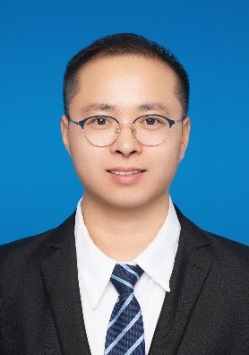 Yukun Huang (黄宇坤)合作论文数School of Chemical Engineering, Zhengzhou University23
 Quanyin Hu (胡全银)合作论文数Pharmaceutical Sciences Division, School of Pharmacy, University of Wisconsin-Madison;Wisconsin Center for NanoBioSystems, University of Wisconsin-Madison20
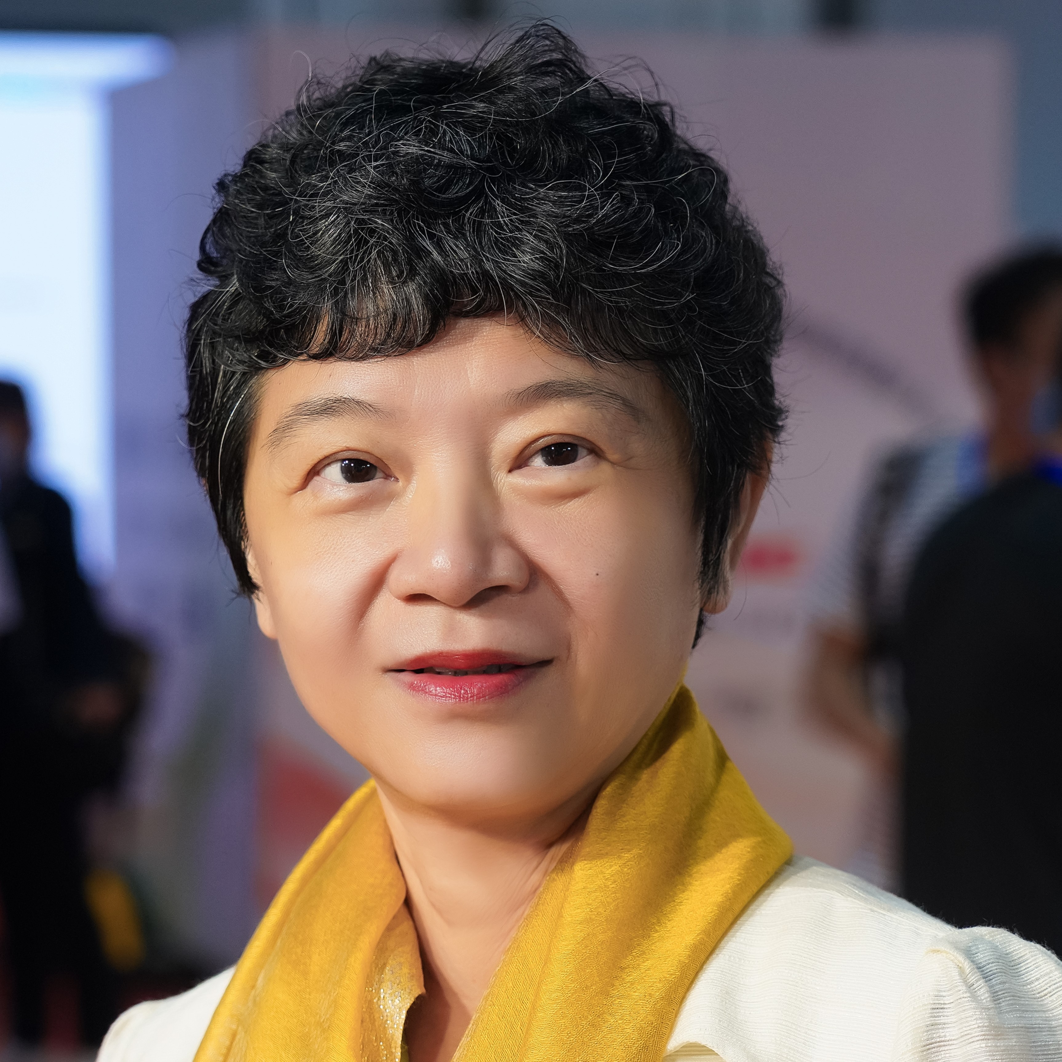 Lei Yao (姚雷)合作论文数School of Design, Shanghai Jiao Tong University20
 Dandan Sheng合作论文数19Mingzhu Liu合作论文数19WenMing Jiang (江文明)合作论文数18YueFeng Hao (郝跃峰)合作论文数18Jiang Gan合作论文数Shanghai Children's Heart Center, Shanghai Jiaotong University School of Medicine18
 Yan Wo (沃雁)合作论文数College of Basic Medical Sciences, Shanghai Jiao Tong University18Fenfen Ma合作论文数17Guang-Zhi Gu合作论文数Department of Pharmaceutics, School of Pharmacy, Fudan University17
 Qizhi Zhang (张奇志)合作论文数School of Pharmacy, Fudan University16Jingxian Feng合作论文数Fudan Univ, Sch Pharm, Minist Educ, Key Lab Smart Drug Delivery, Lane 826,Zhangheng Rd, Shanghai 201203, Peoples R China15
 Tianze Jiang (蒋天泽)合作论文数15
 Chengxuan Yu合作论文数14Jiang Di合作论文数14
 Jian Zhang (张键)合作论文数复旦大学附属中山医院骨科14Mo Chen合作论文数14Jiangyu Cai合作论文数13
 Shiyi Chen (陈诗一)合作论文数School of Economics, Fudan University;Fudan International University School of Finance13Jianhui Yao合作论文数12Huaixuan Sheng合作论文数11
 Luyi Sun合作论文数11Laozhi Xie合作论文数11Mengyin Jiang合作论文数11Yipu Liu合作论文数Shanghai Pudong Hospital & Department of Pharmaceutics, School of Pharmacy, Fudan University, Lane 826, Zhangheng Road, Shanghai, 201203, PR China11Minjun Xu合作论文数Fudan Univ, Sch Pharm, Minist Educ, Key Lab Smart Drug Delivery, Lane 826,Zhangheng Rd, Shanghai 201203, Peoples R China11
 Xiao Gu (谷潇)合作论文数Department of Electronic Engineering, Shanghai Jiao Tong University11Feng Xingye合作论文数11
 Congjian Xu (徐丛剑)合作论文数School of Medicine, Westlake University11Shi-Lei Cao合作论文数11
 Zhongyang Liu合作论文数State Key Laboratory of Functional Materials for Informatics, Shanghai Institute of Microsystem and Information Technology, Chinese Academy of Sciences11Jianpei Xu合作论文数10
 Hongliang Liu (刘洪亮)合作论文数University of Chinese Academy of Sciences;Technical Institute of Physics and Chemistry, Chinese Academy of Sciences10
 Meng Huang合作论文数10
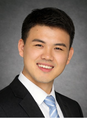 Yu Chen (陈雨)合作论文数Department of Pharmacology and Chemical Biology, Shanghai Jiao Tong University College of Basic Medicial Sciences10
 Wei Lu (陆伟)合作论文数School of Pharmacy, Fudan University;State Key Laboratory of Molecular Engineering of Polymers, Fudan Unversity10
 Chunmei Gao (高春梅)合作论文数College of Chemistry and Environmental Engineering, Shenzhen University10
 Qianqian Zhu合作论文数School of Medicine, Xi’an Jiaotong University10Yimeng Yang合作论文数9




