目的 分析复发缓解型多发性硬化(MS)患者看似正常白质(NAWM)纤维束微结构变化.方法 17例MS患者与年龄、性别匹配健康志愿者参与研究.使用TrackVIS软件持续纤维跟踪成像方法重建白质纤维束,记录白质纤维束的部分各向异性(FA)、表观弥散系数(ADC)和纤维束容积值,进行数据分析.结果 MS及健康对照组各男8例,女9例.与健康对照组相比,MS患者通过胼胝体压部和整个胼胝体神经纤维束的容积下降,两者相比差异有统计学意义(P<0.05).MS患者通过胼胝体膝部、压部及整个胼胝体纤维束的FA值下降(P<0.05).与病灶对侧NAWM相比,通过MS患者病灶的纤维束容积更小,FA值降低,ADC值升高(P<0.05).较之健康对照组,通过患者病灶对侧NAWM的纤维束FA值降低(P<0.05).结论 弥散张量纤维成像技术可用于早期NAWM微小损伤的评估,FA值可作为NAWM纤维束损伤的敏感指标.
Microglia play a critical role in the regulation of CNS immune function, which can be greatly affected by M1/M2 polarization. The role of Notch signaling in Statins induced alteration of M1/M2 polarization in BV2 cells was assessed in this study. M1 markers in LPS and Jagged-1 treated group were significantly increased and such increase was attenuated by simvastatin; however, M2 markers were enhanced. Moreover, simvastatin enhance the expression of Notch signaling molecules, and its regulatory effects were blocked in Notch1 knocked down cells. In conclusion, these findings indicated that simvastatin alters M1/M2 polarization of murine BV2 microglia via Notch signaling.
Objective To establish a simple and ideal method of serum-free primary culture of cortical neurons from newborn rats in vitro.Methods The cortical tissue of newborn rats after birth in 24 hours was digested.Cortical neurons were planted in a culture bottle covered with poly-L-lysine,which containing DMEM high glucose medium and fetal bovine serum as culture medium.Twenty-four hours after the neurons were planted the culture medium was replaced by neurobasal with B27 supplementations.The morphological changes of the cortical neurons were observed under inverted phase-contrast microscope.Immunofluorescence staining for neuron specific enzyme (NSE) was applied to identify the cortical neurons.Results The cortical neurons began to adhere to the culture bottle after 2 h.Cells began to develop small neurites and form network gradually.Up to the 7th day,neurons developed well and formed typical network.The cultured cells were confirmed as neurons by NSE fluorescence staining,and the purity of neurons was (93.57±3.9)%.Conclusion The present technique is a simple and idcal method for culturing primary cortical neurons of newborn rats with high purity in vitro.
Objective:By herpes simplex virus ( HSV) type 1 infecting BV2 cells, to observe the expression of TLR9 and MyD88mRNA.Methods:BV2 cell line of HSV-1 encephalitis was set up .Determine the corilagin concentration and intervention time by MTT , and calculate the TCID50 of virus titer by the Reed-Muench. We randomly divided cells into six groups:HSV-1+corilagin group , HSV-1+PBS group , CpG-ODN+group, corilagin, CPG-ODG+PBS group, PBS+corilagin group, PBS+PBS group (the control group). The expression of TLR9/MyD88mRNA was measured by FQ -PCR and TLR9.Results:Compared with HSV-1+PBS group, HSV-1+Corilagin decreased the TLR9 and MyD88mRNA expressions(P﹤0.05);compared with CpG -ODN +PBS group, CpG-ODN+Corilagin decreased the TLR9 and MyD88mRNA ex-pressions(P﹤0.05); Conclusion:TLR9/MyD88 signaling pathway may be key point in immune response in the HSE by the affected TNF-alpha.Corilagin can reduce TLR9 and MyD88mRNA expressions.
Objective:Through the detection of TLR9,MyD88 protein expression and TNF-α secretion of HSE murine model,to investigate the effect of corilagin on the HSE prognosis.Methods:Injection approach makes HSV-1 from entering into the manufacture of mouse brain tissue of mice with herpes simplex virus encephalitis model,40 mice were randomly divided into normal group and herpes simplex virus groups of 20.Herpes simplex virus group were randomly divided into four groups:control group,corilagin group,HSV-1 + saline group,HSV-1 + corilagin groups of five.Western Blotting to detect the brain tissue of mice TLR9,MyD88 protein.Each brain tissue of mice TNF-α secretion by ELISA analysis to detect changes.H-E stained sections of brain tissue pathological changes observed.Results:(1) After herpes simplex virus type 1 (HSV-1) infection (5 days after the virus injection),the lesions of hemorrhage and necrosis in mice's brains could be observed by microscope,and the injury in much less serious in corilagin group compared to the HSV-1 group.(2)Compared to HSV-1+PBS group,HSV-l+corilagin decrease the TLR9,MyD88 mRNA expression.Compared to HSV-1+PBS group,HSV-l+corilagin decrease the TLR9,MyD88 mRNA expression[(9.36 ± 1.02) Kb vs (14.84 ± 1.83) Kb,(7.5 ± 1.04) Kb vs (11.7 ± 2.63) Kb,P<0.05].(3) Compared to HSV-1+PBS group,HSV-l+corilagin decrease the secretion of TNF-α [(543.97 ± 18.07)pg/mL vs (1036.62 ± 38.84)pg/mL,P>0.05].Conclusions:Corilagin treating HSE may decrease the secretion of TNF-α through reducing the TLR9/MyD88 signaling pathway.
Aims: There is no effective medication to date for herpes simplex virus encephalitis (HSE). In this study, we investigated the anti-inflammatory effect of chlorogenic acid (CGA) on herpes simplex virus (HSV)-1-induced responses in BV2 microglia.Main methods: The cellular model was established with BV2 cells stimulated by HSV-1 and then treated with CGA at different concentrations. Cell viability was assayed by the MTT assay. The mRNA expression of Toll-like receptor (TLR)-2, TLR9 and myeloid differentiation factor88 (Myd88) was assayed by real-time quantitative PCR, and the protein expression was assayed by flow cytometry or Western blotting. Tumor necrosis factor-alpha (TNF-alpha) and interleukin (IL)-6 were measured by ELISA as well as real-time quantitative PCR. Nuclear NF-kappa B p65 protein was assayed by Western blotting.Key findings: The cell survival rate was significantly improved after CGA treatment, and CGA prevented increases in TLR2, TLR9 and Myd88 following HSV-1 challenge in BV2 cells both at the mRNA and protein levels. Moreover, CGA could attenuate HSV-induced INF-alpha and IL-6 release into the supernatant. The mRNA levels of TNF-alpha and IL-6 were also significantly inhibited by CGA. The expression of NF-kappa B p65 increased significantly in the nucleus in HSV-1-stimulated microglia but could be reduced by CGASignificance: CGA inhibits the inflammatory reaction in HSE via the suppression of TLR2/TLR9-Myd88 signaling pathways. CGA may serve as an anti-inflammatory agent and provide a new strategy for treating HSE. (C) 2015 Elsevier Inc All rights reserved.
Objective To study the protection of 1,2,3,4,6-penta-O-galloyl-β-D-glucose (β-PGG) on apoptosis of PC12 cells from Parkinson’s disease models induced by MPP+ and its mechanism.Methods PC12 cells were incubated in high glucose DMEM medium. One week before drug treatment, nerve growth factor was added to the cultures at the final concentration of 50 ng/mL. PC12 cells were divided into control group, MPP+group, and 50μmol/Lβ-PGG groups pretreated for 7, 12, 20, and 30 h. The survivals of PC12 cells in MPP+were observed after pretreatment withβ-PGG for different periods. The death of PC12 cells in MPP+ was evaluated with trypan blue staining method, and the activity of the PC12 cells was determined by MTT assay. The expression of Bax,Fas, FasL, procaspase-3, procaspase-8, and procaspase-9 was analyzed by Western blotting method, and then the activity of caspase-3, caspase-8, and caspase-9 was examined.Results The death rates of PC12 cells in control group were the lowest, while those in MPP+ group were the highest, and those inβ-PGG groups pretreated after 12 h were significantly decreased compared with MPP+ group (P < 0.01). The activities of the PC12 cells in MPP+ group were the lowest, and those inβ-PGG group pretreated for 12 h were further increased, while those inβ-PGG group pretreated for 20 h were the highest. The protein contents of Bcl-2, procaspase-3, procaspase-8, and procaspase-9 inβ-PGG group pretreated for 5 h were increased, and increased to peak at pretreated for 15 h. On the contrary, protein contents of Bax, Fas, and FasL inβ-PGG group pretreated for 5 h were decreased, and decreased to the minimum at pretreated for 30 h. The activities of caspase-3,caspase-8, and caspase-9 of the PC12 cells inβ-PGG group pretreated for 15 h were 36.5%, 40.2%, and 42.2% of those in MPP+ group, respectively.Conclusionβ-PGG has protection against apoptosis of PC12 cells induced by MPP+, and its mechanism is related to inhibiting the apoptosis of PC12 cells induced by MPP+ and then increase survival rate of PC12 cells by increasing the expression of Bcl-2, inhibiting the expression of Bax, Fas, and FasL, and decreasing activities of caspase-3, caspase-8, and caspase-9.
In this study, we tried to explore the molecular mechanism that Corilagin protected against herpes simplex virus-1 encephalitis through inhibiting the TLR2 signaling pathways in vivo and in vitro. As a result, Corilagin significantly prevented increase in the levels of TLR2 and its downstream mediators following Malp2 or HSV-1 challenge. On the other hand, in spite of TLR2 knockdown, Corilagin could still significantly suppress the expression of P38 and NEMO, phosphor-P38, and nuclear factor kappa B. The mRNA and protein expression of TLR2 and its downstream mediators in the brain tissue were also significantly lowered in mice treated with Corilagin. In addition, Corilagin inhibited expression of tumor necrosis factor-α (TNF-α) and interleukin (IL)-6 protein. In conclusion, Corilagin shows the potential to protect against HSV-1-induced encephalitis, and the beneficial effects may be mediated by inhibiting TLR2 signaling pathways.
目的:观察免疫肠内营养支持对老年重症脑血管病患者营养状况及免疫功能的恢复作用。方法将老年重症脑血管病患者随机分为免疫肠内营养组( IEN组)和常规肠内营养组( EN组),每组20例。分别于治疗前,治疗后7 d以及14 d流式细胞仪检测各组患者外周血T细胞亚群、NK细胞及酶联免疫吸附( ELASA)法检测血清中前清蛋白( PA)、( RBP)、白细胞介素( IL)-2、IL-4浓度。结果 IEN及EN支持治疗后14 d,患者的NK、CD3、CD4、CD8、CD4/CD8以及血清蛋白ALB、RBP虽较治疗前有极显著增高(P<0.01),但仍低于正常对照组,且 IEN组与EN组间相比无差异。两组患者的IL-2浓度在治疗后14 d均较治疗前有显著增高(P<0.01),IEN组增高较EN组更显著(P<0.05);在治疗后14 d,IEN组IL-4的浓度下降明显且与EN组相比有显著差异(P<0.05)。 C反应蛋白(CRP)明显下降(P<0.05)。 IEN组与EN组相比,APACHEⅡ评分、MODS评分均降低,机械通气时间缩短短及28 d生存率增高,但两组相比无显著性。结论在改善患者免疫抑制及提高患者的临床预后方面,IEN要较EN更有优势。
目的:观察氯吡格雷在急性脑梗死中的抗炎作用。方法入选短暂性脑缺血发作( TIA)患者40例、急性脑梗死患者66例,后者又按照入院顺序随机、单盲分为A组(氯吡格雷+阿司匹林)和B组(阿司匹林),观察疗程1 w,所有患者测定入院时高敏C反应蛋白(hs-CRP)和P选择素( PS)水平,急性脑梗死患者1 w后复查hs-CRP和PS。观察各组hs-CRP和PS的初始状态及变化情况。结果急性脑梗死患者hs-CRP和PS较TIA患者有明显升高(P<0.001)。急性脑梗死A组和B组hs-CRP和PS在初始状态没有显著差异(P>0.05),1 w后 A组比 B组hs-CRP和Ps显著降低( P<0.001)。结论急性脑梗死存在明显的炎症过程。氯吡格雷可以下调急性脑梗死患者的炎症因子,有良好的抗炎作用。
Under global cerebral ischemia, the effect of different brain temperature on cerebral ischemic injury was studied. Male Sprague-Dawley rats were divided into normothermic (37–38°C) ischemia, mild hypothermic (31–32°C) ischemia, hyperthermic (41–42°C) ischemia and sham-operated groups. Global cerebral ischemia was established using the Pulsinelli four-vessel occlusion model and brain temperature was maintained at defined level for 60 min after 20-min ischemia. The expression of c-fos protein and the levels of malondialdehyde (MDA) and lactate in brain regions were detected by immunochemistry and spectrophotometrical methods, respectively. C-fos positive neurons were found in the hippocampus and cerebral cortex after cerebral ischemia reperfusion. Mild hypothermia increased the expression of c-fos protein in both areas, whereas hyperthermia decreased the expression of c-fos protein in the hippocampus at 24 h reperfusion, and the cerebral cortex at 48 h reperfusion when compared to normothermic conditions. In normothermic, mild hypothermic and hyperthermic ischemia groups, the levels of MDA and lactate in brain tissue were increased at 24, 48 and 72 h reperfusion following 20-min ischemia as compared with the sham-operated group (P<0.01). The levels of MDA and lactate in mild hypothermic group were significantly lower than those in normothermic group (P<0.01). It is suggested that brain temperature influences the translation of the immunoreactive protein product of c-fos after global cerebral ischemia, and MDA and lactate are also affected by hypothermia and hyperthermia.
The human adult brain possesses intriguing plasticity, including neurogenesis and angiogenesis, which may be mediated by the activated sonic hedgehog (Shh). By employing a coculture system, brain microvascular endothelial cells (BMECs) cocultured with astrocytes, which were incubated under oxygen–glucose deprivation (OGD) condition, we tested the hypothesis that Shh secreted by OGD-activated astrocytes promotes cerebral angiogenesis following ischemia. The results of this study demonstrated that Shh was mainly secreted by astrocytes and the secretion was significantly upregulated after OGD. The proliferation, migration, and tube formation of BMECs cocultured with astrocytes after OGD were significantly enhanced, but cyclopamine (a Shh antagonist) or 5E1 (an antibody of Shh) reversed the change. Furthermore, silencing Ras homolog gene family, member A (RhoA) of BMECs by RNAi and blocking Rho-dependent kinase (ROCK) by Y27632, a specific antagonist of ROCK, suppressed the upregulation of proliferation, migration, and tube formation of BMECs after OGD. These findings suggested that Shh derived from activated astrocytes stimulated RhoA/ROCK pathway in BMECs after OGD, which might be involved in angiogenesis in vitro.
Objective To explore the molecular mechanisms underlying the protective effect of curcumin on PC12 cells apoptosis induced by dopamine(DA) oxidative stress.Methods PC12 cells model of DA-induced oxidative stress damage was established.PC12 cells were randomly divided into blank control group,curcumin group(20 μmol/L),DA group(200 μmol/L) and DA(20 μmol/L) + curcumin group(200 μmol/L).After the cells were cultured for 24 h,cell proliferation was detected by MTT.The protein expressions of Bcl-2,Cyt-c and Caspase-3 were detected by Western blotting.Results ① In the presence of curcumin,the PC12 cells inhibition rate,induced by DA was decreased.② Curcumin inhibited down-regulation of anti-apoptotic protein Bcl-2 induced by DA and up-regulated the protein expression of intracellular Bcl-2.③ Curcumin inhibited up-regulation of apoptotic protein Cyt-c induced by DA.④ Curcumin inhibited up-regulation of intracellular cleavage Caspase-3 protein expression induced by DA and curcumin itself had no effect on Caspase-3 expression.Conclusion The protective effect of curcumin on DA-induced apoptosis in PC12 cells may be related to its inhibiting down-regulation of Bcl-2 and up-regulation of Cyt-c and of Caspase-3.
Objective: To observe the characteristics of praxiology of Parkinson disease (PD) rats. Methods: At 1d, 7d, 14d after PD rats’ model were successful; we observed the markers of rotational behavior of Parkinson disease (PD) rats. For example: start time, continuous time, maximum turning, etc. Results: From 1d to 14d, PD rats, start time of the rotational behavior elongated gradually, continuous time shortened by degrees, maximum rotational speed and the number of the rotational behavior were invariable. Conclusions: Improved method of PD is scientific, reliable, and simple. Substantial nigral pathology of PD rats makes the foundation of the rotational behavior. The injury of substantial nigral can be reflected by maximum rotational speed and the number of the rotational behavior of PD rats.
目的 观察信号通路Toll样受体4(TLR4) -p38蛋白激酶(p38MAPK)在小鼠细菌性脑炎模型中的表达及意义.方法 将BABL/c小鼠30只随机分为正常组、细菌性脑炎模型组、SB203580(p38MAPK抑制剂)干预组,分别按干预后3、7 天各分两组.正常组侧脑室注入生理盐水,模型组侧脑室给予脂多糖(LPS); 干预组在给予LPS后即在腹腔注射SB203580.检测脑组织TLR4mRNA和p38MAPKmRNA的表达变化,测定肿瘤坏死因子-α(TNF-α)、白介素-6(IL-6)的含量.结果 与正常组比较,LPS刺激后小鼠脑组织TLR4 mRNA和p38MAPKmRNA表达迅速升高,其相应的脑组织TNF-α、IL-6含量表达也升高,第7天的含量分别为 (65.95±8.82) pg / mg和(70.75±8.65) pg / mg.给予 SB203580 抑制剂后TLR4mRNA和p38MAPKmRNA 表达明显减弱,其相应的脑组织TNF-α、IL-6含量表达与感染组比较也明显降低.结论 LPS刺激小鼠脑组织可引起TLR4-p38 MAPK信号通路的活化并释放炎性细胞因子,而SB203580则对其有明显的抑制作用,证明细菌性脑炎与TLR4-p38MAPK信号通路密切相关.
Objective To investigate the protective effects of AnNao pill on oxygen and glucose deprivation (OGD) in cultured rat cortical neurons.Methods Cultured primary cortical neurons were pretreated or not pretreated with AnNao pill medicated serum and were exposed to the oxygen and glucose deprivation (OGD) model.The results on cell viability rates,the apoptosis rates of neurons,the expression of Bcl-2,caspase-12,glucoseregulated protein (GRP) and cytochrome C (CytC) were compared between OGD and AnNao pill groups.Results Compared to the normal group,the rates of apoptosis [(22.0 ± 2.2) % vs (3.2 ± 0.6) %] and the expression of caspase-12,GRP78 and CytC in the OGD group increased (all P < 0.05) ; The rates of cell viability [(62.4 ±4.6) % vs (100.0 ± 3.1) %] and the expression of bcl-2 decreased (all P < 0.05).Compared to the OGD group,the viability rates in low dose,middle dose and high dose AnNao pill groups increased[(74.9 ±2.7)%,(83.0 ±2.2) %,(75.2 ± 2.8) % vs (62.4 ± 4.6) %] (all P < 0.05).The rate of viability in middle dose AnNao pill group was higher than those in low dose and high dose AnNao pill group (all P < 0.05).the rates of apoptosis [(10.4 ± 0.7) % vs (22.0 ± 2.2) %],the expression of caspase-12 and CytC in the middle dose AnNao pill group were lower than those in the OGD group(all P < 0.05) ; the expression of Bcl-2 were higher(all P < 0.05)than that in the OGD group.Conclusion AnNao pill has a protective effect on rat cortical neurons against OGD and its mechanism may be that the AnNao pill can regulate associated protein of apoptosis and inhibit the apoptosis.
Objective To investigate the effect and significance of TLR2mRNA and p38MAPK mRNA signaling pathway expressed in the LPS-induced BV2 microglia.Methods The routinely cultured BV2microglia in vitro were divided into control group,LPS stimulation group(stimulation lasting 12 and 24 hours)and SB203580 plus LPS treatment group(stimulation lasting 12 and 24 hours).We determined the levels of TNF alpha and IL-6 by ELISA method,and detected the group difference of TLR4mRNA and p38MAPK mRNA by RT-PCR method.Results Compared with those in the control group,the expressions of TLR4mRNA and p38MAPK mRNA in the LPS-induced microglia were significantly increased,which were especially obvious after 24-hour stimulation,TNF-αwas(513.67±14.05)pg/mg,IL-6 was(396.84±15.41)pg/mg.Treatment with SB203580 could effectively inhibit TLR4mRNA and p38 MAPK mRNA expressions in BV2microglia,the concentrations of TNF-αand IL-6 markedly decreased when compared with those in the infected group.Conclusion The TLR4-p38MAPK signaling pathway can be activated by LPS in BV2microglia,leading the releasing of inflammatory cytokines.And the SB203580 significantly inhibits the signaling pathway,demonstrating the close relation between the TLR4-p38MAPK signaling pathway and inflammatory activation of microglia.
To date there has been no valid treatment for herpes simplex encephalitis (HSV). This study explores the protective activity of ethanol extract of Cynanchum paniculatum (bunge) kitagawa for treatment of HSV. Cell models and animal models were established and divided into 4 groups: normal group, virus group, cynanchum paniculatum group and Dexamethasone group. Flow cytometry was employed to detect apoptosis of cell model and TUNEL assay was chosen to detect apoptosis of animal tissues. The survival time of the animal models was observed. ELISA was used to measure TNF-α expression and the Greiss method to measure Nitric Oxide (NO) expression in the mouse brain. As a result, it was found that extract of Cynanchum paniculatum can improve the survival rate of HSV-infected mice. The extract could prevent apoptosis in the neuron cell model and reduce apoptosis rate in brain tissue after HSV infection. With the extract intervention, TNF-α and NO levels in brain tissue were significantly decreased in the animal model. In conclusion, the extract of Cynanchum paniculatum can prevent HSV-inducing impairment in the cell and animal model of HSE.
目的 观察不同剂量阿托伐他汀对脑梗死预后的影响及其血清CRP的变化.方法 将2010年10月~2011年10月收治的80例脑梗死患者随机分为两组,每组40例,观察组常规治疗加上阿托伐他汀40 mg/d,对照组常规治疗加上阿托伐他汀10 mg/d;两组均在治疗前及治疗后4周测定C-反应蛋白( CRP) 浓度,并且进行NIHSS及BI评分.结果 两组治疗后CRP降低,观察组较对照组效果更明显(P<0.05),两组治疗前后NIHSS评分和BI评分均有改善,观察组较对照组改善明显(P均<0.05).结论 阿托伐他汀强化治疗使CRP水平明显下降,并能改善脑梗死患者的预后.
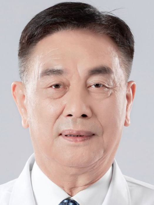 Shenggang Sun (孙圣刚)合作论文数Union Hospital, Tongji Medical College, Huazhong University of Science and Technology81
 ETang Tong (童萼塘)合作论文数41
 Bo Hu (胡波)合作论文数Union Hospital, Tongji Medical College, Huazhong University of Science and Technology29
 Guirong Wei (魏桂荣)合作论文数Union Hospital, Tongji Medical College, Huazhong University of Science and Technology24
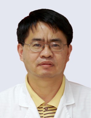 Yunping Chen (陈运平)合作论文数Union Hospital, Tongji Medical College, Huazhong University of Science and Technology22
 Hongyi Xing (邢宏义)合作论文数Neurology Department, Union Hospital, Tongji Medical College, Huazhong University of Science and Technology22E'tang Tong合作论文数20Xiaoqiao Zhang (张小乔)合作论文数18LiFang Zheng (郑丽芳)合作论文数16ChuanYu Liu (刘传玉)合作论文数16
 Xuebing Cao (曹学兵)合作论文数Union Hospital, Tongji Medical College, Huazhong University of Science and Technology14XiaoYing Qiu (邱小鹰)合作论文数13ShanChun Yu (俞善纯)合作论文数13
 Hongge Li (李红戈)合作论文数Union Hospital, Tongji Medical College of Huazhong University of Science and Technology13
 Fei Cao (曹非)合作论文数Union Hospital, Tongji Medical College, Huazhong University of Science and Technology12Ji-Hua Dong (董继华)合作论文数12
 Ling Mao (毛玲)合作论文数Union Hospital, Tongji Medical College, Huazhong University of Science and Technology11
 Xuejun Deng (邓学军)合作论文数Union Hospital, Tongji Medical College, Huazhong University of Science and Technology11MaoYue Zhang (张茂悦)合作论文数10Jing Huang (黄敬)合作论文数10
 Min Zhang (张敏)合作论文数Union Hospital, Tongji Medical College, Huazhong University of Science and Technology10
 Yuan Fang (方瑗)合作论文数Union Hospital, Tongji Medical College, Huazhong University of Science and Technology10Min Bao (包敏)合作论文数9
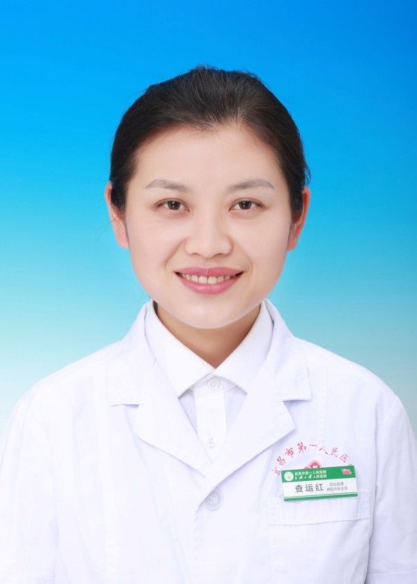 Yunhong Zha (查运红)合作论文数China Three Gorges University9
 Changqin Liu (刘昌勤)合作论文数Union Hospital, Tongji Medical College, Huazhong University of Science and Technology9Yuan-Jin Guo合作论文数9Fang Luo (骆芳)合作论文数8Wang Meng-Die合作论文数8Yu Zhou (周瑜)合作论文数7
 Yuanpeng Xia (夏远鹏)合作论文数Union Hospital, Tongji Medical College, Huazhong University of Science and Technology7
 Yunliang Guo (郭云良)合作论文数School of Basic Medicine, Qingdao University7Ruo-Lian Dai合作论文数7He Wang (王何)合作论文数6
 Yuanjin Guo (郭远瑾)合作论文数Union Hospital, Tongji Medical College, Huazhong University of Science and Technology6
 Junjian Zhang (章军建)合作论文数Zhongnan Hospital of Wuhan University6SuRong Zhou (周素荣)合作论文数5LiaYan Zheng (郑俩燕)合作论文数5ZhiBin Chen (陈志斌)合作论文数5JunMin Li (李俊敏)合作论文数5
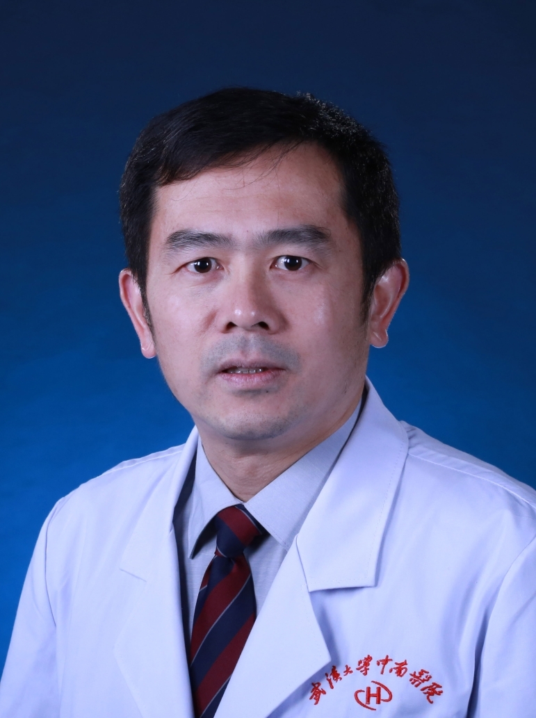 Hong Zhang (张洪)合作论文数Zhongnan Hospital of Wuhan University;Second Clinical Hospital of Wuhan University5
 JiXiang Chen (陈吉相)合作论文数Union Hospital, Tongji Medical College, Huazhong University of Science and Technology5Qin Li合作论文数5Fei Wu (吴非)合作论文数5
 Lei Zhao (赵雷)合作论文数Union Hospital, Tongji Medical College, Huazhong University of Science and Technology5
 Hai Peng (彭海)合作论文数Union Hospital, Tongji Medical College, Huazhong University of Science and Technology5Huaijun Huang (黄怀钧)合作论文数4Su-ning WU (吴苏宁)合作论文数4junling gao (高峻岭)合作论文数4




