Enamel covers teeth, is the hardest tissue in the vertebrate body and has a complex multiscale structure from nanometres to millimetres1. The structure comprises thin, long hydroxyapatite (Ca5(PO4)3OH) nanocrystals2, 50-70 nm wide, many micrometres long, parallel and bundled into approximately 5-µm-wide rods. The rods undulate and cross into a microscale 'decussation pattern' that toughens enamel by deflecting cracks3,4. However, the crystallographic orientation of enamel nanocrystals is poorly understood. Here we show that the misorientation angle of adjacent nanocrystals varies markedly across 12 primate teeth spanning 9 species, 17.8 million years of evolution and diverse diets. Using a method called Polarization Enabled Large Input of Crystal Angles at the Nanoscale (PELICAN)5, we compare nanocrystals in the same (pre)molar locations and show that misorientation increases with food hardness in extant and fossil non-human apes and monkeys. We compare misorientation across three major dietary shifts in human evolution: the transition to meat-eating about 2.0-1.5 million years before present6,7, to agriculture (about 12,000 years before present)8,9, and the Industrial Revolution (about 250 years before present)10. We show that over the past 1.6 million years, in the human lineage misorientation increased with time, especially when meat and stone-ground grains were introduced into human diets, but not with the Industrial Revolution. Thus, besides macro-changes, teeth adapted to dietary change at the nanoscale and crystallographically. This observation suggests that misorientation may contribute to enamel's resilience; thus, bioinspired materials may consider small misorientation angles for added resilience.
Twinning occurs when two crystals share a coherent interface along which their lattices are mirror symmetric. Here, twinning is investigated in biogenic and geologic aragonite (CaCO3) in columnar nacre from the red abalone Haliotis rufescens, sheet nacre from the black-lip pearl oyster Pinctada margaritifera, and geologic aragonite. All samples exhibit the expected single (110) twins, characterized by a 116.2 degrees rotation of the aragonite a-axis. Surprisingly, multiple twins-double, triple, quadruple-are also observed, each involving successive 116.2 degrees rotations of the a-axis around the c-axis. Multiple twinning is concomitant but distinct from the well-known cyclic twinning of aragonite. Multiple twinning is most prevalent in columnar nacre, reaching up to quadruple twins, most frequently occurring between nacre tablets in the same column, whereas sheet nacre and geologic aragonite exhibit up to triple twins. The frequency of twins decreases with increasing twin multiple, consistent with a physical rather than biological origin. This interpretation is supported by observations in geologic aragonite and by simulations of columnar nacre growth, which incorporate only nucleation rate, growth rate, and basic geometric constraints, yet they reproduce the observed twinning behavior in columnar nacre, reinforcing the conclusion that multiple twinning arises from physical growth mechanisms.
Mollusk shells protect the animals that form and inhabit them. They are composites of minerals and organics, with diverse mesostructures, including nacre, prismatic calcite, crossed-lamellar aragonite, and foliated calcite. Twins, that is, crystals mirror symmetric with respect to their coherent interface, occurring as formation or deformation twins, are observed in all mollusk shell mesostructures but never within calcite prisms. Here, nanotwins and microwins within single calcite prisms are observed in different shells. Using Polarization-dependent Imaging Contrast (PIC) mapping with 20-60 nm resolution, twins are observed to be 0.2-3 & mu;m thick layers of differently oriented and colored crystals with respect to the main prism crystal. Multiple twins are interspersed with the prism crystal, parallel to one another, and similarly oriented. When comparing images of calcite prisms and twins obtained by PIC mapping and by Electron Back-Scattered Diffraction (EBSD), the images correspond precisely. All twins are e-twin types, with 127 & DEG; angular distance between c-axes. E-twins are the most common deformation twins in geologic calcite, as also observed here in Carrara marble. Location of all twins near the outer surface of all shells and e-twin type both suggest that twins within calcite prisms in mollusk shells result from deformation twinning.
Calcium carbonate (CaCO 3 ) is abundant on Earth, is a major component of marine biominerals and thus of sedimentary and metamorphic rocks and it plays a major role in the global carbon cycle by storing atmospheric CO 2 into solid biominerals. Six crystalline polymorphs of CaCO 3 are known—3 anhydrous: calcite, aragonite, vaterite, and 3 hydrated: ikaite (CaCO 3 ·6H 2 O), monohydrocalcite (CaCO 3 ·1H 2 O, MHC), and calcium carbonate hemihydrate (CaCO 3 ·½H 2 O, CCHH). CCHH was recently discovered and characterized, but exclusively as a synthetic material, not as a naturally occurring mineral. Here, analyzing 200 million spectra with Myriad Mapping (MM) of nanoscale mineral phases, we find CCHH and MHC, along with amorphous precursors, on freshly deposited coral skeleton and nacre surfaces, but not on sea urchin spines. Thus, biomineralization pathways are more complex and diverse than previously understood, opening new questions on isotopes and climate. Crystalline precursors are more accessible than amorphous ones to other spectroscopies and diffraction, in natural and bio-inspired materials.
Biominerals are organic–mineral composites formed by living organisms. They are the hardest and toughest tissues in those organisms, are often polycrystalline, and their mesostructure (which includes nano‐ and microscale crystallite size, shape, arrangement, and orientation) can vary dramatically. Marine biominerals may be aragonite, vaterite, or calcite, all calcium carbonate (CaCO 3 ) polymorphs, differing in crystal structure. Unexpectedly, diverse CaCO 3 biominerals such as coral skeletons and nacre share a similar characteristic: Adjacent crystals are slightly misoriented. This observation is documented quantitatively at the micro‐ and nanoscales, using polarization‐dependent imaging contrast mapping (PIC mapping), and the slight misorientations are consistently between 1° and 40°. Nanoindentation shows that both polycrystalline biominerals and abiotic synthetic spherulites are tougher than single‐crystalline geologic aragonite. Molecular dynamics (MD) simulations of bicrystals at the molecular scale reveal that aragonite, vaterite, and calcite exhibit toughness maxima when the bicrystals are misoriented by 10°, 20°, and 30°, respectively, demonstrating that slight misorientation alone can increase fracture toughness. Slight‐misorientation‐toughening can be harnessed for synthesis of bioinspired materials that only require one material, are not limited to specific top‐down architecture, and are easily achieved by self‐assembly of organic molecules (e.g., aspirin, chocolate), polymers, metals, and ceramics well beyond biominerals.
We compared the transient precursor phases on the forming surface of fresh corals grown at different pH values: pH 7.2, 7.4, 7.8, and 8.0 1 .Using PhotoEmission Electron Microscopy 2, 3 at the calcium L-edge we identified the transient metastable precursor phases 4-6 as a function of distance from the surface.We expected to find the most and least metastable precursors at the outermost and innermost layers of the surface, but that is not what we found.One of the intermediate phases penetrates deepest into the skeleton.
The mature skeletons of hard corals, termed stony or scleractinian corals, are made of aragonite (CaCO3). During their formation, particles attaching to the skeleton's growing surface are calcium carbonate, transiently amorphous. Here we show that amorphous particles are observed frequently and reproducibly just outside the skeleton, where a calicoblastic cell layer envelops and deposits the forming skeleton. The observation of particles in these locations, therefore, is consistent with nucleation and growth of particles in intracellular vesicles. The observed extraskeletal particles range in size between 0.2 and 1.0 μm and contain more of the amorphous precursor phases than the skeleton surface or bulk, where they gradually crystallize to aragonite. This observation was repeated in three diverse genera of corals, Acropora sp., Stylophora pistillata─differently sensitive to ocean acidification (OA)─and Turbinaria peltata, demonstrating that intracellular particles are a major source of material during the additive manufacturing of coral skeletons. Thus, particles are formed away from seawater, in a presumed intracellular calcifying fluid (ICF) in closed vesicles and not, as previously assumed, in the extracellular calcifying fluid (ECF), which, unlike ICF, is partly open to seawater. After particle attachment, the growing skeleton surface remains exposed to ECF, and, remarkably, its crystallization rate varies significantly across genera. The skeleton surface layers containing amorphous pixels vary in thickness across genera: ∼2.1 μm in Acropora, 1.1 μm in Stylophora, and 0.9 μm in Turbinaria. Thus, the slow-crystallizing Acropora skeleton surface remains amorphous and soluble longer, including overnight, when the pH in the ECF drops. Increased skeleton surface solubility is consistent with Acropora's vulnerability to OA, whereas the Stylophora skeleton surface layer crystallizes faster, consistent with Stylophora's resilience to OA. Turbinaria, whose response to OA has not yet been tested, is expected to be even more resilient than Stylophora, based on the present data.
Glycine, the simplest amino acid, is considered a promising functional biomaterial owing to its excellent biocompatibility and strong out-of-plane piezoelectricity. Practical applications require glycine films to be manufactured with their strong piezoelectric polar 〈001〉 direction aligned with the film thickness. Based on the recently-developed solidification approach of a polyvinyl alcohol (PVA) and glycine aqueous solution, in this work, we demonstrate that the crystal orientation of the as-synthesized film is determined by the orientation of glycine crystal nuclei. By controlling the local nucleation kinetics via surface curvature tuning, we shifted the nucleation site from the edge to the middle of the liquid film, and thereby aligned the 〈001〉 direction vertically. As a result, the PVA-glycine-PVA sandwich film exhibits the highest aver-age piezoelectric coefficient d33 of 6.13 ± 1.13 pC N-1. This work demonstrates a promising kinetic approach to achieve crystallization and property control in a scalable biocrystal manufacturing process.
Abstract The hardest and toughest tissues formed by living organisms are organic-mineral composites termed biominerals 1,2. When they are crystalline, their mesostructure includes the nano- and micro-scale crystallite size, shape, arrangement, and orientation. Mesostructures vary enormously across marine CaCO3 biominerals (aragonite, vaterite, calcite) because they result from divergent evolution: biominerals were formed long after organisms diverged from one another 3,4. Despite such diversity, CaCO3 marine biominerals share a convergent character: adjacent crystals are similarly oriented 5-32. The reason for such convergence is unclear. Here, we show with quantitative, precise measurements at the nanoscale that the slight misorientation is consistently between 1°-40° in diverse biominerals. Can this slight misorientation confer a desirable materials property and therefore an evolutionary advantage to the forming organisms? We test and confirm this hypothesis with nanoindentation in diverse biominerals, geologic aragonite, and in abiotic, slightly misoriented, synthetic spherulites. Molecular dynamics (MD) simulations of bicrystals reveal that aragonite, vaterite, calcite, exhibit toughness peaks when they are misoriented by 10°, 20°, 30°, respectively, demonstrating that slight misorientation alone increases crack deflection and therefore fracture toughness. Slight misorientation, along with other previously known and co-existing toughening mechanisms, was selected repeatedly and convergently, during the course of evolution, to postpone fracture and thus provide organisms with competitive advantage. We anticipate slight misorientation-toughening to be a starting point for more sophisticated materials synthesis and additive manufacturing in many fields. Compared to previously known toughening mechanisms, in fact, the advantages of slight misorientation are that it can and does occur in synthetic materials, it requires one material only and no specific top-down architecture, it is easily achieved by self-assembly of organic molecules (e.g. aspirin, chocolate), polymers, metals, and ceramics 29 well beyond biominerals.
The hardest materials in living organisms are biologically grown crystalline minerals, or biominerals, which are also incredibly fracture-tough. Biomineral mesostructure includes size, shape, spatial arrangement, and crystal orientation of crystallites, observable at the mesoscale (10 nanometer - 10 micron). Here we show that diverse biominerals, including nacre and prisms from mollusk shells, coral skeletons, and tunicate spicules have different mesostructures, but they converged to similar, small (<30 degrees) misorientations of adjacent crystals at the mesoscale. We show that such small misorientations are an effective toughening mechanism. Combining Polarization-dependent Imaging Contrast (PIC) mapping of mesostructures and Molecular Dynamics (MD) simulations of misoriented bicrystals, we reveal here that small misorientations toughen bicrystals, thus explaining why they evolved independently but convergently: preventing fracture is a clear evolutionary advantage for diverse organisms.
Significance Whether reef-building corals form their skeletons ion by ion from solution or by amorphous particle attachment has important implications for isotope incorporation and for understanding coral skeletons’ resilience to ocean warming and acidification. Here we show that all kinds of reef-building corals make their skeletons by attachment of amorphous nanoparticles and fill the spaces between nanoparticles ion by ion. Thus, the dual mechanism of ion- and particle- attachment is general and relevant to reef formation, which provides the infrastructure for one of the most diverse ecosystems on Earth.
 Pupa Gilbert合作论文数Department of Physics, University of Wisconsin–Madison9
 Cayla A Stifler合作论文数Univ Wisconsin Madison, Dept Phys, Madison, WI 53706 USA7
 Sylvie Tambutte合作论文数5
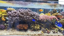 Eric Tambutte合作论文数Centre Scientifique de Monaco4
 Andrew J. Lew合作论文数Dept Chem, MIT3
 Andreas Scholl合作论文数Berkeley Lab, Lawrence Berkeley National Laboratory;Advanced Light Source3
 Markus J. Buehler合作论文数Laboratory for Atomistic & Molecular Mechanics, Department of Civil and Environmental Engineering, School of Engineering, Massachusetts Institute of Technology;Elsevier3
 Laura Müller合作论文数2Virginia Quach合作论文数2
 Alexandra Tits合作论文数2
 Astrid Cantamessa合作论文数2
 Yann Delaunois合作论文数2Benjamin I Fordyce合作论文数2
 Tali Mass合作论文数Dept Marine Biol, Univ Haifa2
 Davide Ruffoni合作论文数Institute for Biomechanics, ETH Zurich2
 Philippe Compère合作论文数University of Liege2
 Alexander A. Venn合作论文数Bermuda Institute of Ocean Sciences (BIOS2
 Fredrick Kyalo Manthi合作论文数Division of Anthropology, American Museum of Natural History;Paleontology Section, Department of Earth Sciences, National Museums of Kenya1Harrison Lisabeth合作论文数1Emily Luffey合作论文数1Anne Voigtländer合作论文数1Ganesh Parida合作论文数1
 Maria Cristina Castillo Alvarez合作论文数1William O. Nachlas合作论文数Dept Geosci, Univ Wisconsin1Jiajie Sui合作论文数Dept Mat Sci & Engn, Univ Wisconsin1
 Laurent Devriendt合作论文数Royal Netherlands Institute for Sea Research (NIOZ), Utrecht University Texel1
 Vanessa Schoeppler合作论文数Tech Univ Dresden, CUBE Ctr Mol Bioengn B, Dresden, Germany1
 Emmanuel Ndiema合作论文数Natl Museums Kenya, Dept Earth Sci, Nairobi, Kenya1
 Mackie C O'Hara合作论文数School of Anthropology and Conservation, University of Kent Canterbury1
 Benjamin Gilbert合作论文数Department of Earth and Planetary Science, University of California, Berkeley1
 Long Gu (顾龙)合作论文数Institute of Modern Physics, Chinese Academy of Sciences1
 Zhaoyong Zou (邹朝勇)合作论文数School of Chemistry, Chemical Engineering and Life Sciences, Wuhan University of Technology;State Key Laboratory of Advanced Technology for Materials Synthesis and Processing, Wuhan University of Technology1
 Yael Politi合作论文数Department of Structural Biology, Weizmann Institute of Science1
 Peter Fratzl合作论文数Max Planck Queensland Centre for the Materials Science of Extracellular Matrices;University of Potsdam1
 Jun Li合作论文数Department of Materials Science and Engineering, University of Wisconsin – Madison1
 Ziyi Zhang合作论文数Computer Sciences and Mathematics at University of Wisconsin -Madison1
 Yan Shao (邵燕)合作论文数Sichuan University Law School1
 Chang-Yu Sun (孫彰佑)合作论文数Department of Materials Science and Engineering, National Cheng Kung University1Albéric Marie合作论文数1
 Ehud Gazit合作论文数Dept of Mol. Microbiology and Biotechnology, Tel Aviv University;George S. Wise Faculty of Life Sciences, Tel Aviv University1
 Xudong Wang合作论文数Department of Materials Science and Engineering, College of Engineering, University of Wisconsin-Madison1
 Debbie Guatelli-Steinberg合作论文数Department of Anthropology, College of Arts and Sciences, The Ohio State University;Department of Evolution, Ecology, and Organismal Biology. The Ohio State University;School of Anthropology and Conservation. University of Kent1
 William Scott Mcgraw合作论文数1
 Daniel R. Green合作论文数New Life Resources , Inc1
 Patrick Mahoney合作论文数Groupe de Recherche en Microélectronique, succ. Centre-Ville1
 Rajesh Vilas Chopdekar合作论文数Western Digital1
 Hans A. Bechtel合作论文数Adv Light Source Div, Lawrence Berkeley Natl Lab1




