Supplementary Figure and Table from Gene Expression Profiling Allows Distinction between Primary and Metastatic Squamous Cell Carcinomas in the Lung
Supplementary Figures 1-7 from <i>Squamous Cell Carcinoma Related Oncogene</i>/DCUN1D1 Is Highly Conserved and Activated by Amplification in Squamous Cell Carcinomas
Supplementary Tables 1-3 from <i>Squamous Cell Carcinoma Related Oncogene</i>/DCUN1D1 Is Highly Conserved and Activated by Amplification in Squamous Cell Carcinomas
Supplementary Methods from <i>Squamous Cell Carcinoma Related Oncogene</i>/DCUN1D1 Is Highly Conserved and Activated by Amplification in Squamous Cell Carcinomas
Purpose: Ectopic expression of squamous cell carcinoma-related oncogene (SCCRO or DCUN1D1) in NIH-3T3 cells induces invasion in vitro and produces highly invasive xenografts in nude mice with a propensity for regional lymphatical metastasis. The aim of this study was to identify the molecular mechanism underlying SCCRO-induced invasion and metastasis.Experimental Design: The molecular mechanism of SCCRO-mediated effects on matrix metalloproteinase-2 (MMP2) levels and activity were assessed using a combination of cell biological and molecular methods, including real-time PCR, reporter assay, RNA interference, and chromatin immunoprecipitation assay. Tumor specimens from primary upper aerodigestive tract carcinomas (n = 89) were examined for levels of SCCRO, MMP2, MMP9, MT1-MMP TIMP1, and TIMP2 mRNA by real-time PCR.Results: Overexpression of SCCRO increases MMP2 levels and activity, which is required for SCCRO-induced invasion. Modified McKay assays reveal that SCCRO does not bind to the MMP2 promoter, suggesting that its transcriptional effects are indirect. Deletion or mutation of the activator protein-2 (AP2) and p53 binding element within the MMP2 promoter abrogates SCCRO-driven activation. Ectopic expression of SCCRO increases AP2 levels and promotes the binding of p53 to the MMP2 promoter. Consistent with these findings, SCCRO and MMP2 are coexpressed (P < 0.0001; r(2) = 0.58; 95% confidence interval, 0.46-0.69) in primary (upper aerodigestive tract) carcinomas (n = 89), and this coexpression is associated with an increased prevalence of regional nodal metastasis (P = 0.04; relative risk, 1.53).Conclusions: SCCRO-induced invasion involves activation of MMP2 transcription in an AP2- and p53-dependent manner. SCCRO is a potential marker for metastatic progression in affected cancers.
Abstract Chromosomal amplification at 3q is common to multiple human cancers, but has a specific predilection for squamous cell carcinomas (SCC) of mucosal origin. We identified and characterized a novel oncogene, SCC-related oncogene (SCCRO), which is amplified along the 3q26.3 region in human SCC. Amplification and overexpression of SCCRO in these tumors correlate with poor clinical outcome. The importance of SCCRO amplification in malignant transformation is established by the apoptotic response to short hairpin RNA against SCCRO, exclusively in cancer cell lines carrying SCCRO amplification. The oncogenic potential of SCCRO is underscored by its ability to transform fibroblasts (NIH-3T3 cells) in vitro and in vivo. We show that SCCRO regulates Gli1—a key regulator of the hedgehog (HH) pathway. Collectively, these data suggest that SCCRO is a novel component of the HH signaling pathway involved in the malignant transformation of squamous cell lineage. (Cancer Res 2006; 66(19): 9437-44)
Abstract Lung neoplasms commonly develop in patients previously treated for head and neck carcinomas. The derivation of these tumors, either as new primary lung cancers or as metastatic head and neck cancers, is difficult to establish based on clinical or histopathologic criteria since both are squamous cell carcinomas and have identical features under light microscopy. However, this distinction has significant treatment and prognostic implications. Gene expression profiling was performed on a panel of 52 sequentially collected patients with either primary lung (n = 21) or primary head and neck (n = 31) carcinomas using the Affymetrix HG_U95Av2 high-density oligonucleotide microarray. Unsupervised hierarchical clustering with Ward linkage and the Pearson correlation metric was performed. To assess robustness, bootstrap resampling was performed with 1,000 iterations. A t test of the normalized values for each gene was used to determine the genes responsible for segregating head and neck from lung primary carcinomas, and those with the most differential expression were used for later analyses. In the absence of a large “test” set of tumors, we used a supervised leave-one-out cross-validation to test how well we could predict the tumor origin. Once a gene expression profile was established, 12 lung lesions taken from patients with previously treated head and neck cancers were similarly analyzed by gene expression profiling to determine their sites of origin. Unsupervised clustering analysis separated the study cohort into two distinct groups which reliably remained segregated with bootstrap resampling. Group 1 consisted of 30 tongue carcinomas. Group 2 consisted of 21 lung cancers and 1 tongue carcinoma. The clustering was not changed even when normal lung or tongue profiles were subtracted from the corresponding carcinomatous lesions, and a leave-one-out cross-validation showed a 98% correct prediction (see Supplementary Data 1). A minimum set of 500 genes required to distinguish these groups was established. Given the ability to segregate these lesions using molecular profiling, we analyzed the lung tumors of undetermined origin. All cases clearly clustered with either lung or tongue tumor subsets, strongly supporting our hypothesis that this technique could elucidate the tissue of origin of metastatic lesions. Although histologically similar, squamous cell carcinomas have distinct gene expression profiles based on their anatomic sites of origin. Accordingly, the application of gene expression profiling may be useful in identifying the derivation of lung nodules and consequently enhances treatment planning.
Purpose: Oncolytic herpes simplex viruses (HSVs) may have significant antitumor effects resulting from the direct lysis of cancer cells. HSVs may also be used to express inserted transgenes to exploit additional therapeutic strategies. The ability of an interleukin (IL)-12-expressing HSV to treat squamous cell carcinoma (SCC) by inhibition of tumor angiogenesis is investigated in this study. Experimental Design: A replication-competent, attenuated, oncolytic HSV carrying the murine IL-12 gene (NV1042), its non-cytokine-carrying analog (NV1023), or saline was used to treat established murine SCC flank tumors by intratumoral injection. The expression of secondary antiangiogenic mediators was measured. Angiogenesis inhibition was assessed by in vivo Matrigel plug assays, flank tumor subdermal vascularity, and in vitro endothelial cell tubule formation assay. Results: Intratumoral injections of NV1042 (2 × 10⁷ plaque-forming units) into murine SCC VII flank tumors resulted in smaller tumor volumes as compared with NV1023 or saline. IL-12 and IFN-γ expression in tumors was 440 and 2.2 pg/mg, respectively, at 24 h after NV1042 injection, but both IL-12 and IFN-γ were undetectable (<0.2 pg/mg) after NV1023 or saline injections. Expression of two antiangiogenesis mediators, monokine induced by IFN-γ and IFN-inducible protein 10, was elevated after NV1042 treatment. Matrigel plug assays of NV1042-transfected SCC VII tumor cells demonstrated significantly decreased hemoglobin content and microvessel density as compared with NV1023 and PBS. Excised murine flank tumors treated with NV1042 had decreased subdermal vascularity as compared with NV1023 and PBS. Both splenocytes and IL-12 expression by NV1042 were required for in vitro inhibition of endothelial tubule formation. Conclusions: IL-12 expression by an oncolytic herpes virus enhances therapy of SCC through antiangiogenic mechanisms. Strategies combining HSV oncolysis with angiogenesis inhibition merit further investigation for potential clinical application.
Background Squamous cell carcinoma related oncogene expression (SCCRO) correlates with vascular endothelial growth factor-A expression. This data is validated in human lung tumors and provides a putative pathway for angiogenesis in a subset of squamous cell carcinomas. Squamous cell carcinoma related oncogene is a novel oncogene identified by positional cloning of a recurrent amplification at 3q26.3. It is over-expressed in 39.8% of lung, head and neck, cervical, and ovarian carcinomas. SCCRO imparts an aggressive phenotype to affected cancers, which may be related to increased angiogenesis due to SCCRO expression. Our previous work has demonstrated a link between SCCRO and vascular endothelial growth factor-A (VEGF-A) expression in vitro, suggesting a mechanism for SCCRO-induced angiogenesis. The present study aims to confirm and validate this link between SCCRO and VEGF-A expression in an ex vivo human tumor cohort. Methods Fresh tissue was collected at Memorial Sloan-Kettering Cancer Center from 34 patients undergoing primary resection of lung squamous cell carcinomas. RNA was extracted from this tissue, reverse-transcribed, and real-time polymerase chain reaction (RT-PCR) was carried out using a BioRad iQ iCycler with SYBR green fluorophore. Microvessel counting was performed on the tumor specimens using CD34 immunohistochemistry. Results The expression of both SCCRO and VEGF-A mRNA varies widely in both tumor and normal tissue. SCCRO and VEGF-A co-expression was significantly correlated (R 2 = 0.63; P < 0.032). Microvessel counts were not associated with expression of SCCRO or VEGF-A and failed to significantly predict survival. VEGF-A expression in this patient group is a predictor of overall survival ( P < 0.032). Conclusions VEGF-A expression correlates with SCCRO expression in these primary human lung squamous cell carcinomas and is a predictor of clinical behavior. This data supports the association of SCRRO and VEGF-A in the induction of angiogenesis.
Purpose: Gene expression profiling has been shown to be a valuable tool for prognostication and identification of cancer-associated genes in human malignancies. We aimed to identify potential prognostic marker(s) in non-small cell lung cancers using global gene expression profiles. Experimental Design: Twenty-one previously untreated patients with non-small cell lung cancer were analyzed using the Affymetrix GeneChip high-density oligonucleotide array and comparative genomic hybridization. Identified candidate genes were validated in an independent cohort of 45 patients using quantitative real-time reverse transcription-PCR and Western blot analyses. Follow-up data for these patients was collected and used to assess outcome correlations. Results: Hierarchical clustering analysis yielded three distinct subgroups based on gene expression profiling. Cluster I consisted of 4 patients with adenocarcinoma and 1 with squamous cell carcinoma (squamous cell carcinoma); clusters II and III consisted of 6 and 10 patients with squamous cell carcinoma, respectively. Outcome analysis was performed on the cluster groups containing solely squamous cell carcinoma, revealing significant differences in disease-specific survival rates. Moreover, patients having a combination of advanced Tumor-Node-Metastasis stage and assigned to the poor prognosis cluster group (cluster II) had significantly poorer outcomes. Comparative genomic hybridization analysis showed recurrent chromosomal losses at 1p, 3p, 17, 19, and 22 and gains/amplifications at 3q, 5p, and 8q, which did not vary significantly between the cluster groups. We internally and externally validated a subset of 11 cluster II (poor prognosis)-specific genes having corresponding chromosomal aberrations identified by comparative genomic hybridization as prognostic markers in an independent cohort of patients with lung squamous cell carcinoma identifying CSNK2A1 and C1-Inh as independent predictors of outcome. Conclusion: CSNK2A1 and C1-Inh are independent predictors of survival in lung squamous cell carcinoma patients and may be useful as prognostic markers.
Lymphatic metastasis represents the single most important clinical prognostic factor in head and neck squamous cell carcinoma (HNSCC), but underlying genetic mechanisms remain ill defined. Genetic differences between primary carcinomas and their corresponding metastases might form a key to understanding the metastatic phenotype. In this study we aimed to characterize such differences using a genome‐wide screening measure.
Interactions between the p53 and PI3K/AKT pathways play a significant role in the determination of cell death/survival. In benign cells these pathways are interrelated through the transcriptional regulation of PTEN by p53, which is required for p53-mediated apoptosis. PTEN exerts its effects by decreasing the phosphorylated AKT fraction, thereby diminishing prosurvival activities. However, the link between these pathways in cancer is not known. In this study, PIK3CA, encoding the p110alpha catalytic subunit of PI3K, is identified as an oncogene involved in upper aerodigestive tract (UADT) carcinomas. Simultaneous abnormalities in both pathways are rare in primary tumors, suggesting that amplification of PIK3CA and mutation of p53 are mutually exclusive events and either event is able to promote a malignant phenotype. Moreover, the negative effect of p53 induction on cell survival involves the transcriptional inhibition of PIK3CA that is independent of PTEN activity, as PTEN is not expressed in the primary tumors. Conversely, constitutive activation of PIK3CA results in resistance to p53-related apoptosis in PTEN deficient cells. Thus, p53 regulates cell survival by inhibiting the PI3K/AKT prosurvival signal independent of PTEN in epithelial tumors. This inhibition is required for p53-mediated apoptosis in malignant cells.
 Bhuvanesh Singh合作论文数Department of Surgery, Memorial Sloan-Kettering Cancer Center, New York, NY12
 Pornchai O-Charoenrat合作论文数Department of Head and Neck Surgery, Royal Marsden Hospital10
 Inderpal S. Sarkaria合作论文数Harold C. Simmons Comprehensive Cancer Center, The University of Texas Southwestern Medical Center;Department of Cardiovascular and Thoracic Surgery, The University of Texas Southwestern Medical Center9Simon G. Talbot合作论文数Plastic and Reconstructive Surgical Service, Memorial Sloan-Kettering Cancer Center9
 Ronald A. Ghossein合作论文数Memorial Sloan Kettering Cancer Center8Pabbathi G. Reddy合作论文数7Ellie Maghami合作论文数6
 Pulivarthi H Rao合作论文数6Ramanathan Yegnanarayana合作论文数5
 Valerie W. Rusch合作论文数Department of Surgery, Memorial Sloan-Kettering Cancer Center;Weill Cornell Medical College5
 Archontoula Stoffel合作论文数5
 Yoshihiro Yonekawa合作论文数4Andrew J Kaufman合作论文数4
 Russell JH. Ryan合作论文数Department of Pathology, Medical School, University of Michigan4Kepal N. Patel合作论文数4Maria E. Dudas合作论文数4Benjamin Lee合作论文数3
 Nicholas D. Socci合作论文数Sloan Kettering Institute, Memorial Sloan Kettering Cancer Center3
 Agnès Viale合作论文数Marie-Josée & Henry R. Kravis Center for Molecular Oncology, Memorial Sloan Kettering Cancer Center3Cherry L Estilo合作论文数2Su Dao合作论文数2
 Jatin P. Shah合作论文数David H. Koch Center for Cancer Care, Memorial Sloan Kettering Cancer Center2Pham Duykhanh合作论文数2Diane L. Carlson合作论文数2
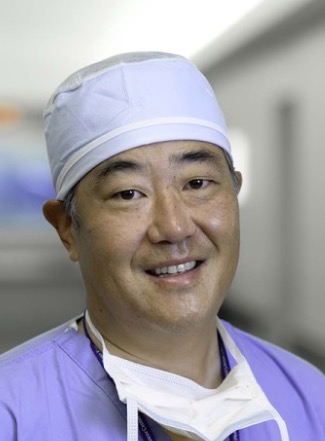 Bernard J. Park合作论文数Memorial Sloan Kettering Cancer Center2
 Richard J. Wong合作论文数Department of Head and Neck Surgery, Cancer Center2Andy Goberdhan合作论文数2Mk Chan合作论文数1
 Mark G. Kris合作论文数David H. Koch Center for Cancer Care, Memorial Sloan Kettering Cancer Center1
 Ting-Chao Chou (周廷潮)合作论文数PD Science, LLC1Cordeiro Christina N合作论文数Johns Hopkins Hospital Bayview Medical Center, Johns Hopkins University1
 Zhenkun Yu (于振坤)合作论文数BenQ Medical Center;The Fourth School of Clinical Medicine, Nanjing Medical University1
 Yuman Fong合作论文数Department of Surgery, City of Hope;Center for International Medicine, City of Hope;Center for Surgical Innovation, City of Hope1
 Ashok R. Shaha合作论文数David H. Koch Center for Cancer Care, Memorial Sloan Kettering Cancer Center1Dennis H. Kraus合作论文数1
 Peter G Sacks合作论文数1
 Benjamin R Lee合作论文数University of Arizona Cancer Center;Department of Urology, College of Medicine, University of Arizona1
 Arnold J. Levine合作论文数The Simons Center for Systems Biology, Institute for Advanced Study;School of Natural Sciences, Institute for Advanced Study1
 Christine Walsh合作论文数School of Medicine, University of Colorado1
 Mukesh Prasad合作论文数Head and Neck Service;Department of Surgery;Memorial Sloan-Kettering Cancer Center;Department of Surgery, Memorial Sloan-Kettering Cancer Center1Volkert B. Wreesmann合作论文数1
 Brendon M. Stiles合作论文数Memorial Sloan-Kettering Cancer Center1Dorothy Wang合作论文数1
 Prasad S. Adusumilli合作论文数Department of Surgery, Memorial Sloan-Kettering Cancer Center;Cellular Therapeutics Center, Memorial Sloan-Kettering Cancer Center1
 Erik Schnaser合作论文数1




