Background:Gastric cancer (GC) is the fifth most commonly diagnosed cancer worldwide and ranks as the third leading cause of cancer-related deaths. In the tumor microenvironment (TME), malignant cells undergo hypoxic adaptation via metabolic reprogramming, which promotes cellular proliferation and lactate accumulation. However, the significance of hypoxia- and lactylation-related genes (HALRGs) in GC remains unclear. This study aimed to investigate the impact of hypoxia and lactylation on GC and to construct relevant prognostic models. Methods:We downloaded single-cell RNA sequencing (scRNA-seq), spatial transcriptome (ST) data, and clinical data. The least absolute shrinkage and selection operator (LASSO) algorithm was used to develop a prognostic model, which was validated in an external cohort. Immune cell infiltration was analyzed using the CIBERSORT algorithm, while the Tumor Immune Dysfunction and Exclusion (TIDE) score and ESTIMATE method were combined to evaluate the prognostic potential of immunotherapy response. Results:We analyzed 23,447 genes and 104,150 cells from scRNA-seq, classifying the cells into seven distinct types. The spatial distribution characteristics of these cell types were further examined using ST data. Inference of copy number variations (InferCNV) analysis was performed on epithelial cells to differentiate between malignant and normal cells, revealing that normal epithelial cells could be further categorized into five subtypes. We then developed a prognostic model based on HALRGs using The Cancer Genome Atlas (TCGA) data, which was validated in the Gene Expression Omnibus (GEO) dataset. Patients were divided into high- and low-risk groups according to HALRG scores. Interestingly, the high-risk group exhibited higher TIDE scores, suggesting a poorer response to immunotherapy. Conclusions:This study uncovered the cellular heterogeneity of GC by integrating scRNA-seq and ST data. The results demonstrated that the HALRG risk model may be effective in predicting the clinical prognosis and has the potential to guide personalized treatment in GC.
This figure addresses the reviewer's questions about the effect of blocking type I IFN on tumor cells and tumor-infiltrating lymphocytes.
This figure shows that CD11b+Gr-1+ cells from anti-PD-1 resistant tumors also show downregulation of MHC class I molecules
Abstract Hepcidin is a circulating peptide hormone made by the liver that is a central regulator of systemic iron uptake and recycling. Here, we report that prostate epithelial cells also synthesize hepcidin, and that synthesis and secretion of hepcidin are markedly increased in prostate cancer cells and tissue. Prostatic hepcidin functions as an autocrine hormone, decreasing cell surface ferroportin, an iron exporter, increasing intracellular iron retention, and promoting prostate cancer cell survival. Synthesis of hepcidin in prostate cancer is controlled by a unique intersection of pathways that involves BMP4/7, IL6, Wnt, and the dual BMP and Wnt antagonist, SOSTDC1. Epigenetic silencing of SOSTDC1 through methylation is increased in prostate cancer and is associated with accelerated disease progression in patients with prostate cancer. These results establish a new connection between iron metabolism and prostate cancer, and suggest that prostatic dysregulation of hepcidin contributes to prostate cancer growth and progression. Cancer Res; 75(11); 2254–63. ©2015 AACR.
This figure shows that PD-L1 expression is not quantitatively altered on the anti-PD-1 resistant tumor cells, compared to parental tumor cells.
This figure shows that the immune suppressive MDSCs and regulatory T cell percentages in parental and anti-PD-1 resistant tumor models.
Background This prospectively randomised, double-blinded, placebo-controlled, multicenter Phase 3 clinical trial was conducted to assess the efficacy and safety profile of nimotuzumab (nimo) plus concurrent chemo-radiotherapy (CCRT) in patients with unresectable locally advanced ESCC. Methods Patients were randomly assigned (1:1) to receive CCRT plus nimotuzumab or placebo. The primary endpoint was overall survival (OS). In addition, interim analysis for short-term response rate was pre-defined. Results A total of 201 patients were randomised into two groups. Eighty patients in the nimo group and eighty-two in the placebo group were evaluable. Three to six months after treatment, 26 (32.5%) patients achieved complete response (CR) in the nimo group, and 10 (12.2%) in the placebo group ( P = 0.002). The ORR of the nimo group was significantly higher than the placebo group (93.8% vs. 72.0%, P < 0.001). The two groups’ grade 3–5 adverse drug reactions were 11.1% vs. 10.9% ( P > 0.05). Conclusions Nimotuzumab, in combination with chemo-radiotherapy, increased the CRR and ORR with a good safety profile. The OS is needed to be followed and finally analysed. Clinical trial registration NCT02409186.
4016 Background: 70% of esophageal carcinoma are unresectable at diagnosis. Despite active clinical research on the treatment of esophageal squamous cell carcinoma (ESCC), the long-term survival rate of advanced patients is still very low, with a 5-year survival rate of 30%-40%. A prospective, randomized-controlled, double-blinded, multicenter, and phase III study (NXCEL1311) was designed to investigate the efficacy and safety of nimotuzumab (anti-EGFR humanized monoclonal antibody;abbreviate,Nimo) plus concurrent chemo-radiotherapy compared with placebo plus chemo-radiotherapy in unresectable locally advanced ESCC. Methods: Unresectable locally advanced ESCC patients were randomized (1:1) to receive Nimo (400 mg, qw) or placebo in combination with concurrent chemo-radiotherapy (paclitaxel+ cisplatin+3DCRT/IMRT) for seven weeks. Patients were followed for five years.The primary endpoints were OS, and the secondary endpoints included ORR, DCR, PFS. Results: 200 patients were assigned to the Nimo group (n = 99) or placebo group (n = 101). An interim analysis was conducted for short term efficacy, i.e secondary endpoints (ORR, DCR) and safety, after completing the 6 months follow-up. The OS events are not enough for analysis. The two groups were comparable on baseline characteristics. Eighty patients in the Nimo group and eighty-two patients in the placebo group were evaluable. The ORR of the Nimo group (75/80, 93.8%) was significantly higher than the placebo group (59/82, 72.0%;Chi-square test, p < 0.001). Twenty-six patients in the Nimo group reached the complete response (CR), and ten placebo group patients were CR. The CR rate in the Nimo group was significantly higher than placebo group (32.5% vs.12.2%, p = 0.002). The DCR of the Nimo group and placebo group were 98.8% (79/80) and 91.5% (75/82), respectively (p = 0.064). Single factor logistic aggression analysis showed that age, sex, target lesion number, and BMI did not affect ORR, CR, and DCR (p > 0.05). Multiple factor correction analysis showed the difference of CR, ORR and DCR between two groups is 20% (95%CI 6.0%̃40.2%), 30% (95%CI 10.6%̃52.1%) and 10% (95%CI -5.2%̃31.1%). The incidence of grade 3-5 drug-related AEs was 11.1%vs.10.9% (p > 0.05). Common drug-related AEs in patients with Nimo plus chemo-radiotherapy treatment were leucopenia, neutrophilic granulocytopenia, thrombocytopenia, hemoglobin, bone marrow inhibition, nutritional anemia, and radioactive inflammation. Conclusions: This interim analysis showed that nimotuzumab in combination with chemo-radiotherapy is safe and can increase the CRR and ORR of the treated patients. The OS needs to be followed and finally analyzed. Clinical trial information: 02409186.
Introduction Oesophageal squamous cell carcinoma (OSCC) is one of the most commonly occurring devastating tumours worldwide, including in China. To date, the standard care of patients with stage IV OSCC is systemic chemotherapy and palliative care, which results in poor prognosis. However, no consensus has been established regarding the role of radiotherapy in targeting the primary tumour in patients with stage IVa OSCC. Thus, the aim of this study is to assess the effectiveness of primary radiotherapy combined with S-1 and nedaplatin (NPD) chemotherapy in the patients with stage IV OSCC. Methods and analysis The study is a multicentre, open-label, randomised controlled trial. A total of 180 eligible patients with stage IV OSCC will be randomised into a study group (90 patients) and a control group (90 patients). Patients in the study group will receive radiotherapy to the primary tumour at a dose of 50.4 Gy combined with 4–6 cycles of S-1 and NPD chemotherapy. In the control group, patients will only receive 4–6 cycles of S-1 and NPD chemotherapy. The primary and secondary outcomes will be measured. The differences between the two groups will be statistically analysed with regard to overall survival, the progression-free survival and safety. All outcomes will be ascertained before treatment, after treatment and after the follow-up period. The results of this study will provide evidence on the role of radiotherapy in patients with stage IV OSCC in China, which will show new options for patients with advanced oesophageal cancer. Ethics and dissemination This study was approved by the Institutional Ethics Committee of The First Hospital Affiliated of Zhengzhou University (approval number: SS-2018–04). Trial registration The trial has been registered at the Chinese Clinical Trial Registry (ChiCTR1800015765) on 1 November 2018; retrospectively registered, http://www.chictr.org.cn/index.aspx.
This study examined the importance of cell-mediated immunity in determining the long-term outcome of patients diagnosed with chronic fatigue syndrome (CSF). A total of 103 patients (74%) of 139 previously enrolled in one of two treatment trials conducted within a university hospital referral centre was reviewed a mean of 3.2 yr after trial entry. Ongoing symptom severity, levels of disability and immunological function were assessed at follow-up. The relationship between immunological function at trial entry and measures of outcome was also evaluated. Sixty-five patients (63%) had improved, while only 6 (6%) reported no current symptoms. Thirty-one subjects (30%) were unable to perform any form of work and 26 (25%) were on a disability benefit directly attributable to CFS. Cell-mediated immune function, as measured at trial entry or follow-up, did not appear to affect outcome. Whilst improvement occurred in the majority of patients with CFS, a substantial proportion (37%) remained functionally impaired. Impairment of cell-mediated immunological function measured during the course of the illness may not be an important factor in determining long-term outcome.
Introduction: Prednisone (10 mg/d) is often used in combination with docetaxel or abiraterone in the treatment of advanced prostate cancer. LATITUDE studies have confirmed that the combination of abiraterone and prednisone (5 mg/d) can be used for the treatment of newly diagnosed high-risk metastatic castration-sensitive prostate cancer, and have achieved satisfactory results. However, it has not been reported that abiraterone combined with prednisone (5 mg/d) in the treatment of metastatic castration-resistant prostate cancer (mCRPC). Patient concerns: Here, we present a case of high-risk advanced prostate cancer with old pulmonary tuberculosis (PTB). The patient developed a relapse of old tuberculosis in both lungs that were discovered following 14 months of continuous application of prednisone (10 mg/d). Diagnosis: The histopathological findings showed prostate adenocarcinoma carcinoma with a Gleason score of 10 (5+5). Further laboratory investigations were suggestive of positive mycobacterium tuberculosis complex DNA in pleural effusion and sputum. Interventions: The patient underwent endocrine therapy, chemotherapy of docetaxel plus prednisone, radiotherapy, and abiraterone combined with prednisone treatment, but he eventually developed into the mCRPC stage. Then, prednisone was reduced to 5 mg/d plus abiraterone, and combined with anti-tuberculosis treatment according to multi-disciplinary diagnosis and treatment. Outcome: Two months later, pleural effusion and atelectasis were relieved, and PSA was remained stable at a low level. The patient achieved complete remission. Conclusion: We cannot, with complete certainty, say that this patient, or any patient, developed old PTB recurrence due to the use of prednisone. Based on the current evidence, endocrine therapy is the foundation, radiotherapy can reduce the tumor load, and early application of abiraterone is beneficial to survival for the high-risk mCRPC. The long-term use of prednisone can be appropriately reduced in mCRPC with old PTB, and a satisfactory curative effect can be achieved. More prospective trials are warranted before a definite recommendation could be drawn.
AbstractBackground and aimWe previously identified forkhead box (FOX) O4 mRNA as a predictor in gastric cancer (GC). However, the underlying mechanism has yet to be elucidated. We aimed to illustrate the mechanism by which FOXO4 regulated glycolysis under hypoxia in GC.MethodsFOXO4 protein expression was investigated by immunohistochemical staining of 252 GC and their normal adjacent tissues. We restored or silenced FOXO4 expression in GC cell lines to explore the underlying mechanisms.ResultsFOXO4 was downregulated in GC. Loss of FOXO4 expression was validated in univariate and multivariate survival analysis as an independent prognostic predictor for overall survival (P < 0.05) and disease‐free survival (P<0.05). Restored FOXO4 expression significantly impaired the glycolysis rate in GC cells, while silencing FOXO4 expression enhanced glycolysis rate. FOXO4 expression was inversely associated with maximum standardized uptake value in mice models and patient samples. Mechanistically, FOXO4 bound to the glycolytic enzyme lactate dehydrogenase (LDH)A promoter and inactivated its activity in a dose‐dependent manner (P < 0.05). Finally, we determined that FOXO4 was a transcriptional target of hypoxia‐inducible factor (HIF) ‐1α, which is central in response to hypoxia.ConclusionsOur data suggested that FOXO4 plays a key role in the regulation of glycolysis in GC, and disrupting the HIF‐1α‐FOXO4‐LDHA axis might be a promising therapeutic strategy for GC.
An amendment to this paper has been published and can be accessed via a link at the top of the paper.
Immunotherapies revolutionized cancer treatment by harnessing the immune system to target cancer cells. However, most patients are resistant to immunotherapies and the mechanisms underlying this resistant is still poorly understood. Here, we report that overexpression of BMP7, a member of the TGFB superfamily, represents a mechanism for resistance to anti-PD1 therapy in preclinical models and in patients with disease progression while on immunotherapies. BMP7 secreted by tumor cells acts on macrophages and CD4 + T cells in the tumor microenvironment, inhibiting MAPK14 expression and impairing pro-inflammatory responses. Knockdown of BMP7 or its neutralization via follistatin in combination with anti-PD1 re-sensitizes resistant tumors to immunotherapies. Thus, we identify the BMP7 signaling pathway as a potential immunotherapeutic target in cancer.
Objective To investigate the relationship between P21-activated kinase 6 (PAK6) expression and radiosensitivity of breast cancer patients undergoing radiotherapy.Methods MDA-MB-453 breast cancer cell line,small interfering RNA (siRNA),and cells transfected with PAK6 siRNA were set as untransfected group,control group and observation group,respectively.Western blot and quantitative real time polymerase chain reaction were used to detect the expression of PAK6 in each group.Every group was given 0,1 ,3 and 5 Gy radiotherapies,respectively,with one radiation dose for once.Thereafter,cell proliferation was detected by cell counting kit-8,and apoptosis was monitored by flow cytometry.Western blotting was used to detect the level of Caspase-3 protein in cells of three groups.Results The PAK6 protein and PAK6 mRNA expression levels of the untransfected group and control group were higher than those of the observation group (P<0. 05 ).The proliferation of breast cancer cells ,with the radiotherapy doses of 0,1 ,3 and 5 Gy,in the untransfected group and the control group were higher than that in the observation group (P<0. 05 );while untransfected group and control group had lower levels of apoptosis than observation group (P<0. 05 ).The expression level of caspase-3 in the observation group was higher than that in the control group and untransfected group (P<0. 05 ). Compared with the untransfected group and control group,the cell inventory score of the observation group was reduced,and the sensitization ratio of radiotherapy was 1.774.Conclusions Interference on PAK6 expression in breast cancer cells can inhibit the proliferation of breast cancer cells,promote the apoptosis of breast cancer cells and increase the radiosensitivity of breast cancer patients.The mechanism may be related to the effects of PAK6 on caspase-3 protein.
Background/Purpose We previously reported that myeloid dendritic cells (mDC) were increased in patients with leukemic cutaneous T-cell lymphoma (L-CTCL) following extracorporeal photopheresis (ECP) using the Therakos UVAR XTS (TM) system. We now assessed monocyte-derived mDCs (Mo-DCs) in L-CTCL patients treated with the CELLEXTM photopheresis system. CD209, a transmembrane receptor, was used to define Mo-DCs. Methods Peripheral blood samples from baseline pre-ECP and at Day 2, 1 month, 3 months, and 6 months post-ECP were analyzed by flow cytometry for Lin(-)HLA-DR(+)CD123(+) plasmacytoid dendritic cells (pDCs), Lin(-)HLA-DR(+)CD11c(+) mDCs, and CD209(+) mDCs. The expression of CD209 mRNA was assessed by real-time PCR. Results At baseline, 7 of 19 patients had lower than normal mDCs, and all patients had lower than normal CD209(+) mDCs in peripheral blood mononuclear cells (0.005% in patients, n = 19, vs 0.50% in healthy donors, n = 7, P < .0001). The CD209(+) mDC numbers only accounted for 3.28% out of total mDCs in patients compared with 66.51% in healthy donors. After treatment, the CD209(+) mDC numbers showed increasing trends in patients. The average absolute numbers of CD209(+) mDCs went up by 4.8-fold at 3 months (n = 10, P = .103) and by 6.4-fold at 6 months (n = 9, P = .100). CD209 mRNA expression went up in two patients responsive to therapy, parallel to CD209(+) mDC numbers. L-CTCL patients achieved 70% overall clinical response rate (7/10) following ECP therapy with the CELLEXTM system. Conclusions Our results suggest that the CELLEXTM photopheresis system is effective for treating L-CTCL patients like the UVAR XTS (TM) system, and in vivo-generated Mo-DCs increase following ECP.
PURPOSE:The limitation of hypofractionated radiation efficacy is due partly to the immunosuppressive tumor microenvironment. Indoleamine 2,3-dioxygenase 1 (IDO1) is an important regulator of tumor immune suppression. We evaluated the effects of IDO1 in hypofractionated radiation using a Lewis lung carcinoma (LLC) mouse model and tested whether IDO1 inhibition could sensitize those tumors to hypofractionated radiation. METHODS AND MATERIALS:Bilateral LLC tumors were established in C57BL/6 mice. Primary tumors were treated with 3 fractions of either 12 Gy or 6 Gy, and the IDO1 inhibitor INCB023843 was given starting on the first day of radiation. Plasma tryptophan and kynurenine levels were quantified by liquid chromatography and tandem mass spectrometry. Tumor-infiltrating immune cells were isolated from the tumors, stained, and quantified by flow cytometry. RESULTS:The combination of INCB023843 and three 12-Gy fractions led to better tumor control and survival than radiation alone; INCB023843 plus three 6-Gy fractions had no benefit. IDO1 expression by tumor-infiltrating immune cells was increased by three 12-Gy doses and inhibited by the addition of INCB023843. Nearly all IDO1+ immune cells were also F4/80+. Percentages of IDO1+F4/80+ immune cells were drastically increased by three 12-Gy fractions and by three 6-Gy fractions, but only INCB023843 combined with three 12-Gy fractions reduced those percentages. IDO1+F4/80+ immune cells were further found to be CD11b+, Gr1-intermediate-expressing, CD206-, and CD11c- (ie, myeloid-derived suppressor cells). Three 12-Gy fractions also increased the percentages of tumor-infiltrating T regulatory cells and CD8+ T cells, but adding INCB023843 did not affect those percentages. CONCLUSIONS:In addition to its immune activation effects, hypofractionated radiation induced "rebound immune suppression" in the tumor microenvironment by activating and recruiting IDO1-expressing myeloid-derived suppressor cells in a dose-dependent manner. Adding an IDO1 inhibitor to hypofractionated radiation reduced the percentages of these cells, overcame the immune suppression, and sensitized LLC tumors to hypofractionated radiation.
Indoleamine 2,3-dioxygenase 1 (IDO1), involved in the catabolism of tryptophan (Trp) to kynurenine (Kyn) is an important regulator of tumor-mediated immunosuppression implicated in resistance to anti-PD1 immunotherapy. We investigated the role of IDO1 in an anti-PD1-resistant lung cancer model (344SQ_R) compared to the parental 344SQ tumors (344SQ_P). IDO1 was overexpressed in tumor-infiltrating leukocytes, and plasma Kyn levels were increased, in 344SQ_R vs. 344SQ_P. The IDO1 inhibitor INCB023843 retarded tumor growth and reduced lung metastases in 344SQ_R. IDO1 was expressed at higher levels in F4/80+Gr1intCD11b+ myeloid-derived suppressor cells (MDSCs) that were prominent in 344SQ_R. The INCB023843 reduced IDO1 expression and percentages of these MDSCs while increasing CD8+ T cells infiltration, hence reactivating antitumor T-cell responses in 344SQ_R. Therefore, IDO1 inhibition holds promise for treating lung cancer that does not respond to anti-PD1 therapy.
 James W. Welsh合作论文数Department of Thoracic Radiation Oncology, Division of Radiation Oncology, The University of Texas MD Anderson Cancer Center;MolecularMatch17
 Maria Angelica Cortez合作论文数Medical School, The University of Texas Southwestern Medical Center;Children's Medical Center Research Institute, The University of Texas Southwestern Medical Center15
 Jonathan E Schoenhals合作论文数Department of Radiation Oncology, University of Texas MD Anderson Cancer Center13
 David Valdecanas合作论文数13Ye Huiping合作论文数10Ailin Li合作论文数10
 Patrick Hwu合作论文数Moffitt Cancer Center9Daniel Gomez合作论文数8
 Ritsuko Komaki合作论文数University of Texas MD Anderson Cancer Center;Department of Radiation Oncology, Baylor College of Medicine;Department of Scientific Research, Baylor College of Medicine8
 James P. Allison合作论文数Department of Immunology, Division of Basic Science Research, The University of Texas MD Anderson Cancer Center8
 Sunil Krishnan合作论文数Vivian L. Smith Department of Neurosurgery, McGovern Medical School, The University of Texas Health Science Center at Houston8
 Stephen Hahn合作论文数7
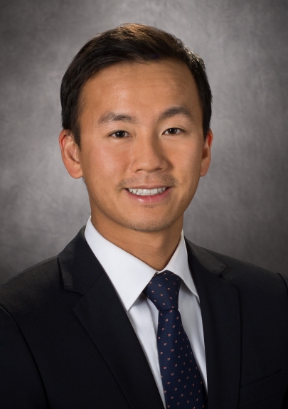 Chad Tang合作论文数Department of Radiation Oncology, Division of Radiation Oncology, The University of Texas MD Anderson Cancer Center7
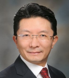 Joe Y. Chang合作论文数Division of Radiation Oncology, University of Texas MD Anderson Cancer Center;Department of Radiation Oncology, University of Texas MD Anderson Cancer Center7Willem W. Overwijk合作论文数7Tse-Hua Tan合作论文数6Hampartsoum B Barsoumian合作论文数Univ Texas MD Anderson Canc Ctr, Dept Radiat Oncol, Houston, TX 77030 USA6
 Tang Ming合作论文数University of Florida College of Medicine, University of Florida College of Medicine6
 Fenglin Zang (臧凤琳)合作论文数6
 Chang Gong Liu合作论文数Department of Experimental Therapeutics, The University of Texas M. D. Anderson Cancer Center6
 Zuohua Feng (冯作化)合作论文数School of Basic Medicine, Tongji Medical College, Huazhong University of Science and Technology6Yufei He合作论文数6Liu Xiuping合作论文数5
 Mauricio S. Caetano合作论文数5Li Ju-Pi合作论文数5
 Cristina Ivan合作论文数Caris Life Sciences5
 George Calin合作论文数Department of Translational Molecular Pathology, Division of Pathology-Lab Medicine Div, The University of Texas MD Anderson Cancer Center;Department of Leukemia, Division of Cancer Medicine, The University of Texas MD Anderson Cancer Center5Chia-Yu Yang合作论文数Chang Gung Univ, Coll Med, Grad Inst Biomed Sci, 259 Wenhua 1st Rd, Taoyuan 333, Taiwan4Chuang Huai-Chia合作论文数4
 Der-Yuan Chen (陳得源)合作论文数Department of Internal Medicine, China Medical University Hospital;Rheumatology and Immunology Center, China Medical University Hospital4
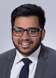 Hari Menon合作论文数Penn State Geisinger Medical Center4Joung-Liang Lan合作论文数4Ferdinandos Skoulidis合作论文数4
 Sharareh Niknam合作论文数3Anping Zheng合作论文数3
 Jie Zhang合作论文数Experimental Radiation Oncology, The University of Texas MD Anderson Cancer Center, Houston, TX, USA.3
 Rishab Ramapriyan合作论文数Univ Texas MD Anderson Canc Ctr, Dept Radiat Oncol, Houston, TX 77030 USA3
 Padmanee Sharma合作论文数Department of Genitourinary Medical Oncology, Division of Cancer Medicine, The University of Texas MD Anderson Cancer Center;Department of Immunology, The University of Texas MD Anderson Cancer Center3
 Ahmed I Younes合作论文数Section of Lymphoma, University of Texas M. D. Anderson Cancer Center3Lu Yue合作论文数3Chiu Li-Li合作论文数3
 Guang Li (李光)合作论文数The First Hospital of China Medical University3
 Ruitai Fan (樊锐太)合作论文数The First Affiliated Hospital, Zhengzhou University3Guimei Zhang (张桂梅)合作论文数Nanchang Hangkong University3
 Marcos R. Estecio合作论文数Department of Epigenetics and Molecular Carcinogenesis, The University of Texas MD Anderson Cancer Center3Mark Wasley合作论文数2Zhongyu Qu合作论文数2




