目的 探讨经皮椎体成形术联合微波消融术治疗多发性骨髓瘤致胸腰椎病理性骨折的效果及安全性.方法 根据治疗方法的不同将60例多发性骨髓瘤致胸腰椎病理性骨折患者分为对照组(n=33)和观察组(n=27),对照组患者进行单纯经皮椎体成形术,观察组患者进行经皮椎体成形术联合微波消融术.比较两组患者的视觉模拟评分法(VAS)评分、Barthel指数评分、病变椎体高度、治疗有效率及术后并发症发生情况.结果 手术后,观察组患者的VAS评分明显低于对照组,Barthel指数评分明显高于对照组,差异均有统计学意义(P﹤0.01).手术后,观察组患者的病变椎体前缘高度、中部高度均高于对照组,差异均有统计学意义(P﹤0.05).观察组患者的治疗有效率高于对照组(P﹤0.05).两组患者的并发症总发生率比较,差异无统计学意义(P﹥0.05).结论 对多发性骨髓瘤致胸腰椎病理性骨折患者进行经皮椎体成形术联合微波消融术治疗,有利于降低患者疼痛程度,提高患者的日常生活活动能力及病变椎体高度,具有较好的治疗效果,且不会增加手术并发症.
目的 在X线与CT引导下对骨病变进行穿刺活检,探讨其在确诊骨病变中临床应用的可行性.方法 350例骨病变患者,胸椎87例,腰椎123例,肢体及骨盆130例,肋骨5例,肩胛骨5例,其中单一椎体132例,多发椎体78例.常规行X线透视下穿刺活检,病变部位局限或位置较深时行CT引导下穿刺活检.结果 其中345例穿刺成功,2例脊柱恶性占位患者因无法耐受疼痛放弃穿刺,3例因取材标本量过少认为穿刺失败.穿刺用时10~25min,平均17.3min.明确诊断330例,阳性率为94.3%.结论 X线与CT引导下行骨病变穿刺活检是一种安全可靠的方式.
背景:80岁以上腰椎管狭窄症患者因其疾病的复杂性与特殊性,加之并发多种内科疾病,需要十分谨慎地选择治疗方法.目的:评价椎间孔镜治疗80岁以上腰椎管狭窄症患者的疗效.方法:选择2012年11月至2017年12月椎间孔镜手术治疗老年腰椎管狭窄症患者52例进行回顾性分析.记录并比较患者术前、术后1个月、3个月、6个月及12个月疼痛视觉模拟评分(VAS)、Oswestry功能障碍指数(ODI)和日本骨科协会(JOA)评分.结果:入选52例患者中,51例患者获得随访,随访时间24~60个月,平均随访时间(36.0±0.4)个月.患者术后1、3、6、12个月、末次随访时疼痛VAS评分、ODI均较术前降低,JOA评分均较术前升高,且差异均有统计学意义(P均<0.001).结论:椎间孔镜技术是治疗80岁以上腰椎管狭窄症患者安全、有效的方法之一.
目的 对比锁定钢板结合微创钢板接骨技术(MIPO)和髓内钉固定治疗老年肱骨近端骨折的疗效.方法 回顾性分析2018年1月至2019年1月期间在航天中心医院骨科手术治疗的老年移位肱骨近端骨折患者51例的临床资料.依据治疗方法分为2组:MIPO组(n=29)和髓内钉组(n=22).对比2组患者围术期指标、术后12 h切口的视觉模拟量表(VAS)评分、并发症发生情况.分别于术后3、6、12个月,采用Constant-Murley评分对2组患者肩关节功能恢复情况进行评价.采用SPSS 13.0软件进行统计分析.2组间比较采用t检验或χ2检验.结果 MIPO组和髓内钉组患者的手术切口长度均较小[(7.3±2.7)和(7.3±2.6)cm]、术中出血量较少[(79.7±25.7)和(78.3±21.4)ml]、手术耗时较短[(64.1±17.2)和(62.5±14.6)min]、术后12 h切口VAS评分较低[(2.47±0.81)和(2.44±0.69)分],2组各指标比较差异均无统计学意义(P>0.05),且术后3、6、12个月2组患者的Constant-Murley评分差异亦无统计学意义(P>0.05).但MIPO组患者术后并发症的发生率显著高于髓内钉组[17.24%(5/29)和9.09%(2/22),P<0.05].结论 锁定钢板结合MIPO技术和髓内钉固定治疗老年肱骨近端骨折都能获得较好疗效,髓内钉固定在控制并发症发生方面比锁定钢板结合MIPO技术更具优势.
目的 对比股骨近端防旋髓内钉(PFNA)和联合拉力交锁髓内钉系统(Inter Tan)治疗老年股骨粗隆间骨折的临床效果.方法 回顾性分析2018年2月至2019年2月期间航天中心医院骨科收治的老年股骨粗隆间骨折患者的临床资料,共49例.依据治疗方法 分为2组:PFNA组(n=24)和Inter Tan组(n=25).对比2组患者的手术时长、出血量、骨折愈合时长、住院时长、并发症发生率.对比2组患者随访6个月时的Harris评分和生活质量(QOL)评分.采用SPSS 13.0软件进行统计分析.2组间比较采用t检验或χ2检验.结果 2组患者术中均无螺钉松动穿出和固定断裂发生.PFNA组的手术时长[(43.04±6.25)和(72.58±6.49)min]和术中出血量[(124.5±18.8)和(238.9±25.6)ml]均显著低于Inter Tan组(P<0.05).PFNA组患者术后并发症发生率显著高于Inter Tan组(16.0%和4.2%,P<0.05).Inter Tan组和PFNA组术后6个月的Harris评分[(89.0±2.5)和(91.5±1.8)分]和QOL评分[(53.9±3.5)和(51.2±3.7)分]差异均无统计学意义(P>0.05).结论 Inter Tan和PFNA对老年股骨粗隆间骨折均具有良好的临床疗效,二者各具优缺点,临床可根据患者实际情况进行选择.
目的 比较双动全髋关节置换与半髋关节置换治疗老年股骨颈骨折的近期临床疗效.方法 回顾性分析2016年6月至2019年6月航天中心医院骨科收治的老年股骨颈骨折患者47例,根据手术方式分为双动全髋关节置换组19例(观察组)和半髋关节置换组28例(对照组),记录2组患者的手术时间、失血量及住院周期,从Harris髋关节评分(HHS)、并发症及术后1年内死亡率等方面进行评估.采用SPSS 21.0软件进行数据分析.2组间计量资料比较采用t检验.结果 全部患者均获得随访,随访时间9~40个月(平均23个月).对照组在手术时间、失血量方面优于观察组(P<0.05),2组患者住院周期比较差异无统计学意义(P>0.05).2组患者术后HHS均较术前提高,且观察组在术后各随访节点HHS均优于对照组(P<0.05),至末次随访时观察组髋关节功能恢复优良率明显占优.术后并发症发生率观察组5.3%(1/19),对照组17.9%(5/28);术后1年内死亡率观察组5.3%(1/19),对照组7.1%(2/28),2组间比较差异无统计学意义(P>0.05).结论 半髋关节置换具有手术时间短、失血量少、操作简单等优点,但在术后髋部疼痛、脱位及再手术率方面较高于双动全髋关节置换,后者还可提供更好的关节功能和假体稳定性,推荐成为老年股骨颈骨折的首选手术方式.
目的 评价"拖尾征"锚定骨水泥椎体后凸成形术(PKP)治疗老年Kummell's病患者的临床疗效.方法 回顾性分析2014年8月至2017年8月在航天中心医院行PKP术治疗的老年Kummell's病患者的临床资料33例.依据手术方法分为2组:采用"拖尾征"锚定骨水泥PKP术治疗者为研究组(n=16);采用常规PKP术治疗者为对照组(n=17).对比2组患者术中及术后并发症发生情况,随访24个月,对比2组患者术前、术后1 d及术后24个月时的Oswestry功能障碍指数(ODI)、视觉模拟量表(VAS)评分、Cobb's角等指标.2组间比较采用t检验或χ2检验.结果 所有患者均顺利完成手术.研究组与对照组患者的手术时间[(55.46±7.63)和(53.56±8.54)min]和骨水泥注入量[(5.8±0.6)和(5.6±0.8)ml]比较,差异均无统计学意义(P>0.05).与对照组相比,研究组患者术后24个月时的Cobb角显著降低[(14.23°±1.85°)和(17.54°±2.02°),P<0.05],而伤椎前缘高度[(1.75±0.42)和(1.39±0.61)cm]和中线高度[(1.69±0.61)和(1.35±0.34)cm]均显著增加(P<0.05).研究组术后并发症发生率显著低于对照组[6.3%(1/16)和41.2%(7/17),P<0.01].结论 "拖尾征"锚定骨水泥PKP术与常规PKP术治疗老年Kummell's病均能取得短期良好手术效果,但长期随访发现通过"拖尾征"锚定骨水泥可有效减少骨水泥移位的发生风险.
Objective To evaluate the technical feasibility and clinical effects of kyphoplasty accomplished with minimal incision for the treatment of severe osteoporotic vertebral compression fracture(sOVCF).Methods Sixty-five patients with severe osteoporotic vertebral compression fractures treated with kyphoplasty accomplished with minimal incision were included in the study. A total of 73 vertebrae were involved,including 8 T10,7 T11,19 T12,23 Ll,5 L2and 3 L4.All the damaged vertebral bodies were compressed more than 2/3 and were treated with kyphoplasty accomplished with minimal incision.All patients were followed up for more than one year.Mean duration of surgery,the times of X-ray used during operation were recorded.Visual Analogue Scale(VAS)score,Oswestry Dability Index(ODI),anterior height of vertebral body,Cobb angle were measured before operation,3 days after operation,and 1 year after operation.Complications such as cement leakage,pulmonary embolism and so on were counted.Results All patients were performed the operation successfully,except for 8 cases of cement leakage including 5 cases of intervertebral disc leakage and 3 cases of leakage to paraspinal soft tissue.But there were no symptoms of nerve and spinal cord compression, no pulmonary embolism and other server complications.The mean duration of surgery was (25.4±8.7)min/vertebra and the mean times of X-ray used during operation was (12.4±8.3)times.The VAS score,ODI and Cobb angle of patients 3 days and 1 year after operation were significantly lower than those before operation, and the anterior height of vertebral body were significantly higher than those before operation (P<0.05 ).Conclusion The kyphoplasty accomplished with minimal incision in treatment of very serious osteoporotic vertebral compression fractures can improve the safety of the operation, reduce puncture and intraoperative fluoroscopy,save the time of operation and recovery the height of vertebral and Cobb angle in different degree in the same time.It can quickly relieve pain and gain satisfactory curative effects.
Objective To investigate the influencing factors on deep venous thrombosis (DVT) after lumbar posterior surgery. Methods ?The clinical data about the patients who underwent lumbar posterior surgery in our hospital from June 2014 to January 2017 were retrospectively analyzed. According to different surgical methods,these patients were divided into four groups: balloon catheter extension vertebroplasty group, laminectomy group, TLIF single segment fusion group, TLIF multi segmental fusion group (fusion segment≥3). All the patients underwent bilateral lower limb venous ultrasonography to estimate the incidence of DVT at preoperative 1 day,postoperative 1 day and postoperative 1 week. Multivariate regression analysis was used to analyze the related risk factors and protective factors of DVT,Moreover the D-dimer levels after operation were detected,and its clinical value in diagnosis of DVT after lumbar posterior surgery was evaluated through ROC curve. Results The incidence rate of DVT after lumbar posterior surgery was the highest in TLIF multi segmental fusion group (P<0. 05). Logistic regression showed that patient's age,body mass index,cardiovascular complications, diabetes,postoperative stay in bed for a long time,he increase of D-dimer levels were the risk factors of DVT (P<0. 05),however, active and passive exercise in both lower limbs after surgery and application of antithrombotic drugs were the protective factors of DVT formation (P<0. 05). Moreover ROC curve analysis showed that D-dimer content ≥19. 5μg/ml was the cut-off value of patients with DVT after lumbar posterior surgery. Conclusion The related factors with deep venous thrombosis after lumbar posterior surgery are numerous,it is important to much attention to the high risk population of DVT, and to prevent the incidence of DVT by taking corresponding preventive measures. Moreover D-dimer can be regarded as an important observation index to evaluate the incidence of DVT after lumbar posterior surgery.
Objective To evaluate the applicability and safety of posterior single open-door vertebral canaloplasty combined with anterior decompression,fusion and internal fixation for the management of cervical ossification of the posterior longitudinal ligament,the authors retrospectively reviewed 68 consecutive procedures of this type performed at their respective institutions and studied the surgical effects and surgical complications,surgical indications.Methods Retrospectively analyzed 68 patients with the disease of cervical ossification of the posterior longitudinal ligament operated by combining posterior-anterior approachs surgical treatment from March in 1999 to April in 2006,All the patients were followed up minimum of 1 years(mean 18 months).JOA scores system and Nurick grading system were used to evaluate the effect and safety of the treatment.Results JOA scores were improved from preoperative(6.7±1.1) to postoperative(15.1±0.6),T-test was used to compare preoperative and postoperative JOA scores.The statistical results were significantly different(P0.001,α=0.05).The improvement rate of posterior-anterior unites to cure the cervical ossification of posterior longitudinal ligament was(81.5±5.6)%;Nurick grading were improved from(2.8±1.2) to(0.7±0.5),and were statistically analyzed with the method of rank-sum test of the ordered Variable data.The statistical results were significantly different(P0.001,α=0.05).Approaching segment degeneration increased in 3 cases.There were no cases of spinal cord or nerve root abiding injury,strut graft extrusion,or anterior plate or screw fracture,or cervical kyphosis,and other severe complacations.Conclusion The combined anterior-posterior decompression and instrumentation procedure provides the thoroughly decompression for cervical spinal cord,corrects cervical kyphosis,and keeps the stabilization of the cervical spine.Furthermore,it lowers the rate of injuring spinal cord,poor prostecdtive efficacy,and diminishs anterior plate failure or strut graft extrusion and pseudoarticulation.A better surgery outcome and higher safety are obtained with this combined approach.
 Xing Wei (韦兴)合作论文数Aerospace Center Hospital9
 Shen Liu (刘申)合作论文数5Shu-Ming LI (李树明)合作论文数4GuangZe Song (宋光泽)合作论文数4YongHui Liang (梁永辉)合作论文数4XinTian Li (李新天)合作论文数3QingSong Li (李青松)合作论文数3Feng Gu (顾锋)合作论文数3
 Xiaosui Tang (唐小穗)合作论文数Health Science Center, Peking University3Bo-Ran LIANG (梁勃冉)合作论文数2Zheng-Jie TANG (唐政杰)合作论文数2LiGang Shi (石立刚)合作论文数2JingYuan Zhao (赵京元)合作论文数2WenJie Guo (郭文杰)合作论文数2Sun Guangcai (孙广才)合作论文数1Yi-Zhen HE (何宜蓁)合作论文数1Jing CHEN (陈晶)合作论文数1LiLi Feng (冯丽丽)合作论文数1
 Honghui Lu (鹿洪辉)合作论文数Dongzhimen Hospital, Beijing University of Chinese Medicine1
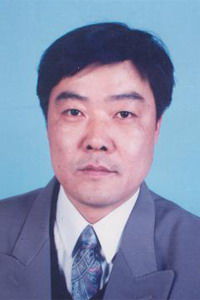 Yong Shen (申勇)合作论文数河北医科大学第三医院脊柱外科1
 Ai Guo (郭艾)合作论文数Beijing Friendship Hospital, Capital Medical University1




