Ethnopharmacological relevance: The clinical application of the traditional Chinese medicinal formula Jiedu Xiaozheng Yin (JXY) for gastrointestinal tumors, particularly colorectal cancer (CRC), is well-established, yet the precise biological mechanism underlying its efficacy in CRC treatment remains elusive. Aims of the study: This study endeavors to unravel the intricate mechanism through which JXY modulates colorectal cancer stem cells, thus elucidating the pathways by which it exerts its potent anti-tumor effects. Materials and methods: In this study, the regulatory impact of JXY on the signaling pathway and function of CRC cells was analyzed through Network pharmacology. The ethyl acetate extract of JXY was detected the major compounds using HPLC and then treated the HCT-116 cells for RNA-Sequencing (RNA-Seq). Protein expression and stemness of HCT-15 and HCT-116 cells following JXY extract treatment were assessed using Western blot analysis and matrigel spheroid assays. Additionally, the (3-catenin transcriptional activity was evaluated using a TOPflash reporter assay with or without Lithium chloride (LiCl) stimulation. Patient-derived organoids of CRC (CRC PDOs) were cultured using a stemness maintenance medium, and their viability was measured using ATP assays after treatment of JXY extract. Furthermore, the anti-tumor efficacy of JXY extract was assessed using a xenograft mice model derived from HCT-15 cells. Results: Network pharmacology emphasized the influence of JXY on cancer stem cells and the Wnt signaling pathway. HPLC analysis confirmed that the JXY extract contained the three most prevalent pharmaceutical compounds among the four herbs documented in the Chinese Pharmacopoeia (rosmarinic acid, quercetin, and kaempferol). RNA-Seq results further elucidated the effect of JXY extract, particularly targeting cancer stem cells and the Wnt signaling pathway. Furthermore, JXY extract inhibited spheroid formation in CRC cells and downregulated CRC CSC markers (CD133, DCLK1, and C-MYC). Additionally, JXY extract suppressed the (3-catenin expression and transcriptional activity as well as the Wnt pathway target proteins, including C-MYC and Cyclin D1. Consistent with findings from cell lines, JXY extract suppressed the growth of CRC PDOs exhibiting stemness characteristics. And JXY extract demonstrated a significant inhibitory effect on tumor growth, C-MYC, and (3-catenin protein levels in xenograft tumors. Conclusions: These results highlight the novel function of JXY extract in targeting CRC CSCs by regulating Wnt signaling pathway, underscoring its potential as a therapeutic agent for treating CRC.
The Wnt signaling pathway is a key driver of stemness and progression which contributes to mortality in colorectal cancer (CRC). R-spondins bind to LGR receptors to inhibit ubiquitin E3 ligases, thus protecting Frizzled from degradation and activating downstream Wnt signaling. Herein, we identify Palmdelphin ( PALMD ) as a functional marker of CRC stem cells, interspersed between intestinal and colonic crypt base epithelial cells, and predictive of aggressive CMS4 CRC and poor survival. Gene knockdown and overexpression studies revealed that PALMD initiates paracrine activation of Wnt/β-catenin signaling via interacting with and facilitating the secretion of R-spondin2 ( RSPO2 ), resulting in enhanced stemness and tumor growth in vitro and in vivo . Physiologic or pharmacologic inhibition of the PALMD-RSPO2 axis using R-spondin2-specific antibody or 6-methyl-1,3,8-trihydroxyanthraquinone (emodin), respectively, attenuates PALMD- mediated Wnt reporter activation, self-renewal, and tumorigenesis in cell and patient-derived organoid models. Together, these findings identify PALMD as a previously unknown player in Wnt signaling in CRC, and underscore the pro-tumorigenic role of R-spondin2 in this context. ### Competing Interest Statement YN Y, ZC, JP, and NW are inventors on patent CN114231623B.
Background: Patient-derived organoids (PDOs) are ex vivo models that retain the functions and characteristics of individualized source tissues, including a simulated tumor microenvironment. However, the potential impact of undiscovered differences between tissue sources on PDO growth and progression remains unclear. Objective: This study aimed to compare the growth and condition of PDO models originating from surgical resection and colonoscopy and to provide practical insights for PDO studies. Methods: Tissue samples and relevant patient clinical information were collected to establish organoid models. PDOs were derived from both surgical and colonoscopy tissues. The growth of the organoids, including their state, size, and success rate of establishment, was recorded and analyzed. The activity of the organoids at the end stage of growth was detected using calcein-AM fluorescence staining. Results: The results showed that the early growth phase of 2/3 colonoscopy-derived organoids was faster compared to surgical PDOs, with a growth difference observed within 11-13 days of establishment. However, colonoscopy-derived organoids exhibited a diminished growth trend after this time. There were no significant differences observed in the terminal area and quantity between the two types of tissue-derived organoids. Immunofluorescence assays of the PDOs revealed that the surgical PDOs possessed a denser cell mass with relatively higher viability than colonoscopy-derived PDOs. Conclusion: In the establishment of colorectal patient-derived organoids, surgically derived organoids require a slightly longer establishment period, while colonoscopy-derived organoids should be passaged prior to growth inhibition to preserve organoid viability.
目的 观察芪灵扶正清解方对抑郁模型大鼠行为学及海马神经元微结构的影响,探讨其对自噬相关蛋白、基因的调节及其抗抑郁机制.方法 30只SD大鼠随机分3组:正常组、模型组、芪灵组.正常组不干预,其余2组以慢性不可预知温和应激联合孤养建立抑郁大鼠模型.各组大鼠进行糖水消耗实验、敞箱实验行为学观察;HE染色观察海马细胞形态;透射电镜观察海马细胞超微结构及自噬变化;蛋白免疫印迹法(Western blot)和实时荧光定量聚合酶链式反应(Real-time PC R)检测Ulk1、Beclin1、P62、LC 3蛋白和mRNA的表达;免疫组织化学染色法(IHC)检测海马组织Beclin1表达.结果 行为学实验提示:与正常组比较,模型组大鼠糖水消耗量明显降低(P<0.05);直立次数、水平穿越的格数和理毛时间减少,粪便粒数和中央格停留时间增加(P<0.05);与模型组比较,芪灵组大鼠糖水消耗量明显增加(P<0.05);直立次数、水平穿越的格数和理毛时间增加,粪便粒数和中央格停留时间减少(P<0.05).HE染色提示:与正常组比较,模型组海马CA3区细胞数量减少,排列紊乱,部分细胞核模糊;CA1区细胞形态不规则,数量稀疏,体积明显变大,排列紊乱,细胞核模糊;与模型组比较,芪灵组海马CA1区、CA3区细胞形态相对规则,细胞数量相对较多,排列相对齐整,细胞体积相对正常,细胞核较清晰.透射电镜显示:与正常组比较,模型组大鼠海马区神经细胞形态不规则,线粒体结构损伤严重,细胞自噬的表征数量较为丰富;与模型组比较,芪灵组神经细胞形态相对规则,线粒体损伤相对轻微,细胞自噬的表征数量较少.Western blot与qPCR实验提示:与正常组比较,模型组大鼠的Ulk1、Beclin1、LC3-Ⅱ蛋白和mRNA的表达增加(P<0.05),P62蛋白和mRNA的表达减少(P<0.05);与模型组比较,芪灵组大鼠的Ulk 1、Beclin 1、LC3-Ⅱ蛋白和mRNA的表达减少(P<0.05),P62蛋白和mRNA的表达增加(P<0.05).IHC实验提示:与正常组比较,模型组大鼠海马CA3区Beclin1表达增加(P<0.05);与模型组比较,芪灵组大鼠CA3区Beclin1表达减少(P<0.05).结论 CUMS联合孤养诱导的抑郁模型大鼠海马神经元CA1区、CA3区首先出现损伤性改变,CA3区可能首先出现自噬异常激活;芪灵扶正清解方能改善抑郁模型大鼠的行为学指标,对其海马神经细胞具有一定的神经保护作用,其抗抑郁机制可能与调节海马细胞异常自噬有关.
机体多种疾病的产生都离不开免疫,免疫系统在维持人的正常生理活动方面发挥了必不可少的作用.中医药在提高机体免疫力方面的地位随着中医药的不断发展而不断提高.当归补血汤作为一种经典名方,能够修复机体的免疫损伤,提高机体的免疫力.文章从当归补血汤及其主要成分的概述和作用两个方面分析近年来当归补血汤及其主要成分在机体免疫系统中的作用,为今后免疫功能的治疗和当归补血汤的应用提供参考和建议.
Background:Fuzheng Yiliu decoction (FZYLD), a Chinese formula consisting of four herbs, can be clinically used as an adjuvant therapy after surgery or palliative treatment for advanced liver cancer.Methods:This study identified the endogenous and exogenous metabolites of FZYLD in rat serum to characterize the underlying mechanism of its antitumor activity, as well as relieving cancer-related weakness. An orthotopic transplantation rat model of HepG2 cells was established and administered with FZYLD by gastric perfusion for 14 days. Cardiopulmonary function and tail suspension test were used to evaluate the bodily weakness of hepatocellular carcinoma (HCC) rats. Tumor weight and size were measured to calculate inhibition ratios. Serum of different concentrations of FZYLD was used to culture the 2-[N-(7-nitrobenz-2-oxa-1, 3-diaxol-4-yl) amino]-2-deoxyglucose-labeled HepG2 cells. IC50 value was measured using MTT assay. Endogenous and exogenous metabolites in rat serum were detected using nuclear magnetic resonance or LC-MS/MS spectroscopy.Results:FZYLD improved cardiopulmonary function, decreased immobility time in tail suspension test, and yielded tumor inhibition ratios of up to 27%. Serum endogenous markers, such as lipoproteins (high- and low-density lipoproteins), glucose, and valine, and lactic acid metabolic disturbance were recovered, to some extent, in HCC rats. Exogenous metabolites, diosgenin, apigenin-7-O-acetyl-β-D-glucoside, calycosin-7-glucoside, calycosin, ganoderic-acid-A, formononetin, and methylnissolin, became detectable in the blood. FZYLD-containing serum substantially inhibited the proliferation of HepG2-cells. IC50 value was found to be 24.31%. Further, we confirmed that FZYLD could revert energy and lipid metabolism disorders and that its constituents could be bioactive components that induce apoptosis in cancer cells.Conclusion:The present study explained the mechanism of the effect of FZYLD on body empty, fatigue, and low immunity in patients with cancer, offering an efficient way for research of natural compounds in traditional Chinese medicine.
Doublecortin‑like kinase 1 (DCLK1) has been identified as a novel biomarker of cancer stem cells among several different cancer types, including colon, breast, pancreas, kidney, liver, stomach and esophageal cancers. Studies have demonstrated that DCLK1 regulates tumorigenesis and epithelial‑mesenchymal transformation via several important pathways, such as Notch, Wnt/β‑catenin, RAS and multiple microRNAs. The function and biological mechanisms, including their association with the molecular structure and isoforms of DCLK1, are gradually being elucidated. However, the currently available knowledge regarding DCLK1 in terms of developing effective anti‑cancer drugs remains incomplete. In the present review, the molecular characteristics, biomarker function and biological mechanisms of DCLK1 are summarized and DCLK1 is proposed as a potential anti‑tumor target via the glucose metabolism pathway.
目的 探讨解毒消癥饮(JXY)对人淋巴管内皮细胞(HLEC)体外淋巴管生成能力的影响.方法 MTT和Western blot法检测各浓度JXY对人肝癌细胞HepG2细胞活力和血管内皮生长因子C(VEGF-C)表达的影响.将HLEC细胞分为对照组、VEGF-C组、JXY低剂量组(简称低剂量组)、JXY中剂量组(简称中剂量组)、JXY高剂量组(简称高剂量组).对照组每孔加入100μL ECM完全培养基,VEGF-C组予外源性因子VEGF-C干预,低、中、高剂量组分别予VEGF-C联合不同浓度JXY(0.05、0.1、0.2 mg/mL)进行干预,每组均干预24 h.采用MTT法检测各组细胞活力,Hoechst 33258染色法检测细胞凋亡情况,划痕和迁移实验检测细胞损伤修复和迁移能力,管腔形成实验检测体外淋巴管生成,Western blot法检测血管内皮生长因子受体3(VEGFR-3)、基质金属蛋白酶9(MMP-9)和MMP-2蛋白表达.结果 JXY可显著抑制肝癌细胞的活力和VEGF-C蛋白的表达(P<0.01),抑制HLEC细胞体外增殖(P<0.01),促进细胞凋亡,抑制划痕损伤修复、细胞迁移及淋巴管腔的体外生成(P<0.01),下调了淋巴管生成相关蛋白VEGFR-3、MMP-9、MMP-2的表达(P<0.01).结论 JXY显著抑制肿瘤细胞分泌的VEGF-C进而抑制HLEC的体外淋巴管生成,其作用机制可能与下调肿瘤微环境中的VEG-FR-3、MMP-9和MMP-2的表达相关.
Colorectal cancer (CRC) is a major source of morbidity and mortality, characterized by intratumoral heterogeneity and the presence of cancer stem cells (CSCs). Bufalin has potent activity against many tumors, but studies of its effect on CRC stemness are limited. We explored bufalin’s function and mechanism using CRC patient-derived organoids (PDOs) and cell lines. In CRC cells, bufalin prevented nuclear translocation of β-catenin and down-regulated CSC markers (CD44, CD133, LGR5), pluripotency factors, and epithelial–mesenchymal transition (EMT) markers (N-Cadherin, Slug, ZEB1). Functionally, bufalin inhibited CRC spheroid formation, aldehyde dehydrogenase activity, migration, and invasion. Network analysis identified a C-Kit/Slug signaling axis accounting for bufalin’s anti-stemness activity. Bufalin treatment significantly downregulated C-Kit, as predicted. Furthermore, overexpression of C-Kit induced Slug expression, spheroid formation, and bufalin resistance. Similarly, overexpression of Slug resulted in increased expression of C-Kit and identical functional effects, demonstrating a pro-stemness feedback loop. For further study, we established PDOs from diagnostic colonoscopy. Bufalin differentially inhibited PDO growth and proliferation, induced apoptosis, restored E-cadherin, and downregulated CSC markers CD133 and C-Myc, dependent on C-Kit/Slug. These findings suggest that the C-Kit/Slug axis plays a pivotal role in regulating CRC stemness, and reveal that targeting this axis can inhibit CRC growth and progression.
This paper reviewes the clinical and scientific research history of professor XUE Chong-cheng and summarizes his academic thoughts and main achievements. Professor XUE Chong -cheng created the first personality and constitution inventory in China to lay the foundation for the discipline of TCM psychology; discovered meridian sensory conduction in phantom limbs and proposed the extension of the central model; invented a method for treating mental illness with a low-dose electroacupuncture convulsion therapy instrument; and interpreted the TCM medical model as a Time-Space-Social- Psychological-Biological medical model.
The approval of immune checkpoint inhibitors has expanded treatment options for renal cell carcinoma (RCC), but new therapies that target RCC stemness and promote anti-tumor immunity are needed. Previous findings demonstrate that doublecortin-like kinase 1 (DCLK1) regulates stemness and is associated with RCC disease progression. Herein, we demonstrate that small-molecule kinase inhibitor DCLK1-IN-1 strongly inhibits DCLK1 phosphorylation and downregulates pluripotency factors and cancer stem cell (CSC) or epithelial-mesenchymal transition (EMT)-associated markers including c-MET, c-MYC, and N-Cadherin in RCC cell lines. Functionally, DCLK1-IN-1 treatment resulted in significantly reduced colony formation, migration, and invasion. Additionally, assays using floating or Matrigel spheroid protocols demonstrated potent inhibition of stemness. An analysis of clinical populations showed that DCLK1 predicts RCC survival and that its expression is correlated with reduced CD8+ cytotoxic T-cell infiltration and increases in M2 immunosuppressive macrophage populations. The treatment of RCC cells with DCLK1-IN-1 significantly reduced the expression of immune checkpoint ligand PD-L1, and co-culture assays using peripheral blood monocytes (PBMCs) or T-cell expanded PBMCs demonstrated a significant increase in immune-mediated cytotoxicity alone or in combination with anti-PD1 therapy. Together, these findings demonstrate broad susceptibility to DCLK1 kinase inhibition in RCC using DCLK1-IN-1 and provide the first direct evidence for DCLK1-IN-1 as an immuno-oncology agent.
目的 分析中医食疗在癌因性疲乏中的研究现状和发展动态,为临床实践与研究提供借鉴.方法 以癌因性疲乏、食疗等为主题词在PubMed、Embase、Cochrane Library、Web of Science、CNKI、CBM、WanFang Data、VIP中检索文献,并辅以追溯检索,采用文献计量法和内容分析法对纳入文献进行分析.结果 共纳入文献32篇,其中随机对照试验24篇;文献发文量呈逐年上升趋势,发表期刊来源较多;相关文献共计被引24篇,共引频次197次;共有13篇文献受基金项目支持;药食同源品种31种,其中黄芪、甘草、人参、茯苓、当归为最常见的5种;食疗干预措施主要为在对照组的基础上加具有补益气血、滋阴功效的中医食疗方,共21种;主要是观察患者的疲乏、生活质量和免疫功能情况.结论 中医食疗对癌因性疲乏患者的研究已受到国内的关注,并掌握了相应的食疗方应用规律,但该领域研究的深度与广度还有很大的提升空间,值得更加深入的研究探讨.
Aim. To evaluate the effects and medication safety of Duhuo Jisheng Decoction (DHJSD) alone or as a combination therapy with other interventions on the related clinical index in postmenopausal osteoporosis condition. Methods. Search in CNKI, WanFang, CBM, VIP, PubMed, EMBASE, and Cochrane Library databases and randomized controlled trials where at least one group received any form of DHJSD for postmenopausal osteoporosis condition. Risk of bias was based on the Cochrane handbook, the quality of evidence was assessed by the GRADEpro online, and analyses were performed by RevMan 5.3 software. Results. Eight studies were enrolled with 650 participants. DHJSD alone or with other interventions had a significant effect on BMD of the lumbar spine (MD = 0.46, 95%CI (0.24, 0.68), P<0.0001), E2 (SMD = 0.49, 95%CI (0.30, 0.68), P<0.0001), and clinical effectiveness (OR = 5.07, 95%CI (3.07, 8.35), P<0.0001). However, no effect at BGP (MD = −0.84, 95%CI (−1.69, 0.00), P=0.05) was seen. Conclusion. The pooled estimate suggested that DHJSD combined with conventional medical therapies has a certain clinical curative effect on postmenopausal osteoporosis. However, considering the unsatisfactory quality of included trials, more high-quality trials are needed to elucidate this issue.
Objective:Up to now,morbidity and mortality of hepatocellular carcinoma (HCC) have been increasing all over the world especially in China.5-fluorouracil (5-FU) is a common used chemotherapy drug for many types tumor in clinic including HCC.However with the bigger dosage of 5-FU applying the more severely toxicity and side-effects have limited it's widely clinical use.Therefore,there is a strong demand to find some new drugs of low side-effect for HCC treatment.Accumulating attention has been paid to Traditional Chinese Medicine (TCM) because of the multi-targets,low-toxicity and the whole system regulation for patients.Maybe it is a potential effective way on finding some TCM to enhance anti-tumor effect with low dose 5-FU.Jiedu Xiaozheng Decoction (JXD),a polyherbal formula of TCM,has been used in clinical treatment for digestive tract tumor especially HCC for more than a decade.The previous studies found that JXD inhibited tumor growth by inducing apoptosis,blocking cell cycle and anti-angiogenesis.In this study,we evaluated anti-cancer effects of JXD ethyl acetate extract (EE-JXD) combined with 5-FU in vitro and in vivo in order to explore the biological mechanism and provide more experimental basis for clinical application of JXD.Methods:EE-JXD was prepared according to the describe in the previous study.Then EE-JXD was diluted to 200 mg/mL with dimethyl sulfoxide for in vitro experiment and was dissolved in normal saline to a final concentration of 6 mg/mL in vivo study.HepG2 cell line was cultured in RPMI-1640 medium supplemented with 10% fetal bovine serum,1% penicillin and streptomycin at 37 ℃ in an incubator containing 5% CO2.In vitro,cells viability was detected by MTT assay after HepG2 cells were treated with different concentrations of EE-JXD (0.05,0.1,0.2,0.4 mg/mL),5-FU (0.05,0.1,0.2,0.4 mg/mL),and EE-JXD+5-FU (0.05+0.05,0.1+0.1,0.2+0.2,0.4+0.4 mg/mL) for 24 hours.And then the HepG2 cells were divided into control group,EE-JXD group (0.1 mg/mL),5-FU group (0.1 mg/mL) and EE-JXD+5-FU group (0.1mg/mL+0.1 mg/mL).Cells apoptosis of four groups were evaluated by fluorescence assay and flow cytometry respectively after treated for 24 hours.In fluorescence assay,the HepG2 cells were stained with Hoechst 33258 at room temperature and apoptosis was observed under a fluorescence microscope.The treated HepG2 cells were processed and analyzed using an Annexin V-FITC/propidium iodide (PI) assay with flow cytometry for quantita tive determination of early and late stage apoptotic cells.Thirty-two male BALB/c (nu/nu) mice (body weight,18-22 g) were used to establish the subcutaneous xenotransplanted tumor model of HepG2 cell line.HepG2 cells in density of 5×10⁶ cells/mL were subcutaneously injected in the right flank of each mouse.After seven days thirty-two mice were randomly assigned to four groups:EE-JXD group,administered 15 g/(kg ·d) EE-JXD (i.g.);5-FU group,administered 10 mg/(kg· d) 5-FU (i.p.),a low dose confirmed by pre-experiment;EE-JXD+5-FU group,administered 15 g/(kg· d) EE-JXD and 10 mg/(kg· d) 5-FU;and control group (normal saline).Tumor volume and body weight were measured every three days.Tumor weight was weighed after the tumor was stripped at day 30.The apoptosis rate of tumor was evaluated by terminal dexynucleotidyl transferase (TdT)-mediated dUTP nick end labeling (TUNEL).Results:EE-JXD,5-FU and EE-JXD+5-FU inhibited proliferation of HepG2 cells in a concentration-dependent manner.The combination of JXD and 5-FU had a strong inhibitory effect on the proliferation of HepG2 (P<0.01,compared with the control group).Apoptosis was obvious in both 5-FU and EE-JXD groups in fluorescence assay.Apoptosis were enhanced when EE-JXD combined with 5-FU.Further assay using Annexin V-FITC/PI double staining showed HepG2 cells treated with 5-FU,EE-JXD and EE-JXD+5-FU to induce apoptosis in 5%,7.8% and 12.9% after 24 hours.In contrast,mice treated with either 5-FU,EE-JXD or 5-FU plus EE-JXD showed slower progression of established tumors.Control mice reached a total tumor weight of (0.43±0.06) g within 30 days.At this time point,the tumor weight of mice treated with 5-FU was significantly smaller (P< 0.01) at (0.08±0.01) g.Significant slowdown in tumor progression compared with control mice was observed in the group of mice receiving 5-FU and EE-JXD,reaching only (0.05±0.01) g tumor volume with in 30 days (P< 0.01).During 30 days' treatment,the body weight between the control group and the treatment groups were similar (P>0.05).Apoptosis index was significantly higher in the 5-FU group compared to the control group (P< 0.05),and was further increased when EE-JXD combined with 5-FU (P< 0.01).Conclusion:The present study shows that low-dose 5-FU combined with EE-JXD has a strong anti-cancer effect than 5-FU alone.The effect is more obvious in vivo than in vitro experiments.No adverse events appeared in mice treated with both 5-FU and EE-JXD,indicating that 5-FU combine with EE-JXD at the indicated dose is safe.The results suggest that EE-JXD may potentially be combined with 5-FU for HCC and warrants additional investigation.
Acute pancreatitis (AP) is a self-digestive disease and one of the clinical acute abdominal diseases. Severe AP is associated with severe morbidity and mortality, and its comprehensive treatment involves medication, surgery and complementary therapies. In China, Traditional Chinese medicine (TCM) is one of the most popular complementary and alternative choices for AP Current both basic and clinical researches show that Qingyi decoction has effective effect on AP. This article reviewed the possible mechanisms of widely used Chinese Qingyi decoction and its main activity ingredients (rhubarb and emodin) in the adjuvant treatment of AP. As it is rich, low cost and safe as well as other TCM, Qingyi decoction would be used to deal with various pathogenesis, which can achieve potentially better clinical benefits and more choices for AP treatment in the future. *Correspondence to: Xiao-dong Xu, Cuiyingmen no.82, Lanzhou University Second Hospital, Chengguan District, Lanzhou 730030, China, E-mail: 13893273850@163.com Feng-xian Wei, Lanzhou University Second Hospital, Lanzhou 730030, China, E-mail: weifx08@126.com
目的 建立测定芪灵扶正清解颗粒中黄芪甲苷含量的方法 .方法采用Nova-pak C18色谱柱(250mm×4.6mm,4μm),乙腈-水(35:65)为流动相.结果 黄芪甲苷在20.08μg·mL-1~1004.00μg·mL-1范围内与峰面积呈良好的线性关系(r=0.9999),平均加样回收率为100.8%,RSD为0.78%.结论 该法操作简便,结果准确,重复性好.
It is well documented that A proliferation-inducing ligand (APRIL), a member of the tumor necrosis factor superfamily, plays a crucial role in the occurrence and development of tumors. In the present study, we evaluated the synergistic effect of APRIL knockdown and Jiedu Xiaozheng Yin (JXY), a Traditional Chinese Medicinal recipe, on the inhibition of hepatocellular carcinoma (HCC) cell proliferation and elucidated the underlying mechanism. The results demonstrated that both APRIL knockdown using small interfering RNA (siRNA) and JXY treatment could trigger cell cycle arrest and cell apoptosis, and suppress HCC cell proliferation through an NF-κB-related pathway. Synergism was further demonstrated between APRIL knockdown and JXY treatment. In conclusion, these results indicate that APRIL is a target gene for HCC and combination of siRNA-APRIL and JXY application holds great promise as a novel approach for the treatment of APRIL-positive HCC.
目的 探讨扶正抑瘤方对H22原位肝癌小鼠CD4+ CD25+调节性T淋巴细胞(Tregs)的影响.方法 建立小鼠原位肝癌模型,随机分为模型组(荷瘤小鼠灌胃生理盐水)和药物组(荷瘤小鼠灌胃扶正抑瘤方18 g· kg-1·d-1),另设空白组(正常小鼠灌胃生理盐水),每组8只.连续给药2周后处死,测量肿瘤体积,计算抑瘤率;流式细胞术检测外周血CD3+、CD4+、CD8+T淋巴细胞及CD4+CD25+ Tregs的百分比;Western blot检测瘤组织中又头样转录因子3(Foxp3)和诱导的T细胞共刺激分子(ICOS)的蛋白表达量.结果 药物组的瘤体体积明显小于模型组(P<0.05),抑瘤率达62.56%;模型组CD3+、CD4+T淋巴细胞含量较空白组明显减低(P<0.05),CD4+ CD25+ Tregs较空白组明显升高(P<0.05).药物组给药后CD3+、CD4+T淋巴细胞含量较模型组有明显升高(P<0.05),CD4+ CD25+ Tregs则明显下降(P<0.05),Foxp3和ICOS蛋白水平也较模型组明显下降(P<0.05).结论 扶正抑瘤方可以通过调控CD4+ CD25+ Tregs的数量和功能,提高H22原位肝癌小鼠的细胞免疫功能,发挥抑制肿瘤生长的作用.
 ZhiYun Cao (曹治云)合作论文数46CHEN Xuzheng (陈旭征)合作论文数37Lianming Liao (廖联明)合作论文数福建中医药大学中西医结合研究院26Zhizhen Liu合作论文数21
 Jing Cai (蔡晶)合作论文数福建中医药大学中西医结合学院17
 Zongyang Yu (余宗阳)合作论文数中国人民解放军联勤保障部队第九〇〇医院肿瘤科16
 Xuenong Ouyang (欧阳学农)合作论文数南京军区福州总医院13Haixia Hu合作论文数11Shuanghong Shen (沈双宏)合作论文数10Zheng Liang-Pu (郑良朴)合作论文数10ZHANG You-quan (章尤权)合作论文数8YuMin Le (乐宇民)合作论文数8TianMin Wu (吴天敏)合作论文数7
 Jiumao Lin (林九茂)合作论文数7Jinnong Li (黎金浓)合作论文数7LI Can-dong合作论文数6Cai Jing合作论文数6
 Juan Hu (胡娟)合作论文数6Ruiguo Wang (王瑞国)合作论文数福建中医药大学药学院6XiFei Liu (刘锡棐)合作论文数5
 Nathaniel Weygant合作论文数Dept Med, Univ Oklahoma5Qin Lu (陆琴)合作论文数5
 Jinshui Chen (陈金水)合作论文数The First Affiliated Hospital, Fujian Medical University5Hong Chen (陈虹)合作论文数Tianjin Medical University;Logistics University of People's Armed Police Force5
 Chongshan Lv (吕崇山)合作论文数School of Medicine, Xiamen University5Hailan Feng合作论文数4Yuning Yang合作论文数4
 Jinyan Zhao (赵锦燕)合作论文数4YaZhou Lin (林亚洲)合作论文数4YuHai Wang (王玉海)合作论文数4Yun Chen (陈云)合作论文数4
 Junshan Huang (黄俊山)合作论文数Fujian Academy of Traditional Chinese Medicine4Wensheng Pang (庞文生)合作论文数4
 Chunjiang Tan合作论文数4
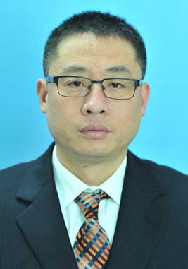 Liwu Chen (陈立武)合作论文数福州中医药大学第二临床医学院4
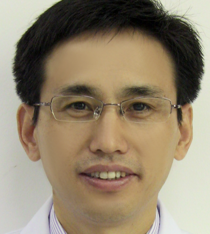 Xuejun Li (李学军)合作论文数The First Affiliated Hospital of Xiamen University4Hong-Zhi Ye合作论文数4Qiwen Dai合作论文数3Wei Lin合作论文数3Hong-cheng ZHANG (张鸿程)合作论文数3ZiXing Yan (严子兴)合作论文数3YuLian Deng (邓玉莲)合作论文数3LinYing Zhou (周琳瑛)合作论文数3LianYun Chen (陈莲云)合作论文数3ZhenHua Ge (葛振华)合作论文数3ChunXuan Xu (许春萱)合作论文数3QiuCheng Lin (林求诚)合作论文数3Lin Chen (陈林)合作论文数3




