目的 探讨原发性肝癌患者肝静脉压力梯度(hepatic vein pressure gradient,HVPG)与门静脉压力梯度(portal pressure gradient,PPG)相关性.方法 161 例原发性肝癌患者在TIPS术中测量下腔静脉压力(inferior vena cava pressure,ICVP)、肝静脉自由压(free hepatic vein pressure,FHVP)、肝静脉楔压(wedged hepatic vein pressure,WHVP)和门静脉压力(portal vein pressure,PVP),计算HVPG(HVPG=WHVP-FHVP)和PPG(PPG=PVP-IVCP).结果 161例患者HVPG为(20.18±9.22)mmHg,PPG为(26.44±6.82)mmHg,2 者无相关性(r=0.112);PPG明显高于HVPG(P<0.05).HVPG与PPG相差在 5 mmHg以上者 90 例,占 55.9%,HVPG与PPG相差在 5 mmHg以内者 71 例,占 44.1%.球囊阻断肝静脉造影有肝内静脉-静脉侧支分流(intrahepatic venous-venous collateral shunt,HVVC)者 42 例(26.09%),HVPG为(10.91±6.11)mmHg,PPG为(28.43±6.11)mmHg,2 者呈弱相关(r=0.384);PPG显著高于HVPG(P<0.05).球囊阻断肝静脉造影无HVVC者119例,HVPG为(23.45±7.81)mmHg,PPG为(25.74±6.94)mmHg,2者呈弱相关(r=0.249);PPG明显高于HVPG(P<0.05).结论 原发性肝癌合并门静脉高压(portal hypertension,PHT)HVPG与PPG总体相关性差,大部分患者的HVPG不能准确代表PPG,并且前者低于后者;有HVVC形成是严重低估HVPG值的重要原因.
Objective To investigate the role of P-I-R classification and Laennec grading in evaluating histological changes in patients with hepatitis B cirrhosis after receiving antiviral therapy, as well as the association of these two evaluation systems with clinical prognosis. Methods A total of 218 patients from 14 centers were consecutively screened from October 2013 to October 2014, and these patients were diagnosed with liver cirrhosis based on pathology(Ishak score ≥5), received antiviral therapy for 72 weeks, completed two liver biopsies, and met the P-I-R classification criteria. The 218 patients were divided into non-hepatocellular carcinoma(HCC) group with 186 patients and HCC group with 32 patients. The chi-square test and the Fisher’s exact test were used for comparison of categorical data between groups. For the comparison of HCC after antiviral therapy, the non-parametric Mann-Whitney U test was used for continuous variables, and for the comparison of P-I-R classification and Laennec grading, the non-parametric Kruskal-Wallis H test was used for continuous variables. Univariate and multivariate Cox regression analyses were used to calculate hazard ratio(HR) and 95% confidence interval(CI), and the Kaplan-Meier method was used to calculate the cumulative incidence rate of HCC. Results After 72 weeks of antiviral therapy, there was a significant difference in P-I-R classification between the non-HCC group and the HCC group(P<0.001). There were significant differences in the distribution of Laennec grading and P-I-R classification before and after antiviral therapy(P<0.001). After antiviral therapy, the 218 patients were divided into 4A group with 33 patients, 4B group with 71 patients, and 4C group with 114 patients according to Laennec grading, and there were significant differences between these three groups in platelet count(PLT)(H=36.429, P<0.001), liver stiffness measurement(LSM)(H=13.983, P=0.004), Ishak score(χ~2=23.060, P<0.001), and HAI score(P<0.001). After antiviral therapy, the 218 patients were divided into R group with 70 patients, I group with 52 patients, and P group with 96 patients according to P-I-R classification, and there were significant differences between these three groups in PLT(H=7.193, P=0.028), LSM(H=6.238, P=0.045), Ishak score(χ~2=7.986, P<0.001), HAI score(P=0.002), and HCC(P<0.001). There was a significant difference in the incidence rate of HCC between the P and R groups based on P-I-R classification(HR=24.21, 95%CI: 0.46-177.99, P=0.002). After adjustment for other confounding factors, P-I-R classification was an independent predictive factor for HCC(HR=12.69, 95%CI: 4.63-34.80, P=0.002). Conclusion Both P-I-R classification and Laennec grading can reflect the features and changes of fibrosis before and after antiviral therapy, and P-I-R classification is more sensitive to fibrosis changes after antiviral therapy. P-I-R classification(after treatment) can be used to assess the risk of HCC in patients after antiviral therapy.
ObjectiveTo investigate the clinical efficacy and safety of percutaneous cryoablation combined with percutaneous ethanol injection (PEI) in elderly patients with early-stage hepatocellular carcinoma aged 70 years or older. MethodsA retrospective analysis was performed for the clinical data of 92 elderly patients with hepatocellular carcinoma who were admitted to The Fifth Medical Center of Chinese PLA General Hospital from January 2014 to January 2018, among whom 46 underwent cryoablation alone (CRYO group) and 46 underwent cryoablation combined with PEI (combination therapy group). The two groups were compared in terms of clinical outcome, adverse reactions, and changes in liver function parameters after treatment, and the patients were followed up to observe tumor recurrence and survival. The t-test was used for comparison of normally distributed continuous data between two groups, and the Mann-Whitney U test was used for comparison of non-normally distributed continuous data between two groups; the chi-square test was used for comparison of categorical data between two groups. The Kaplan-Meier method was used for survival analysis, and the log-rank test was used for comparison of survival curves. The Cox regression analysis was used to identify the independent risk factors for survival and prognosis. ResultsThere was no significant difference in the response rate of initial ablation between the combination therapy group and the CRYO group (89.1% vs 73.9%, P>0.05). There were no significant differences between the CRYO group and the combination therapy group in overall survival time and tumor-free survival rate after surgery (P>0.05), and compared with the CRYO group, the combination therapy group had significantly lower 1-, 2-, and 3-year local tumor progression rates (20%/21%/21% vs 30%/46%/46%, χ2=4.187, P<0.05). The multivariate Cox regression analysis showed that cryoablation alone might be an independent risk factor for local tumor progression(HR=2.206,95%CI: 1.003-4.850, P=0.049). There was no significant difference in the incidence rate of adverse reactions between the two groups (P>0.05), but 3 patients in the CRYO group experienced serious adverse reactions, while no patients in the combination therapy group experienced such reactions. ConclusionFor elderly patients with early-stage hepatocellular carcinoma, cryoablation combined with PEI is safer and more effective than cryoablation alone and can significantly reduce local tumor progression rate.
背景 乙型肝炎病毒(hepatitis?B?virus,HBV)感染是引起肝纤维化、肝硬化的重要原因,口服核苷(酸)类似物抗病毒治疗获得持续的生化学和病毒学应答可以使肝纤维化好转,但部分慢性乙肝患者的肝纤维化仍可进展.目的 分析慢性乙型肝炎患者应用恩替卡韦抗病毒治疗后肝纤维化逆转的影响因素.方法 本研究为进一步分析一项前瞻性随机双盲安慰剂对照临床研究数据,选取2013年10月-?2014年10月解放军总医院第五医学中心等共14家医院收治的慢性乙肝初治患者,给予恩替卡韦治疗,并在治疗基线和第72周进行配对肝活检.按照治疗72周后是否获得肝纤维化逆转(Ishak纤维化评分下降F≥1分)分为逆转组和无逆转组,采用多因素logistic回归方法筛选肝纤维化逆转因素.结果 357例患者进行了配对肝活检,被纳入分析,平均年龄(42.4±10.1)岁,男性246例(68.9%),HBeAg阳性209例(58.5%).治疗72周后获得肝纤维化逆转者165例(46.2%),未获得逆转者192例(53.7%).多因素logistic回归分析显示基线肝弹性检测(liver?stiffness?measurement,LSM)值(9.4?~?17.0?kPa组,OR=0.509,95%?CI:0.301?~?0.860,P=0.012;>17.0?kPa组,OR=0.472,95%?CI:0.252?~?0.882,P=0.019)和血小板(platelet,PLT)>85?×?109?L?1(OR=2.683,95%?CI:1.068?~?6.742,P=0.036)与肝纤维化逆转独立关联,年龄和HBV?DNA定量与肝纤维化逆转无关.结论 肝纤维化经抗病毒治疗后可以获得组织学逆转;恩替卡韦抗病毒治疗期间,基线较低LSM值和较高PLT的患者更易获得肝纤维化逆转.
目的 通过生物信息学方法,挖掘影响肝细胞癌(HCC)患者生存预后的关键基因,对比在外周血单个核细胞(PBMC)和肿瘤组织中关键基因水平,探索关键基因作为新型标志物判断HCC患者生存预后的价值.方法 检索GEO和TCGA数据库中HCC数据,通过STRING构建蛋白质相互作用(PPI)网络和GEPIA生存验证,在Cytoscape筛选关键基因模块,对差异表达基因(DEGs)进行分析,应用Cox比例风险回归模型进行生存预后分析.结果 在PBMC中分析得到DEGs 225个,其中上调基因105个,下调基因120个;通过关键模块筛选和验证,得到6个关键基因,即GPSM2、PPIL1、POLR2H、CRNKL1、U2SURP和TRA2B,这些基因在肿瘤组织中的高水平与总体生存率显著相关;应用TCGA数据库临床数据行预后分析,结果表明GPSM2高水平与III期HCC总体生存期缩短独立相关(HR=1.556,95%CI:1.153~2.100);最后,通过比对PBMC与肿瘤组织中这6个关键基因的表达水平,发现其中GPSM2、TRA2B和U2SURP在两者中的表达水平趋势相同.结论 GPSM2在HCC肿瘤组织和PBMC中的高水平与Ⅲ期HCC患者预后独立相关,可能是HCC筛查和判断预后的新型标志物,甚至可成为治疗HCC的靶基因.
患者女性,28岁,因"发现肝占位1月余"入我院.既往体健.查体:神志清,精神可,腹部平坦,中上腹部偏左侧可触及6 cm包块,距肋弓约6 cm ,质韧,无触痛,全腹软,无压痛、反跳痛.化验:癌胚抗原、甲胎蛋白(电化学发光)、糖类抗原125、糖类抗原19-9均正常.腹部超声提示:1、肝内多发实性占位(Ca ,考虑转移可能) ,2、左上腹腔内实性占位(考虑Ca ,建议结合增强影像学检查) ,3、轻度脂肪肝.上腹部增强CT:影像所见:平扫(见图1)肝内见多发结节状及团块状混杂稍低密度影,较大者位于肝左外叶,局部外突,大小约12.3cm×10.2 cm ,动态增强扫描动脉期(见图2 )环形不均匀强化,门脉(见图3)及延迟期(见图4)呈环形强化、中心呈稍低密度影.腹腔左侧肾前方见类圆形稍低密度影,大小约6.5cm×5.7cm,增强扫描环形强化,病灶与邻近胃关系密切.腹腔内左肾及脾脏前方可见范围约15 .2 cm × 8 .8 cm不均质回声团,边界清,CDFI示可见血流信号.诊断意见:肝及腹腔内多发占位,考虑恶性肿瘤,转移不除外.经患者及家属同意后,行肝脏占位穿刺取病理.
HDV的重叠感染可加速慢性乙型肝炎患者的肝病进程,尤其肝细胞癌(HCC)的发生风险明显增高.但HDV的致癌机制有待进一步探索,治疗方式及效果也亟待突破.综述了HDV相关HCC流行病学新特点、致病机制的新认识和诊疗进展等,对推进研发更精准的HDV检测手段、更有效的治疗药物及减少HDV相关HCC具有重要意义.
目的 探讨恩替卡韦联合复方鳖甲软肝片治疗慢性乙型肝炎肝纤维化的临床疗效.方法 选取2014年4月—2016年3月于我院治疗的64例慢性乙型肝炎肝纤维化患者作为研究对象,按照随机、双盲、安慰剂对照原则将其分为对照组(恩替卡韦联合安慰剂治疗)和治疗组(恩替卡韦联合复方鳖甲软肝片治疗),例数分别为37例和27例.治疗期间定期进行血常规、血生化、HBV DNA、肝脏硬度(liver stiffness measurement,LSM)的检查,并观察2组患者治疗前和治疗72周时肝穿刺活检的疗效.结果 入组64例患者,其中59例完成临床疗效观察.与治疗前相比,治疗后2组患者ALT、AST、TBIL、HBV DNA水平及LSM和肝纤维化评分均较治疗前明显下降(P均<0.05);在改善肝纤维化程度方面,治疗组有效率为66.7%,明显高于对照组的45.8%,差异有统计学意义(P<0.05).结论 恩替卡韦联合复方鳖甲软肝片治疗可改善慢性乙型肝炎肝纤维化患者的肝脏炎症和纤维化程度.
Objective To investigate the clinical value of hepatitis B virus core-related antigen (HBcrAg) in predicting the natural course of chronic hepatitis B (CHB) and liver fibrosis regression. Methods A total of 138 CHB patients who were admitted to 302 Hospital of PLA, The First Affiliated Hospital of Zhengzhou University, and Fuzhou Infectious Disease Hospital from January 2013 to December 2015 were enrolled and divided into HBeAg-positive group with 69 patients and HBeAg-negative group with 69 patients. Of all patients, 109 with an Ishak score of ≥3 were treated with entecavir for 72 weeks. Liver biopsy specimens and serum were collected at baseline and after 72 weeks of treatment to observe histopathology and HBcrAg level. The t-test was used for comparison of normally distributed continuous data between two groups, and the Wilcoxon rank-sum test was used for comparison of non-normally distributed continuous data between two group. The chi-square test was used for comparison of categorical data between groups. A Spearman correlation analysis was also performed. The area under the receiver operating characteristic curve (AUC) was used to analyze the value of HBcrAg in the diagnosis of liver fibrosis. Results In HBeAg-positive CHB patients, serum HBcrAg level was negatively correlated with liver fibrosis stage (r=-0.342, P=0.004); in HBeAg-negative CHB patients, serum HBcrAg level was positively correlated with liver fibrosis stage and inflammation (r=0.439 and 0.437, both P<0.001). In HBeAg-positive patients, serum HBcrAg level had an AUC of 0.705 in predicting advanced liver fibrosis and 0.701 in predicting liver cirrhosis (both P<0.05); in HBeAg-negative CHB patients, serum HBcrAg level had AUCs of 0815, 0.815, 0.726, and 0.675 in predicting mild liver fibrosis, marked liver fibrosis, advanced liver fibrosis, and liver cirrhosis, respectively (all P<0.05). After antiviral therapy, the group with a high serum HBcrAg level was more likely to experience liver fibrosis regression than that with a low level (53.7% vs 32.7%, χ2=4.888, P=0.027). The patients with liver fibrosis regression had a significantly greater reduction in serum HBcrAg level than those without regression[1.5 (0.4-3.2) log IU/ml vs 0.8 (0.1-1.8) log IU/ml, Z=-1.724, P=0.042]. Conclusion Serum HBcrAg can be used as a new marker in predicting liver fibrosis stage and liver fibrosis regression in clinical practice.
HBV通过激发宿主免疫反应,引起肝损伤,导致肝纤维化、肝硬化、肝癌.肝纤维化进展是肝脏疾病相关死亡的关键过程,归纳了HBV基因型、HBV基因(包括基本核心启动子区/前C区、前S区、X区)变异、HBV基因剪接体、HBV RNA、HBcAg、HBsAg与肝纤维化进展的相关性研究.认为上述HBV生物学特性和肝纤维化进展密切相关.
目的:观察肝癌合并门静脉癌栓分级对索拉非尼治疗效果的影响.方法:收集2014年7月 2016年2月本院收治的晚期肝癌合并门静脉癌栓216例患者的临床资料,均接受肝动脉栓塞治疗,其中联合索拉非尼治疗42例;密切随访患者预后,用生存期分析探索门静脉癌栓分级对索拉非尼治疗效果的影响.结果:索拉非尼联合肝动脉栓塞治疗42例的生存期为(9.83±4.63)个月,中位生存期为9个月;其中门静脉癌栓Ⅰ或Ⅱ级27例的生存期为(11.44±4.47)个月、中位生存期为11个月,门静脉癌栓Ⅲ或Ⅳ级15例的生存期为(6.93±3.41)个月、中位生存期为6个月.单纯肝动脉栓塞治疗174例的生存期为(7.60±3.49)个月,中位生存期为8个月;其中门静脉癌栓Ⅰ或Ⅱ级119例的生存期为(8.72±3.38)个月、中位生存期为8个月,门静脉癌栓Ⅲ或Ⅳ级55例的生存期为(5.16±2.25)个月、中位生存期为5个月.索拉非尼联合肝动脉栓塞治疗肝癌合并门静脉癌栓Ⅰ或Ⅱ级患者的生存期,非常显著高于单纯肝动脉栓塞治疗的同级患者(P<0.01).结论:门静脉癌栓分级与索拉非尼治疗效果显著相关,门静脉癌栓Ⅰ或Ⅱ级患者采用索拉非尼治疗获益明显.
Objective To investigate the risk factors for vascular invasion of primary liver cancer (PLC).Methods A retrospective analysis was performed for the clinical data of 211 patients with liver cancer who were hospitalized in 302 Hospital of PLA from January 2013 to June 2014.The logistic regression model fitting was used for the data of 133 patients,and the data of the other 78 patients was used for the verification of this model.Univariate and multivariate logistic regression analyses were used to identify the influencing factors for vascular invasion of PLC;the logistic regression model was established and the receiver operating characteristic (ROC) curve was plotted to determine the optimal cut-off value of this model.Results The univariate logistic regression analysis showed that tumor diameter (odds ratio [OR]=1.594,95% confidence interval [CI]:1.376-1.846,P =0),neutrophil-lymphocyte ratio (NLR) (OR =2.783,95% CI:1.847-4.195,P=0),platelet-to-lymphocyte ratio (PLR) (OR=1.016,95% CI:1.008-1.024,P=0),fibrinogen (Fb) (OR=2.295,95 % CI:1.608-3.274,P =0),and lymph node metastasis (OR =11.664,95 % CI:3.744-36.338,P =0) were risk factors for vascular invasion of PLC;the multivariate logistic regression analysis showed that tumor diameter (OR =1.506,95% CI:1.250-1.815,P =0),PLR (OR=1.499,95% CI:0.173-0.998,P=0.022),NLR (OR =2.491,95% CI:1.411-4.397,P=0.002),and Fb(OR =1.486,95% CI:1.008-2.193,P =0.046) were used for regression model fitting.The area under the ROC curve was 0.927 (95% CI:0.881-0.973),and the ROC curve showed that the model had the highest sensitivity of 92.9%,the highest specificity of 86.5%,and a prediction accuracy rate of 82.79%.Conclusion The regression equation containing tumor diameter,PLR,NLR,and Fb established in this study has a high prediction accuracy of vascular invasion of PLC and provides a reference for early warning of vascular invasion of PLC.
2017 年 2 月 24—25 日,"中国研究型医院学会肝病专业委员会 2017 年学术年会"在北京万达嘉华酒店举行.本次会议由中国研究型医院学会肝病专业委员会主办,中国肝炎防治基金会、解放军第三〇二医院和《传染病信息》杂志社共同承办.来自全国近 10 个省和直辖市的专家为大会呈现了慢性肝病多个临床和科研方向的精彩报告,来自全国 20 余个省和直辖市的 300 余名代表参加了本次大会.中国研究型医院学会肝病专业委员会选举产生了首届青年委员会,肝病专委会主任委员杨永平兼任首届青委会主任委员,南方医科大学南方医院祁小龙当选副主任委员兼秘书长.
Objective To explore the susceptible factors of fever and infection among hepatocellular carcinoma (HCC) patients who received cryo-ablation (CA) therapy,and to propose intervention countermeasures accordingly.Methods The fever and infection status of totally 428 HCC patients who received CA therapy was examined systematically;then the influencing factors were analyzed by single factor analysis and logistic regression analysis;and finally,nursing strategies were proposed accordingly.Results Totally 229 patients developed fever (53.5%),and 6 suffered from complicated infection (1.4%) after CA therapy;tumor size,CA area,Child-Pugh classification and preoperative leukocyte level were factors that contributed to the fever of HCC patients who received CA therapy.Conclusions We should attach importance to the susceptible factors of fever and infection among HCC patients who received CA therapy,and scientific intervention and nursing are the key to reducing the possibilities of fever and infection among patients who received CA therapy.
The National Health and Family Planning Commission of the People's Republic of China published evidence-based clinical practice guidelines on diagnosis, treatment and management of hepatocellular carcinoma (V2017) in June 2017. In this guidelines, the diagnosis, staging, and treatment of hepatocellular carcinoma are updated. This article briefly describes and interprets only new or changed recommendations.
Objective To investigate the clinical features of HBsAg-negative HBV-related hepatocellular carcinoma (HCC).Methods The patients who were newly diagnosed with HCC from January 2005 to January 2012 were enrolled.According to the HBV-related serological markers,the patients were divided into HBsAg-negative HBV-related HCC group and hepatitis B-related HCC group.A retrospective analysis was performed for the clinical and laboratory examination data at initial diagnosis,including sex,age,hepatitis B virus markers,total bilirubin (TBil),albumin (Alb),alpha-fetoprotein (AFP),HBV DNA,body mass index (BMI),drinking history,history of diabetes,therapies,and follow-up results.The t-test was used for comparison of normally distributed continuous data between groups;the non-normally distributed data were expressed as median and interquartile range (Q1 and Q3),and the Wilcoxon rank sum test was used for comparison of non-normally distributed continuous data between groups;the chi-square test or Fisher's exact test were used for comparison of categorical data.The log-rank test was used to compare survival curves between the two groups,and the Kaplan-Meier method was used to calculate survival rates.Results Compared with the hepatitis B group and all patients,the HBsAg-negative group had a significantly higher mean age (59.42 ± 11.13 years) and significantly higher proportions of patients with age > 60 years (46.85%),low body weight (12.24%),and overweight (20.98%) (all P <0.01).Compared with all patients and the hepatitis B group,the HBsAg-negative group had a significantly lower male-to-female ratio (2.86∶1),a significantly lower proportion of patients regularly undergoing physical examination every year (10.41%),a significantly lower HBV DNA positive rate (1.77%),and significantly lower proportions of patients with smoking and drinking histories (all P <0.01).Of all patients in the HBsAg-negative group,64.08% had positive anti-HBs,anti-HBe,and anti-HBc,21.13% had positive anti-HBe and anti-HBc,and 14.79% only had positive anti-HBc.The HBsAg-negative group had a significantly lower AFP level than all patients and the hepatitis B group (P =0.039).Compared with the hepatitis B group and all patients,the HBsAg-negative group had a significantly higher proportion of multiple lesions,a significantly greater mean maximum tumor diameter,and a significantly higher number of patients with extrahepatic metastasis (all P < 0.001).Compared with the hepatitis B group,the HBsAg-negative group had a higher proportion of patients with an Eastem Cooperative Oncology Group Performance Status score of 0 at initial diagnosis (17.69%) and a lower proportion of patients with early-to-medium Barcelona Clinic Liver Cancer stage (stages A and B) (30.26% vs 51.60%,P <0.001);the number of patients with stages C and D in the HBsAg-negative group was 1.83 times that in the hepatitis B group (P <0.001).The HBsAg-negative group had a significantly lower median survival time (15.22 months) than all patients and the hepatitis B group (P =0.024、0.031).Compared with all patients and the hepatitis B group,the HBsAg negative group had significantly lower 1-and 3-year survival rates (1-year survival rate:56.0% vs 61.7% and 66.2%,x2 =4.93,P =0.026;3-year survival rate:24.0% vs 45.2% and 44.1%,x2 =6.867,P =0.009).Conclusion HBsAg-negative HBV-related HCC is commonly seen in patients aged >60 years with positive anti-HBs,anti-HBe,and anti-HBc.The population aged >60 years with HBV infection sbould be the high-risk population for HCC,and it is recommended to perform HCC screening every 6 months to increase the early diagnostic rate of HBsAg-negative HBV-related HCC.
目前,肝细胞癌(简称肝癌)诊断与治疗尚存在诸多难点与瓶颈 :①早期诊断难 ;②复发转移率高,5 年复发率近 60% ;③肝癌个性化治疗的愿景与临床实践的大环境不匹配 ;④有效药物和干预手段少.但是,人们对肝癌的防治已从阴雨中走向阳光 :①大规模乙肝疫苗免疫接种是降低乙肝相关性肝癌发生的最有效手段 [1] ;②肝癌分子生物学研究的进展,使患者人群细化分类,为有的放矢地进行防治创造条件 ;③越来越多的医学工作者开始从事肝癌治疗有效性的对比研究 ;④ 继索拉非尼治疗进展期肝细胞癌取得肯定疗效后 [2,3],以PD-1 阻滞剂为代表的免疫治疗再次成为肝细胞肝癌(hepatocellular carcinoma,HCC)治疗热点 [4,5].
Objective To evaluate the efifcacy and safety proifles of tenofovir disoproxil fumarate (TDF) vs adefovir dipivoxil (ADV) for Chinese patients with chronic hepatitis B (CHB).Methods In this randomized, double-blind, controlled study, patients with CHB received TDF or ADV (ratio for 1︰1) once daily for 48 weeks were assigned, randomly. The primary efficacy end point was plasma HBV DNA level < 20 IU/ml at week 48. The levels of HBV DNA decrease, ratios of ALT normalization, HBsAg loss and seroconversion and HBeAg loss and seroconversion, while the adverse events and adverse effects were also compared between patients in TDF group and ADV group.Results Total of 267 patients were enrolled, among whom, there were 141 patients in TDF group and 139 patients in ADV group. The baseline characteristics were comparable between the two groups (P all > 0.05). At week 48, virological responses occurred in more HBeAg positive patients with TDF treatment than patients with ADV treatment (55.3%vs. 20.0%;χ2= 20.750,P < 0.001) and in more HBeAg negative patients with TDF treatment than patients receiving ADV treatment (86.2%vs. 62.7%;χ2= 20.750,P = 0.003). There were no signiifcant difference in ratio of ALT normalization in both HBeAg positive (84.5%vs. 80.5%;χ2= 0.406,P = 0.524) and negative groups (84.2%vs. 80.7%;χ2= 0.243,P = 0.622). In HBeAg positive group, there were no significant difference in ratio of HBeAg loss (5.6%vs. 12.8%;χ2= 2.335,P = 0.126) and HBsAg loss (1.4%vs. 3.8%;χ2= 0.182,P = 0.670). No case in HBeAg negative group lost HBsAg after 48 weeks therapy. Safety proifles including incidence of adverse events and adverse effects were compared between TDF and ADV groups with no significant difference (P all > 0.05).Conclusions Among Chinese CHB patients with HBeAg positive and negative, TDF had superior antiviral efifcacy with a similar safety proifle compared with ADV through 48 weeks.
目的 探讨肝脏神经内分泌肿瘤(liver neuroendoerine tumor,LNET)的磁共振成像(magnetic resonance imaging,MRI)特征.方法 回顾性分析19例LNET患者的临床病理特征及MRI特点.结果 LNET主要来源于消化系统,以胰腺最多见;其组织病理分型以G3型(63.16%)为主,G2型占26.32%,G1型占10.53%.MRI表现主要分为4类:Ⅰ型:动脉期轻度强化或环形强化,门脉期造影剂减退,延迟期呈稍低信号影并可见假包膜环形强化;Ⅱ型:动脉期轻度环形强化或周边斑片状不均匀强化,门脉期及延迟期强化范围增大,呈稍高信号;Ⅲ型为混合类型即同时存在Ⅰ型和Ⅱ型的特点;Ⅳ型为特殊类型:病变为囊实性病变.其中以Ⅰ、Ⅱ型为主.结论 MRI特点:Ⅰ型多见,Ⅱ型次之,其动脉期环形强化、延迟期假包膜形成为相对特征性表现,但诊断仍需依据病理检查.
肝细胞癌(hepatocellular carcinoma,HCC)居世界癌症死因第三位,中国第二位。手术切除率仅占20%~30%。因此,发展了多种局部消融治疗,如射频消融(RFA)、冷冻消融、微波、无水酒精、激光等在HCC的治疗中发挥重要作用[1]。冷冻消融HCC是近年发展的一项局部消融,与广泛应用的RFA及其他热消融相比,具有如下优势:产生较大的毁损面积、清晰可辨的治疗靶区。本文对冷冻消融HCC的适应证、技术、病人管理及安全性、疗效作以评述。
 Chunping Wang (王春平)合作论文数Chinese PLA General Hospital25
 Xudong Gao (高旭东)合作论文数Chinese People's Liberation Army General Hospital14Min Lou (楼敏)合作论文数12
 Yinying Lu (陆荫英)合作论文数Chinese PLA General Hospital;Beijing Syngentech Co., Ltd.12YongYi Feng (冯永毅)合作论文数11Zhen Ceng (曾珍)合作论文数11
 JianHui Qu (曲建慧)合作论文数解放军总医院第五医学中心肝脏肿瘤诊疗与研究中心11Shu-hui SU (苏淑慧)合作论文数10Xiujuan Chang (常秀娟)合作论文数10XueMei Ma (马雪梅)合作论文数9Linjing An (安林静)合作论文数9
 Zhengsheng Zou (邹正升)合作论文数Chinese PLA General Hospital9Yan Chen (陈艳)合作论文数8
 Wenlin Bai (白文林)合作论文数Chinese PLA General Hospital7
 Lin Zhou (周霖)合作论文数Chinese PLA General Hospital7Yihui Rong (荣义辉)合作论文数7
 Shaojie Xin (辛绍杰)合作论文数Chinese PLA General Hospital7XinZhen Wang (王新真)合作论文数6GuiLin Xu (许桂林)合作论文数6
 Baosen Li (李保森)合作论文数Chinese People's Liberation Army General Hospital6
 Jumei Chen (陈菊梅)合作论文数.The 302 Hospital of PLA,Beijing ,China6
 Jun Zhao (赵军)合作论文数Chinese PLA General Hospital5Liangping Hu (胡良平)合作论文数5Yinyin Li合作论文数4
 Xianzhi Zhou (周先志)合作论文数Air Force Medical University4
 Huaming Wang (王华明)合作论文数Chinese PLA General Hospital4Deyong Chen (陈德永)合作论文数4
 Fusheng Wang (王福生)合作论文数Chinese People's Liberation Army General Hospital4
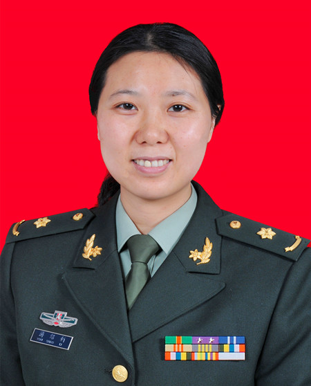 Shaoli You (游绍莉)合作论文数Chinese PLA General Hospital4QI Dong-ying (齐冬颖)合作论文数3Hong Wang (王鋐)合作论文数3Ze Liu (刘泽)合作论文数3Xin Zhang (张昕)合作论文数3
 Minna Zhang (张敏娜)合作论文数解放军总医院第五医学中心感染性疾病诊疗与研究中心3Zhe Yu合作论文数2Jiagan Huang合作论文数2LUO Jing (罗婧)合作论文数2ZHANG Wei (张伟)合作论文数2Caihong Lv合作论文数2Bai-long ZHANG (张百龙)合作论文数2Kun DONG (董琨)合作论文数2Cheng-wu WANG (王成武)合作论文数2YiChen Wu (吴贻琛)合作论文数2WeiHua Chang (常伟华)合作论文数2HongJun Jia (贾红军)合作论文数2ShiLan Zhang (张世兰)合作论文数2LuJun Wang (王陆军)合作论文数2Yun Li (李筠)合作论文数2




