Diabetic chronic wounds, especially in the moist and mechanically dynamic oral environment, pose a significant therapeutic challenge due to neutrophil extracellular traps (NETs)-mediated inflammation, biofilm infections, and extracellular matrix remodeling defects. To address this challenge, we develop a protein-based adhesive bioshield, functioning through the combined action of physical blocking and electrostatic interaction, that simultaneously acts as a bacterial barrier and NETs scavenger, thereby reactivating the focal adhesion signaling. The protein's high-density lysine residues establish a robust adhesion network that resists the dynamic oral environment while creating a persistent antibacterial bioshield. Notably, the high net charge of adhesive bioshield enables rapid NETs neutralization by electrostatically binding cell-free DNA (cfDNA), which reduces local NETs levels and inflammatory responses. This NETs clearance subsequently alleviates inflammation-mediated suppression of focal adhesion signaling, which coordinately accelerates diabetic wound healing through enhanced epithelial migration, endothelial proliferation, and angiogenesis. Overall, this study presents a charge-driven therapeutic strategy that targets NETs-mediated inflammation, offering an alternative approach for managing diabetic wounds in dynamic and wet environments.
Dental defects, ranking among the most prevalent diseases globally, pose a serious threat to human health, with extensive defects involving dentin leading to complications such as pulp and periodontal diseases, as well as maxillofacial dysfunctions, significantly impairing quality of life. Current clinical treatments primarily rely on rigid materials such as metals, composite resins, and ceramics for macroscopic filling. However, their inherent limitations, differences in compositional and structural characteristics from natural dentin, mismatched mechanical properties, and interfacial adhesion instability, fail to meet the clinical demand for long‐term and stable restoration of natural dentin. In situ dentin regeneration, inspired by the complex composition and hierarchical structure of natural dentin, aims to induce the autonomous repair of dentin. This approach effectively overcomes the traditional limitations, shifting from traditional passive filling to active regenerative repair. Based on the growth direction and mineralization pattern of the repair layer, current research focuses on three strategies: “inward growth”, “outward growth”, and “synchronized inward‐outward growth”. This review primarily focuses on the roles and clinical applications of key bioactive materials in these strategies, providing a feasible basis for future material and performance optimization of dentin in situ regeneration.
Significant efforts to utilize artificial soft materials in developing tissue engineering scaffolds show considerable promise for maxillofacial bone regeneration. However, most biological materials cannot simultaneously satisfy the multiple requirements of robust adhesive strength under masticatory environments, pathological immune regulation, and efficient cell-specific targeting therapy, ultimately compromising the repair process. Herein, a tissue-adhesive hydrogel loaded with mesenchymal stem cells-derived nanovesicles (PEG-pp@nMSC@MT) is introduced with inflammation regulation and cell-specific targeting as an all-in-one tool for maxillofacial bone repair. Relying on the quick amidation reaction between active ester groups of PEG-SG and amine groups of PEG-NH2 polymers, the uniform networks are rapidly formed with easy injection, favorable biocompatibility, and robust adhesive strength, which is capable of resisting frequent masticatory force in oral cavity. The incorporation of a matrix metalloproteinase 2 (MMP2)-cleavable peptide allows the hydrogel to respond to elevated protease levels at defect sites, enabling on-demand release of encapsulated nanovesicles while concurrently attenuating excessive MMP2 activity. Of note, the efficient transport of therapeutic melatonin to the intended BMMSCs enhances their osteogenic and immunomodulatory functions. Collectively, the reduction of MMP2, secretion of anti-inflammatory factors by BMMSCs, and immunomodulatory effects of nMSC@MT synergistically promote macrophage polarization toward M2 phenotype and facilitate bone regeneration. The therapeutic effect of PEG-pp@nMSC@MT displays superior biodegradability and osteo-inductive capacity compared with Bio-Oss, a first-line grafting material used in maxillofacial bone defect in current clinical treatment. Thus, this innovative hydrogel platform combines precise immunomodulation with cell-specific targeting, representing a promising therapeutic strategy for the effective repair of maxillofacial bone defects.
Conventional electrochemical sensing techniques detect predefined molecular biomarkers for disease diagnosis, while compromised by the biases in the calibration process due to the complexity and volatility of peripheral biofluids. Meanwhile, abundant electrochemical information at the electrode-biofluid interfaces remains to be discovered to gain comprehensive metabolic signatures for diagnostic applications. Here, we propose an electrochemomics (EC-omics) approach to comprehensively profile the dynamics of electrochemical properties of biomolecules in peripheral biofluids during disease onset. As a proof of concept, we customized a portable electrochemical profiling platform, where the high sensitivity and low background noise of the carbon nanotube/bacterial cellulose (CNT/BC) electrodes enabled a holistic and unbiased capturing of the electrochemical features in biofluids. We applied the EC-omics platform to profile saliva for periodontitis diagnosis. The obtained saliva EC-omics database is compatible with various intelligent algorithms, which could accurately discriminate periodontitis (93%), surpassing the untargeted nuclear magnetic resonance data (89%) and significantly outperforming the periodontitis-related molecular biomarkers (70%) and peak intensity features (57%). Additionally, our study demonstrated the feasibility of EC-omics in human urine and mouse serum analysis, suggesting its potential to expand our understanding of the complex metabolic networks of biofluids and further foster a broader range of novel diagnostic tools across various sensing paradigms for decentralized healthcare.
Chiral nanostructures exhibit unique immunomodulatory properties by engaging stereospecific recognition mechanisms within biological systems, enabling precision immune engineering. Yet the underlying mechanisms of chiral-dependent effects on immune cells-specifically, how distinct chiral configurations activate intracellular cascades to elicit anti-tumor effects-remain poorly understood. Here, we engineered chiral silver-shelled gold nanoparticles (Au@Ag-L/D NPs) with specifically tailored morphologies and plasmon-enhanced photoresponsivity, demonstrating that chiral particles with weak optical dissymmetric factors can sufficiently elicit enantiomer-dependent immune reprogramming. Notably, D-enantiomers preferentially bind to scavenger receptors to disrupt lysosomal autophagy, effectively repolarizing tumor-associated macrophages from pro-tumorigenic M2 to antitumor M1 phenotypes. Combined with near-infrared light-triggered hyperthermia and ROS generation, chirality-guided M1 polarization amplifies anti-tumor immunity while enabling localized tumor ablation. In vivo, D NPs with photoirradiation enhanced tumor suppression in colorectal cancer models through concerted immunological microenvironment remodeling and multi-modal tumor eradication. This study thus established precise chirality-immunomodulation coordination, by combining phototherapy with immunotherapy to synergistically enhance tumor ablation and reverse immunosuppression.
Human parvovirus B19 (B19V) infection is a significant but underrecognized complication, commonly linked not only to aplastic anemia but also pancytopenia, especially in hematopoietic stem cell transplantation (HSCT) recipients. While B19V’s tropism for erythroid progenitor cells (EPCs) is well-documented, its restriction to EPCs fails to fully explain the pathogenesis of pancytopenia. In this study, we used PrimeFlow RNA assay, and single-cell full-length transcriptome sequencing (scFAST-seq) to show that B19V could infect hematopoietic stem cells (HSCs) and initiate viral transcription, resulting in increased apoptosis, impaired self-renewal and multilineage differentiation of HSCs, which may contribute directly to pancytopenia. Further analysis revealed that B19V could activate the JAK2/STAT5 signaling pathway in HSCs to promote viral persistence. Pharmacological inhibition with baricitinib markedly reduced the viral load and partially restored hematopoietic differentiation capacity in vitro. Taken together, our findings reveal B19V as a previously unrecognized HSC-tropic virus that disrupts function of HSCs and may drive pancytopenia. Targeting the JAK2/STAT5 signaling by baricitinib shows promising therapeutic potential for reversing virus-induced bone marrow failure. This work not only deepens our understanding of viral tropism and pathogenesis in the hematopoietic niche, but also opens up new possibilities for treating bone marrow damage after transplantation and in other hematological diseases.
Saliva assay is a promising potential strategy for widespread screening and prompt surveillance of oral cancer to improve the prognosis and reduce the financial burden. But current saliva detection methods are hampered from clinical application by their restricted target diversity, complex procedures, and costly equipment. In this work, we introduce an enzymatic responsive serial-nanofluids strategy, enabling simultaneous trace-level detection of multiple humoral markers in saliva. Enzymatic-responsive nanochannels were engineered respectively by modifying the outer surfaces of AAO arrays with polypeptides featuring target-cleavage sites. Thus, the macromolecular markers in saliva could cleave corresponding polypeptide, opening the ion pathway and enhancing the ion flux of nanochannels. Upon the open of a set of nanochannel-arrays tandemly connected, serial-nanofluids are generated and ionic currents are coupled to enable collaborative detection of multiple targets. Employing oral cancer markers of MMP-1 and MMP-3 as models, we developed a mobile nanosensor that can concurrently monitor their trace variation low to 2.14 × 10- 1 3 g/mL. Furthermore, the clinical trial indicated that our nanosensor could noninvasively, conveniently, and effectively distinguish healthy individuals from oral cancer patients, and those with lymph node metastasis. This strategy offers a robust framework for multi-marker detection in clinical diagnostics, facilitating high-frequency and large-scale cancer screening through saliva test.
Failure of timely bone regeneration compromises structural integrity and delays functional recovery; therefore immune regulation of the early repair microenvironment is crucial for successful healing. M1 (pro-inflammatory) phenotype macrophages play pivotal roles in vascularisation during the early phase of bone regeneration and are typically activated by interferon-gamma (IFN-γ) or lipopolysaccharide (LPS) as well as by metabolite-derived signals. Lactate, a metabolite known to regulate a series of pathophysiological processes, has not yet been fully investigated for its specific immunomodulatory role in the microenvironment of bone injury healing. Our in vitro experiments demonstrated that lactate induced macrophage polarisation to the M1 phenotype and accelerated angiogenesis, with the HIF1α-NOD1-calcium influx axis identified as a key mediator. In vivo validation further confirmed the positive effects of lactate intervention in promoting vascularised bone regeneration at the early stage of injury. Thus, this study uncovers how lactate modulates immune response in association with M1 macrophages and indicates its potential as a therapeutic strategy for promoting vascularised bone healing.
Osteoarthritis (OA) arises from impaired epigenetic coordination of inflammatory and metabolic cues, leading to compromised cartilage homeostasis. Such coordination is partly governed by ribonucleic acid (RNA) epigenetic mechanisms, however, the role of the predominant RNA modification N6-methyladenosine (m6A) in this process remains unclear. Herein, we identify an epigenetic-metabolic pathway in which Wilms' Tumor 1-Associating Protein (WTAP)-mediated m6A modification stabilizes low-density lipoprotein receptor-related protein 1 (LRP1) and redirects lipid metabolism toward chondrogenesis. Loss-of-function assays demonstrate that WTAP is required for the chondrogenic differentiation of BMSCs, as its depletion suppresses the expression of multiple cartilage-associated genes. Mechanistically, WTAP enhances m6A methylation and stabilizes Lrp1 transcripts, a key regulator of cholesterol metabolism and matrix synthesis, thereby driving lipid metabolic reprogramming toward chondrogenesis. Structure-based screening identified silibinin and estradiol benzoate as LRP1-specific agonists that activate the WTAP-LRP1 pathway to promote cartilage repair in vivo. Collectively, our findings establish m6A-dependent metabolic reprogramming as a pivotal epigenetic mechanism of cartilage regeneration with therapeutic potential for promoting chondrogenesis.
Orthodontic treatment corrects various craniofacial malformations primarily through triggering active alveolar bone remodeling, however the potential impacts of this intervention on the systemic immune state have long been insufficiently explored. Here, we show that mechanical force applied during orthodontic treatment can trigger systemic inflammation dominated by adaptive immune responses. This response not only impairs bone repair at distant body sites but also causes temporary liver dysfunction resembling autoimmune conditions characterized by the expansion of CD69⁺ B cells. Mechanistically, IgM produced by these activated B cells acts as a key pathogenic driver of systemic pathology. Inhibiting the activation of local B cells effectively reversed this immunopathology, underscoring the central role of B cell-derived IgM. We also verified that Piezo1⁺ macrophages function as vital mechanosensors, linking orthodontic mechanical stimulation to B cell activation and subsequent IgM release. Multi-organ metabolomic profiling revealed significant amino acid metabolic dysregulation, which further aggravated systemic inflammation. Crucially, supplementation with lysine and alanine alleviated orthodontic treatment-induced inflammation, restored liver function and promoted distal bone repair. Together, these findings uncover an unrecognized systemic immune-metabolic axis during orthodontic therapy and suggest that targeting B cell-IgM responses or metabolic dysregulation may provide therapeutic opportunities to enhance treatment safety and outcomes.
Understanding hydrogen dissolution mechanisms in bridgmanite (Bgm), the most abundant mineral in the lower mantle, is essential for understanding water storage and rheological and transport properties in the region. However, interpretations of O-H bands in Fourier transform infrared spectroscopy (FTIR) spectra of Bgm crystals remain uncertain. We conducted density functional theory (DFT) calculations on vibrational characteristics of O-H dipoles and performed polarized FTIR measurements to address this issue. DFT calculations for four substitution models-Mg vacancies, Si vacancies, Al3+ + H+ substitution for Si4+, and Al substitution with Mg vacancies-reveal distinct O-H bands with different polarizations. Deconvolution of polarized FTIR spectra on Mg0.88Fe2+0.035Fe3+0.065Al0.14Si0.90O3 and Mg0.95Fe2+0.033Fe3+0.027Al0.04Si0.96O3 crystals shows five major O-H bands with distinct polarizations along principal crystallographic axes. These experimental and calculated results attribute O-H bands centered at 3,463-3,480, 2,913-2,924, and 2,452-2,470 cm-1 to Mg vacancies, Si vacancies, and Al3+ + H+ substitution for Si4+, respectively. The total absorbance coefficient of bridgmanite was calculated to be 82,702(6,217) L/mol/cm2. Mg and Si vacancies account for 43%-74% of the total water content, making them dominant hydrogen dissolution mechanisms in Bgm. The band frequencies for the Mg and Si vacancies in Bgm are drastically different from those in olivine and ringwoodite, corresponding to the significant changes in O-H bond strengths and in the Si and Mg coordination environments from upper-mantle to lower-mantle minerals. These results highlight the need to incorporate hydrogen dissolution mechanisms in Bgm for understanding electrical conductivity and rheology of the lower mantle.
Present main-stream medical ultrasonic nebulizers contain Pb(Zr,Ti)O3-based piezoceramics, which could expose the patients to toxic lead when the acidic liquid medicine, compounded with ultrasonic vibration, corrodes the piezoceramics. Enormous amount of lead ions up to 150 ppm can be extracted when lead-containing piezoceramics were exposed to typical acidic liquid medicines. Thus, a series of nebulization in a course of treatment could easily raise a child's blood lead level well above the blood lead reference value of 0.035 ppm, causing adverse and likely irreversible health effects. Herein, we introduce the lead-free medical ultrasonic nebulizers based on high-performance (K,Na)NbO3-based lead-free piezoceramics. It's designed for facile and safe at-home treatment, offering a healthy, safe, and lead-free solution for patients. It also marks the first commercialization of (K,Na)NbO3-based lead-free piezoceramics as a replacement for Pb(Zr,Ti)O3, helping expedite the lead-removing process of piezoelectric materials in general, in line with sustainable development goals worldwide.
The electrocaloric effect of ferroelectrics holds great promise for solid-state cooling, potentially replacing traditional vapor-compression refrigeration systems. However, achieving adequate electrocaloric cooling capacity at room temperature remains a formidable challenge due to the need for a high intrinsic electrocaloric effect. While barium titanate ceramic exhibits a pronounced electrocaloric effect near its Curie temperature, typical chemical modifications to enhance electrocaloric properties at room temperature often reduce this intrinsic electrocaloric effect. Herein, a structural design is introduced for barium titanate-based ceramics by incorporating isovalent cations. This leads to a well-ordered local structure that decreases the Curie temperature to room temperature while preserving a sharp phase transition, enabling a large dielectric constant and tunable polarization. This design achieves a remarkable electrocaloric strength of ~1.0 K·mm/kV, surpassing previous reports. Atomic-resolution structural analyses reveal that the presence of multiscale nanodomains (from ~10 nm to >100 nm), and the dipole polarization distribution with gradual dipole rotation enable rapid phase transition and facile polarization rotation, accounting for the giant electrocaloric response. This work provides a strategy for achieving a strong intrinsic electrocaloric effect in ferroelectrics near room temperature and offers key insights into the microstructure landscapes driving this enhanced electrocaloric effect.
The persistent challenge of diabetic wound healing arises from chronic inflammation, which disrupts the immunometabolic microenvironment. This dysregulated microenvironment exhibits remarkable chirality, but how the stereochemical specificity governs host immunometabolism networks remains elusive. In this study, we engineered chiral gold nanoparticles (L/D-AuNPs) to modulate immunometabolic interactions, thereby significantly promoting diabetic wound healing. To align with the chiral characteristics of the vimentin intermediate filaments (VIFs), L/D-AuNPs with mirror-symmetric windmill-like configurations were synthesized using a seed-mediated growth method. In diabetic murine wounds, L-AuNPs demonstrated superior therapeutic efficacy, enhancing wound healing rate by 29 % compared to the control group. Mechanistic studies revealed that L-AuNPs upregulated the expression of the intermediate filaments vimentin, enhancing fibroblast migration. Concomitant mechanotransduction activated the TGF-β/Slug signaling axis, driving nicotinamide metabolic reprogramming in fibroblasts, which subsequently polarized macrophages toward an anti-inflammatory M2 phenotype. This immunometabolic crosstalk was further validated by single-cell transcriptomic profiling of diabetic patient skin specimens. Based on this finding, we combined L-AuNPs with exogenous nicotinamide, which further accelerated wound regeneration through a significant synergistic effect. This study reveals that chiral AuNPs can promote tissue repair by modulating immunometabolic interactions, providing a novel therapeutic strategy for diabetic wound healing.
Severe bone defects pose a formidable clinical challenge in orthopedics, urgently demanding the development of advanced biomaterials to restore structural and functional integrity. While current regenerative materials, such as collagen-containing products, demonstrate a certain degree of biocompatibility, they are still hampered by limitations that include poor mechanical performance, restricted barrier effects, and arduous preparation methods. Here, we report a rapid-curing methodology to engineer recombinant resilin bioshield with tunable modulus, superior bioactivity, and rapid assembly kinetics. The resilin bioshield is rapidly formed within minutes via a tyrosine-mediated photo-crosslinking strategy, achieving spatially programmable assembly. Enzymatic integration of alkaline phosphatase into the resilin matrix drives in situ mineralization, yielding densely packed hydroxyapatite (HAP) nanocrystals. Remarkably, this process enables controlled modulus tuning of the bioshield across three orders of magnitude, achieving an exceptional maximum modulus of 145 MPa while retaining excellent flexibility, thus surpassing conventional guided bone regeneration materials. Beyond its mechanical superiority, the mineralized resilin bioshield not only directs cellular behavior by enhancing adhesion and spreading but also robustly drives the osteogenic differentiation of mesenchymal stem cells, thereby accelerating functional bone regeneration. As a result, our work provides an alternative approach for creating high-performance barrier membranes for guided bone regeneration.
Chirality is a pivotal determinant in stem cell differentiation, yet discerning the individual effects of chirality across different scales within native three-dimensional (3D) environments remains challenging. Here, a strategy is employed using nanostructures with controlled chirality to precisely assess the impact of molecular and supramolecular chirality on mesenchymal stem cell (MSC) osteogenic differentiation. We synthesized two pairs of enantiomers, l/d-phenylalanine (l/D-Phe) and l/d-1-naphthylalanine (L/D-1-Nap) derivatives, which could form four distinct chiral fibrous hydrogels with different molecular and supramolecular chiralities: L-supP and D-supP (supP indicates supramolecular right-handed helix), and L-supM and D-supM (supM denotes supramolecular left-handed helix). Both experimental and computational analyses reveal that the supramolecular supM/supP helicity is governed by conformational changes in aromatic side chains, switching between outward and inward orientations. Intriguingly, MSCs encapsulated within these chiral fibers displayed osteogenic differentiation that was predominantly influenced by higher-order supramolecular chirality rather than molecular chirality. Specifically, supM-nanofibrils significantly promoted the MSC commitment to the osteoblast lineage, whereas supP-nanofibrils lacked this osteoinductive potential. Additionally, we observed subtle positive and negative modulations of MSC osteogenic differentiation by l- and d-enantiomeric molecular chiralities, respectively. Our study presents a strategy for chiral hydrogel design and delineates how supramolecular chirality surpasses molecular chirality in directing MSC osteogenesis within 3D hydrogels, highlighting the potential of chiral biomaterials in bone tissue engineering.
Organohydrogels have significant applications in numerous fields. The current synthetic strategies generally rely on the intricate and complex design of lipophilic or hydrophilic polymers to achieve the goal of oil-water interpenetration. Herein, sub-nanowires organohydrogels with a dual-phase structure are fabricated by simply mixing hydroxyapatite sub-nanowires with organic solvent and aqueous phase. The sub-nanowires in the oil phase provide structural support, while surfactants in the sub-nanowires exist at the interface between oil and water, thus forming the water-in-oil structure. The organohydrogels possess commendable mechanical properties, an inherent self-healing ability, and a specific temperature-responsive behavior. Moreover, the organohydrogels are compatible with a variety of organic solvents and polymers, reserving the promise for wide-range applications in the future.
Tooth enamel, as the hardest and the most resilient bioceramic material (~95.5 wt% apatite minerals) in human body, forms complex, highly ordered, hierarchical hetero-phase array structure over millions of years of evolution. This multiscale complex structure endows tooth enamel with excellent mechanical stability (especially the resistance to fracture, wear, and impact), high chop efficiency, and superb durability. However, in the complex oral environment, several factors such as oral bacteria, acidic foods, and mechanical collisions, can cause the dissolution of apatite crystals and even the damage of the enamel, resulting in a series of lesions such as dental caries that severely affects human health and life quality. Therefore, the urgent need for restoring to the normal function of natural teeth by repairing enamel has motivated researchers to develop advanced synthetic strategies for constructing artificial enamels. In this review, based on the understanding of the hierarchical heterogeneous structure-mechanical property-function relationship of natural human tooth enamel, we firstly introduced several synthetic strategies of biomimetic enamel nanocomposites such as cell-based tissue engineering, organic matrix-guided crystal growth, microgel-based microenvironment mineralization, amorphous precursor mineralization, and physicochemical methods, as well as presenting their microstructures and mechanical properties published in recent years. Finally, we discussed the biological safety of these artificial enamel nanocomposites and their dental repair applications.
Aim or purpose: This study aimed to determine whether chiral biomaterials could resolve impaired diabetic wound healing by reprogramming immunometabolic crosstalk, providing insights for oral soft tissue repair strategies. Materials and methods: Chiral hydrogels incorporating L-/D-configured nanoparticles were applied to full-thickness dorsal wounds in diabetic male db/db mice (12-week-old, n=8/group), approved by Ethics Committee (No.DLASBD0594). RNA sequencing, metabolomics, and multiplex cytokine profiling were performed to decode fibroblast-macrophage dynamics. RT-qPCR and ELISA assessed macrophage polarization (CD206+ M2), while Seahorse analysis quantified mitochondrial metabolism. Statistical significance was determined by ANOVA (p<0.05). Results: L-chiral hydrogels accelerated diabetic wound closure by 40% vs. controls (p<0.01), driving M2 macrophage dominance (2.3-fold increase) and enhancing fibroblast oxidative phosphorylation activity. Mechanistically, L-configuration activated TGF-β/Slug axis, synchronizing anti-inflammatory signaling with metabolic adaptation. Multi-omics results revealed chiral-dependent lipid redistribution, suppressing IL-1β while elevating pro-resolving mediators. Conclusions: Chiral topology orchestrates immunometabolic harmony in diabetic wounds, overcoming persistent inflammation and metabolic stagnation. By mimicking oral mucosal repair mechanisms, this strategy bridges diabetic systemic complications with localized tissue regeneration, offering a blueprint for chiral biomaterial design in oral mucositis and peri-implant soft tissue defects. The findings position chirality as a master regulator of host-material dialogue, with translational potential for diabetes-related oral and systemic wound management.
 Xuliang Deng (邓旭亮)合作论文数Department of VIP Dental Service, Peking University School of Stomatology98
 Xuehui Zhang (张学慧)合作论文数School of Stomatology, Peking University45
 Mingming Xu (徐明明)合作论文数Peking University School of Stomatology27
 Bing Han (韩冰)合作论文数Peking University Hospital of Stomatology24
 Boon Chin Heng合作论文数21
 Shengjie Jiang合作论文数16XiaoYang Hu (胡晓阳)合作论文数12Ying Huang合作论文数12
 Lei Jiang (江雷)合作论文数Key Laboratory of Bionic Materials and Interface Science, Technical Institute of Physics and Chemistry, Chinese Academy of Sciences;University of Chinese Academy of Sciences11Ying He合作论文数10
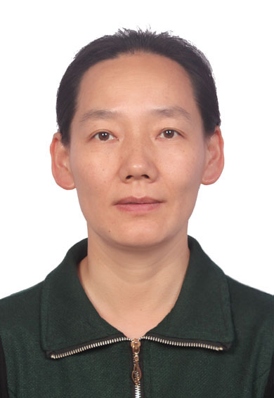 Qing Cai (蔡晴)合作论文数College of Materials Science and Engineering, Beijing University of Chemical Technology10
 Yuanhua Lin (林元华)合作论文数School of Materials Science and Engineering, Tsinghua University10
 Xiaoping Yang (杨小平)合作论文数State Key Laboratory of Organic-Inorganic Composites, School of Materials Science and Engineering, Beijing University of Chemical Technology;College of Mechanical and Electrical Engineering, Beijing University of Chemical Technology;National Carbon Fiber Engineering Technology Research Center, School of Materials Science and Engineering, Beijing University of Chemical Technology10Qiannan Sun合作论文数9
 Yunfan Zhang合作论文数Programming Languages Lab, Software Engineering Institute, School of Computer Science, Peking University9
 Xinzhi Wang (王新知)合作论文数Peking University School of Stomatology8
 Xiang-Yu Kong (孔祥玉)合作论文数Technical Institute of Physics and Chemistry, Chinese Academy of Sciences7Jingjing Deng合作论文数7
 Liping Wen (闻利平)合作论文数Technical Institute of Physics and Chemistry, Chinese Academy of Sciences;University of Chinese Academy of Sciences7
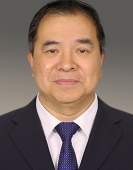 Lin Guo (郭林)合作论文数School of Chemistry, Beihang University7
 Yaru Guo (郭亚茹)合作论文数National Center for Stomatology, Peking University School and Hospital of Stomatology6Zhewen Hu (胡哲文)合作论文数6Xiaohan Dai合作论文数6song meng合作论文数6Yunyang Bai合作论文数Peking Univ, Dept Geriatr Dent, Sch & Hosp Stomatol, Beijing 100081, Peoples R China6Liying Peng合作论文数6Zuohui Xiao合作论文数6Qi Ma合作论文数6Xiaoju Mo合作论文数5Siqin Ma合作论文数5Yusi Guo合作论文数5
 Chuanliang Feng (冯传良)合作论文数School of Materials Science and Engineering, Shanghai Jiao Tong University5Wenhsuan Lu合作论文数4Xiaopei Chi合作论文数4Jinqi Wei合作论文数4Huimin Zheng合作论文数4
 Mengting Si合作论文数4Congcong Zhu合作论文数Chinese Acad Sci, Tech Inst Phys & Chem, Key Lab Bioinspired Mat & Interfacial Sci, Beijing 100190, Peoples R China4
 Jinxing Zhang (张金星)合作论文数Department of Physics, Beijing Normal University4
 Jinying Liu (刘进营)合作论文数School of Materials, Henan University4Wang Ying合作论文数4
 Hewei Zhao (赵赫威)合作论文数School of Chemistry, Beihang University4
 Chengyun Ning (宁成云)合作论文数School of Materials Science and Engineering, South China University of Technology4
 Hongjie Zhang (张洪杰)合作论文数Department of Chemistry, Tsinghua University;Changchun Institute of Applied Chemistry, Chinese Academy of Sciences4
 Fan Wang (王帆)合作论文数State Key Laboratory of Rare Earth Resource Utilization, Changchun Institute of Applied Chemistry, Chinese Academy of Sciences4
 Yang Shen (沈洋)合作论文数School of Materials Science and Engineering, Tsinghua University4
 Shutao Wang (王树涛)合作论文数Technical Institute of Physics and Cehmistry, Chinese Academy of Sciences;University of Chinese Academy of Sciences4
 Lili Chen (陈莉莉)合作论文数Hospital of Stomatology, Sun Yat-sen University4




