The lack of physiologically relevant in vitro models remains a limitation in liver transplantation research. Progress in organ-on-a-chip technologies enables the generation of clinically translatable data in vitro. A vascularized liver tissueoid-on-a-chip (LToC) model is engineered to replicate human liver tissue's structural and functional features for modeling liver regeneration and allograft rejection. The LToC comprises a microfluidic device containing donor-matched human hepatic progenitor cells and intrahepatic portal vein endothelial cells embedded in a fibrin matrix and maintained in dynamic culture for 49 days. The system supports self-assembly into a perfusable microvascular network and liver lobule-like architecture, with >95% cell viability, stable vascular integrity, and active hepatic function (albumin, urea, complement factors, and hepatocyte growth factor secretion). The mature tissueoid includes hepatocytes (CK18+, albumin+, CYP2D6+), cholangiocytes (CK19+, EPCAM+), Kupffer cells (CD68+), stellate cells (PDGFR-β+), and endothelial cells (CD31+). Perfusion with allogeneic T cells induces cellular rejection, characterized by decreased viability, endothelial disruption, hepatic marker loss, HLA-I upregulation, and a proinflammatory cytokine response (IL-6, TNF-α, IL-1β, IFN-γ, granzyme A and B, and perforin). The LToC provides a physiologically relevant platform for studying immune-mediated liver injury, tissue regeneration, and allograft rejection, with potential applications in immunosuppressive drug testing and personalized transplant medicine.
Bulk scaffold-supported current wound dressings often fail to heal the skin wounds, particularly chronic and infected wounds, because it lacks the microarchitecture and bioactivity needed for rapid, scar-minimized repair. This review highlights the rise of modular biomaterials, especially microgels, porous microspheres, and microporous annealed particles (MAPs), scaffolds as next-generation platforms for wound healing. Here, we showcased key modular fabrication strategies, like emulsification, microfluidics, electrospraying, and light-based 3D printing. Then we showed how modularity enables different aspects like injectability, defect conformability, microporosity, and improved cell infiltration, and vascularization compared with conventional bulk scaffolds. We further review applications of MAPs, porous microsphere, and injectable microgels for controlled therapeutic delivery, immunomodulation, and infection management. Finally, we outline translational considerations and emerging trends, including scalable manufacturing and AI/ML-guided biomaterial design, to accelerate personalized wound care.
Burn wounds are a common traumatic injury that impair cellular function and hinder the healing process, often resulting in significant skin loss. While autologous skin grafting is considered the gold standard for treating burns, its widespread use is limited due to donor site morbidity and the requirement for large amounts of tissue. Traditional wound dressings and treatments often fail to ensure complete recovery. Being initially FDA-approved to treat multiple sclerosis, 4-aminopyridine (4-AP) has also been shown to accelerate burn wound closure by modulating keratinocytes and fibroblasts when administered systemically. However, prolonged systemic use of 4-AP can lead to significant side effects. In this study, we aimed to repurpose 4-AP for treating skin burn wounds by delivering it topically using a laponite-gelatin gel formulation. This method allows for non-invasive and localized drug delivery on the burn wound site. We evaluated the physical properties of the 4-AP gel, including shear thinning behavior, drug release kinetics, cytocompatibility, and functional wound closure (48hr) using a scratch assay (>90%). Moreover, our in vivo experiments showed that the 4-AP loaded gel accelerates wound healing by reducing inflammation, thereby enhancing re-epithelialization and angiogenesis, and promoting the transformation of fibroblasts into myofibroblasts. Burn closure in the 4-AP gel group was consistently higher than the control gel from day 6 through day 21, reaching near-complete healing by day 21, whereas control-treated wounds remained partially open. This enhanced closure correlated with accelerated collagen remodeling, as the 4-AP gel significantly increased collagen type I and III deposition and their ratio compared to controls (438% vs. 267%; 288% vs. 215%; ratio 1.7 vs. 1.2; ∗P < 0.05 to ∗∗∗P < 0.0002), indicating improved matrix maturation during burn healing. This novel application of the 4-AP gel could offer a promising alternative to current burn wound therapies, potentially leading to improved outcomes for burn patients.
Over the past decade, piezoelectric biomaterials have emerged as powerful platforms for tissue repair and regeneration by enabling self-generated, minimally invasive bioelectric stimulation. Among them, skin wound healing has become a prominent area of research due to the tissue's accessibility and clinical relevance. Specifically, the microcurrents powered by piezoelectric biomaterials closely mimic the endogenous electrical cues present in healthy skin. These bioelectrical signals regulate multiple stages of wound healing, including cell migration and proliferation, angiogenesis, extracellular matrix formation, and immune modulation. This review will provide a comprehensive overview of piezoelectric biomaterials for skin wound healing and examine the physiological and biochemical roles of the naturally occurring electric fields in healthy skin and injured skin. By integrating these perspectives, we highlight how piezoelectric scaffolds can power skin tissue repair by recapitulating native bioelectric signaling. We further discuss current challenges, translational opportunities, and future directions toward the development of next-generation electroactive wound dressings for the treatment of acute and chronic wounds.
Severe hypoxia within thick bioengineered tissues critically impairs cell viability and function, limiting their application in organ-scale engineering and regenerative medicine. Current methods for oxygen delivery often fall short of providing sustained oxygenation before neovascularization. Here, we introduce a smart self-oxygenating tissue (SSOT) platform that leverages a bio-ionic liquid (BIL)-functionalized biocompatible hydrogel electrolyte for localized and controlled oxygen generation via electrolysis. Comprehensive characterization of the system confirmed stability and electrochemical properties, with molecular dynamics simulations demonstrating that BIL enhances oxygen release. In vitro, the SSOT platform maintains cell viability and promotes vascularization under severe hypoxic conditions. Diabetic wound healing studies using mouse models showed that an SSOT patch accelerates wound closure in chronic and non-chronic wounds. These findings highlight the potential of electrolysis-driven methods for providing on-demand and sustained oxygen delivery, essential for the development of functional living tissues and ultimately organs. Hypoxia of bioengineered tissue limits cell viability and is a key consideration in tissue engineering. Here, a smart self-oxygenating tissue based on a bio-ionic liquid functionalized hydrogel electrolyte enables oxygen release, and accelerates wound healing in mouse models.
Peripheral nerve injury (PNI) is a common problem worldwide. PNI can lead to loss of sensory and motor functions, chronic neuropathic pain, and mental health issues, significantly impacting the patients' quality of life. Recent studies revealed that, beyond the topical injury site at peripheral nerves, PNIs can also induce dysfunctions in the central nervous system by causing maladaptive plasticity, which will result in exaggeration and exacerbation of the pathological condition caused by the primary injuries. The typical therapy strategies for traumatic PNI treatment are using sutures, nerve autografts, or conduits in cases requiring surgical intervention as well as applying physical-based rehabilitation to facilitate functional recovery. However, the functional restoration is generally unsatisfactory due to insufficient regeneration and long-lasting maladaptive neuroplasticity in the nervous system. In this review, we summarized various neurotrophic factors and neuroprotective agents that have been extensively studied as adjuvant therapies to enhance recovery efficiency after PNIs in the last two decades. Particularly, with the rapid development of biomaterials and bioengineering, controllable drug delivery techniques have shown great potential to maintain the drug bioactivity and, consequently, prolonging the therapeutic effects. Additionally, we explored the virus-based gene delivery technique, which has been used to transduce neural cells for enhancing nerve regeneration. Finally, we discussed current challenges, including inadequate motor function restoration, poorly defined rehabilitation protocols, unresolved chronic inflammation, and limited understanding of macrophage dynamics. We also offered perspectives on integrating various approaches to develop effective and comprehensive treatment strategies for PNIs.
Burn wounds are a common traumatic injury that impair cellular function and hinder the healing process, often resulting in significant skin loss. While autologous skin grafting is considered the gold standard for treating burns, its widespread use is limited due to donor site morbidity and the requirement for large amounts of tissue. Traditional wound dressings and treatments often fail to ensure complete recovery. Being initially FDA-approved to treat multiple sclerosis, 4-aminopyridine (4-AP) has also been shown to accelerate burn wound closure by transforming keratinocytes and fibroblasts when administered systemically. However, prolonged systemic use of 4-AP can lead to significant side effects. In this study, we aimed to repurpose 4-AP for treating skin burn wounds by delivering it topically using a laponite-gelatin gel formulation. This method allows for non-invasive and localized drug delivery on burn wound site. We evaluated the physical properties of the 4-AP gel shear thinning behavior, drug release kinetics, biocompatibility, and functional wound closure using a scratch assay. Moreover, our in vivo experiments showed that the 4-AP loaded gel accelerates wound healing by enhancing re-epithelialization and hair follicle regeneration and promoting fibroblast to myofibroblast transformation, which supports extracellular matrix remodeling after skin burns. This novel application of the 4-AP gel could offer a promising alternative to current burn wound therapies, potentially leading to improved outcomes for burn patients.
3D bioprinting has emerged as a transformative technology in tissue engineering, significantly impacting the creation of patient-specific tissues to enhance clinical outcomes. Despite its rapid advancement, translating this technology from bench to bedside remains a critical clinical need. New bioprinting approaches, such as handheld printers or robotic arm-driven in-situ biofabrication techniques, have emerged as promising alternatives. These advancements enable the reconstruction of damaged tissue directly on living anatomical structures, offering adaptability and precise matching to the affected area. The integration of biomaterials, tissue engineering principles, and digital technologies, particularly robotics, has garnered substantial interest from both academic and industrial sectors, highlighting its potential for clinical applications. However, challenges persist, including refining bioink formulations, adjusting mechanical properties, facilitating in situ crosslinking, and accurately mimicking the extracellular matrix. This review explores the cutting-edge frontier of in situ 3D bioprinting for tissue regeneration, utilizing both handheld and robotic arm-assisted 3D printers. It systematically examines the relative advantages, disadvantages, challenges, and prospects of this technology as it transitions from bench side to bed side.
Tissue engineering techniques and particularly in situ bioprinting using handheld devices and robotic systems have recently demonstrated promising outcomes to address volumetric muscle loss injuries. Nevertheless, these approaches suffer from insufficient printing precision and/or lack of quantitative analysis of the thickness and uniformity of bioprinted constructs (BPCs) - which are critical for ensuring cell viability and growth. To address these limitations, in this study, we present a framework for robotic bioprinting and complementary vision-based algorithms to quantitatively analyze thickness and uniformity of BPCs with curved geometries. The performance of the proposed robotic bioprinting and complementary algorithms has been thoroughly evaluated using various simulation and experimental studies on BPCs with constant and variable thicknesses. The results clearly demonstrate the remarkable and accurate performance of the proposed method in calculating the thickness and its variations along the geometry of the BPCs.
Controlling cellular organization in hydrogels is of great interest in tissue engineering and regenerative medicine. In the body, cell organization is regulated by aligned extracellular matrices, such as collagen fibers. However, generating patterned extracellular matrix fibers in hydrogels, such as microfabricated gels, is not easily accomplished. Here, filamented-light (FLight)-based 3D microfabrication is used to fabricate microgels with precise internal architecture to direct cellular organization. It is demonstrated that fibrillated rod-shaped microgels encapsulating C2C12 muscle cells promote highly aligned myotube formation, offering potential as mini-injectable muscle tissues for minimally invasive muscle loss therapies. Furthermore, photoreceptor cells encapsulated in rod-shaped microgels generated structures that mimicked the outer retina. Moreover, these microgels can be used as injectable scaffolds, both in vitro and in vivo, where they facilitate angiogenesis when conjugated with QK peptide. Overall, this technique can be used to generate microgels with precise internal architecture thus providing a potentially significant tool for engineering tissue-like structures.
Volumetric muscle loss (VML) presents a significant challenge in tissue engineering due to the irreparable nature of extensive muscle injuries. In this study, we propose a novel approach for VML treatment using a bioink composed of silk microfiber-reinforced silk fibroin (SF) hydrogel. The engineered scaffolds are predesigned to provide structural support and fiber alignment to promote tissue regeneration in situ. We also validated our custom-made handheld 3D printer performance and showcased its potential applications for in situ printing using robotics. The fiber contents of SF and gelatin ink were varied from 1 to 5 %. Silk fibroin microfibers reinforced ink offered increased viscosity of the gel, which enhanced the shape fidelity and mechanical strength of the bulk scaffold. The fiber-reinforced bioink also demonstrated better cell-biomaterial interaction upon printing. The handheld 3D printer enabled the precise and on-demand fabrication of scaffolds directly at the defect site, for personalized and minimally invasive treatment. This innovative approach holds promise for addressing the challenges associated with VML treatment and advancing the field of regenerative medicine.
Smart biomaterials have significantly impacted human healthcare by advancing the development of medical devices designed to function within human tissue, mimicking the behavior of natural tissues. While the intelligence of biomaterials has evolved from inert to active over the past few decades, smart biomaterials take this a step further by making their surfaces or bulk respond based on interactions with surrounding tissues, imparting outcomes similar to natural tissue functions. This interaction with the surrounding tissue helps in creating stimuli-responsive biomaterials, which can be useful in tissue engineering, regenerative medicine, autonomous drug delivery, orthopedics, and much more. Traditionally, material engineering focused on refining the static properties of biomaterials to accommodate them within the body without evoking an immune response, which was a major obstacle to their unrestricted operation. This review highlights and explains various engineering approaches currently under research for developing stimuli-responsive biomaterials that tune their outcomes based on responses to bodily factors like temperature, pH, and ion concentration or external factors like magnetism, light, and conductivity. Applications in soft and hard tissue engineering, 4D printing, and scaffold design are also discussed. The advanced application of microfluidics, like organ-on-a-chip models, extensively benefits from the intrinsic smart properties of biomaterials, which are also discussed below. The review further elaborates on how smart biomaterial engineering could revolutionize biosensor applications, thereby improving patient care quality. We delineate the limitations and key challenges associated with biomaterials, providing insights into the path forward and outlining future directions for developing next-generation biomaterials that will facilitate clinical translation.
In this study, we introduce a new concept for reconstruction of Volumetric Muscle Loss (VML) injuries and propose the spatial robotic embedded bioprinting technique. As opposed to the traditional layer-by-layer printing, we leverage the support-free nature of embedded bioprinting to print spatial and complex structures of fascicles in a fusiform muscle. To demonstrate feasibility of this concept, we first propose our robotic bioprinting framework including a robotic arm integrated with a custom-designed bioprinting injector. Complementary motion planning algorithms uniquely designed for this printing task are further proposed. Moreover, the effect of embedded bioprinting parameters, as well as the supporting bath and injecting materials compatibility on the uniformity and quality of the printed constructs has been analyzed. Finally, we perform a case study by printing a fusiform muscle-shape construct using the proposed concept and algorithms, and evaluate the quality of the printed structure.
The structure and design flexibility of aerogels make them promising for soft tissue engineering, though they tend to come with brittleness and low elasticity. While increasing crosslinking density may improve mechanics, it also imparts brittleness. In soft tissue engineering, resilience against mechanical loads from mobile tissues is paramount. We report a hybrid aerogel that consists of self-reinforcing networks of micro- and nanofibers. Nanofiber segments physically entangle microfiber pillars, allowing efficient stress distribution through the intertwined fiber networks. We show that optimized hybrid aerogels have high specific tensile moduli (~1961.3 MPa cm 3 g −1 ) and fracture energies (~7448.8 J m −2 ), while exhibiting super-elastic properties with rapid shape recovery (~1.8 s). We demonstrate that these aerogels induce rapid tissue ingrowth, extracellular matrix deposition, and neovascularization after subcutaneous implants in rats. Furthermore, we can apply them for engineering soft tissues via minimally invasive procedures, and hybrid aerogels can extend their versatility to become magnetically responsive or electrically conductive, enabling pressure sensing and actuation.
In situ bioprinting has been identified as a promising tissue engineering technique for treating volumetric muscle loss (VML) injuries. However, the success of this procedure significantly depends on the uniform and precise deposition of cells contributing to the regeneration of muscles. To address this critical need, in this work, we present design and quantitative evaluation of a novel autonomous in situ bioprinting surgical robotic framework that can be used with a generic bioprinting material. The proposed framework consists of three main components: (i) a bioprinting tool integrated with a seven-degree-of-freedom robotic manipulator to perform a precise autonomous bioprinting procedure; (ii) a unique 3D visual measurement framework comprised of a high-accuracy structured light camera with complementary 2D/3D computer vision algorithms-to enable online and accurate measurement and reconstruction of the bioprinted constructs; and (iii) a quantitative evaluation module with novel assessment metrics-to characterize and evaluate the performance of the bioprinting process toward finding optimal bioprinting parameters. To ensure the biological functionality of a printed construct using our robotic system, we performed 90 experiments and identified optimal bioprinting parameters using the proposed novel assessment metrics.
Glioblastoma (GBM) is an aggressive form of cancer, comprising ≈80% of malignant brain tumors. However, there are no effective treatments for GBM due to its heterogeneity and the presence of the blood-brain barrier (BBB), which restricts the delivery of therapeutics to the brain. Despite in vitro models contributing to the understanding of GBM, conventional 2D models oversimplify the complex tumor microenvironment. Organ-on-a-chip (OoC) models have emerged as promising platforms that recapitulate human tissue physiology, enabling disease modeling, drug screening, and personalized medicine. There is a sudden increase in GBM-on-a-chip models that can significantly advance the knowledge of GBM etiology and revolutionize drug development by reducing animal testing and enhancing translation to the clinic. In this review, an overview of GBM-on-a-chip models and their applications is reported for drug screening and discussed current challenges and potential future directions for GBM-on-a-chip models.
Chronic wounds resulting from diabetes, pressure, radiation therapy, and other factors continue to pose significant challenges in wound healing. To address this, this study introduces a novel hybrid fibroin fibrous scaffold (FFS) comprising randomly arranged fibroin fibers and vertically aligned cryogel fibers (CFs). The fibroin scaffold is efficiently degummed at room temperature and simultaneously formed a porous structure. The aligned CFs are produced via directional freeze-drying, achieved by controlling solution concentration and freezing polymerization temperature. The incorporation of aligned CFs into the expanded fibroin fiber scaffold leads to enhanced cell infiltration both in vitro and in vivo, further elevating the hybrid scaffold's tissue compatibility. The anti-inflammatory peptide 1 (AP-1) is also conjugated to the hybrid fibrous scaffold, effectively transforming the inflammatory status of chronic wounds from pro-inflammatory to pro-reparative. Consequently, the FFS-AP1+CF group demonstrates superior granulation tissue formation, angiogenesis, collagen deposition, and re-epithelialization during the proliferative phase compared to the commercial product PELNAC. Moreover, the FFS-AP1+CF group displays epidermis thickness, number of regenerated hair follicles, and collagen density closer to normal skin tissue. These findings highlight the potential of random fibroin fibers/aligned CFs hybrid fibrous scaffold as a promising approach for skin tissue filling and tissue regeneration.
Copper-cystine-based high aspect ratio structures (CuHARS) possess exceptional physical and chemical properties and exhibit remarkable biodegradability in human physiological conditions. Extensive testing has confirmed the biocompatibility and biodegradability of CuHARS under diverse biological conditions, making them a viable source of essential Cu2+. These ions are vital for catalyzing the production of nitric oxide (NO) from the decomposition of S-nitrosothiols (RSNOs) found in human blood. The ability of CuHARS to act as a Cu2+ donor under specific concentrations has been demonstrated in this study, resulting in the generation of elevated levels of NO. Consequently, this dual function makes CuHARS effective as both a bactericidal agent and a promoter of angiogenesis. In vitro experiments have shown that CuHARS actively promotes the migration and formation of complete lumens by redirecting microvascular endothelial cells. To maximize the benefits of CuHARS, they have been incorporated into biomimetic electrospun poly(ε-caprolactone)/gelatin nanofiber aerogels. Through the regulated release of Cu2+ and NO production, these channeled aerogels not only provide antibacterial support but also promote angiogenesis. Taken together, the inclusion of CuHARS in biomimetic scaffolds could hold great promise in revolutionizing tissue regeneration and wound healing.
Diabetic foot ulcers (DFUs) are a significant challenge in the clinical care of diabetic patients, often necessitating limb amputation and compromising the quality of life and life expectancy of this cohort. Minimally invasive therapies, such as modular scaffolds, are at the forefront of current DFU treatment, offering an efficient approach for administering therapeutics that accelerate tissue repair and regeneration. In this study, we report a facile method for fabricating granular nanofibrous microspheres (NMs) with predesigned structures and porosities. The proposed technology combines electrospinning and electrospraying to develop a therapeutic option for DFUs. Specifically, porous NMs were constructed using electrospun poly(lactic-co-glycolic acid) (PLGA):gelatin short nanofibers, followed by gelatin cross-linking. These NMs demonstrated enhanced cell adhesion to human dermal fibroblasts (HDF) during an in vitro cytocompatibility assessment. Notably, porous NMs displayed superior performance owing to their interconnected pores compared to nonporous NMs. Cell-laden NMs demonstrated higher Young's modulus values than NMs without loaded cells, suggesting improved material resiliency attributed to the reinforcement of cells and their secreted extracellular matrix. Dynamic injection studies on cell-laden NMs further elucidated their capacity to safeguard loaded cells under pressure. In addition, porous NMs promoted host cell infiltration, neovascularization, and re-epithelialization in a diabetic mouse wound model, signifying their effectiveness in healing diabetic wounds. Taken together, porous NMs hold significant potential as minimally invasive, injectable treatments that effectively promote tissue integration and regeneration.
 Jingwei Xie合作论文数Department of Surgery, College of Medicine, University of Nebraska Medical Center30
 Il Kim合作论文数BK21 PLUS Center for Advanced Chemical Technology, Pusan National University27
 Alec McCarthy合作论文数College of Medicine, University of Nebraska Medical Center26
 Shixuan Chen (陈世萱)合作论文数Department of Histology and Embryology, Southern Medical University15
 Renjith P Johnson合作论文数The WCU Center for Synthetic Polymer Bioconjugate Hybrid Materials, Pusan National University13
 Yajuan Su (粟雅娟)合作论文数Integrated Circuit Advanced Process R&D Center, Institute of Microelectronics, Chinese Academy of Sciences;University of Chinese Academy of Sciences11
 Ali Khademhosseini合作论文数Omeat;Stealth Startup11
 Hongjun Wang合作论文数Department of Biomedical Engineering, Charles V. Schaefer, Jr. School of Engineering and Science, Stevens Institute of Technology;Center for Healthcare Innovation, Stevens Institute of Technology9
 Nafiseh Moghimi合作论文数†Department of Chemistry, University of Waterloo7
 Young-Il Jeong合作论文数Department of Microbiology, Pusan National University College of Nature Science7Hong Suk Suh合作论文数7
 Saji Uthaman合作论文数Yenepoya Research Centre7Meenakshi Kamaraj合作论文数6Rimesh Augustine合作论文数6S M Shatil Shahriar合作论文数6Navatha Shree Sharma合作论文数College of Medicine, University of Nebraska Medical Center6
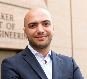 Farshid Alambeigi合作论文数Walker Department of Mechanical Engineering, Cockrell School of Engineering, University of Texas at Austin6Kang Dae Hwan合作论文数6Chung-Wook Chung合作论文数5
 Akshay Joshi合作论文数Ctr Biomed Engn, Indian Inst Technol Delhi5
 Min Heo合作论文数Pusan National Univ.5Anik Karan合作论文数5huiju park合作论文数5Carlson M A合作论文数5
 Vadim Jucaud合作论文数5
 Najafabadi Alireza Hassani合作论文数Brigham and Women's Hospital, Harvard Medical School;Harvard-MIT Division of Health Sciences and Technology, Massachusetts Institute of Technology;Department of Chemistry, Amirkabir University of Technology;Harvard-MIT Division of Health Sciences and Technology, Amirkabir University of Technology5In-Kyu Park合作论文数4Marvin Mecwan合作论文数Terasaki Institute for Biomedical Innovation4
 Reihaneh Haghniaz合作论文数These authors contributed equally to this work.4
 Kalpana Mandal合作论文数Terasaki Inst Biomed Innovat4
 Mohsen Akbari合作论文数Faculty of Medicine, The University of British Columbia4
 Yu S Zhang合作论文数Department of Medicine, Harvard Medical School;Harvard Stem Cell Institute;Brigham and Women's Hospital4Hye Ri Lee合作论文数4Shelbie Weihs合作论文数3Lorenzo Saldana合作论文数3
 Syed Muntazir Andrabi合作论文数3
 Demi Brown合作论文数Department of Surgery - Transplant and Mary & Dick Holland Regenerative Medicine Program, University of Nebraska Medical Center3
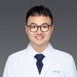 Ze-Yu Luo (罗泽宇)合作论文数Department of Medicine Brigham and Women's Hospital, Harvard Medical School3
 Jaeho Kim合作论文数Department of Physiology, School of Medicine, Pusan National University3
 Saurabh Sharma合作论文数Terasaki Inst Biomed Innovat3
 Surjendu Maity合作论文数Terasaki Institute for Biomedical Innovation3
 Danial Khorsandi合作论文数Terasaki Institute for Biomedical Innovation3
 Hossein Heidari合作论文数University of California3
 Inkyu Park合作论文数Department of Mechanical Engineering, College of Engineering, Korea Advanced Institute of Science and Technology3Teusink Matthew J合作论文数3
 Hongyu Chen (陈虹宇)合作论文数Department of Chemistry, School of Science, Westlake University3
 Sunil Kumar Boda合作论文数Helmholtz-Institute for Biomedical Engineering-Biointerface Laboratory, RWTH Aachen University3
 Fatemeh Zehtabi合作论文数University of Manitoba, University of Manitoba3
 Bin Duan合作论文数Division of Cardiology, Department of Internal Medicine, University of Nebraska Medical Center3




