围手术期血糖水平异常现象是影响患者预后的重要因素.目前,已有较多关于外科手术围手术期血糖管理指南,但对于口腔颌面外科手术围手术期血糖异常情况尚未有规范的管理方案.根据已发表的相关指南、共识及文献,并结合颌面外科的特点及我们的经验,本文主要从术前高血糖的筛查、评估,术中血糖安全管理,术后血糖水平的监测及治疗等方面规范了标准化血糖管理操作规程,为颌面外科围手术期血糖管理提供参考.
目的:探讨死亡与并发症病例讨论会(mortality and morbidity conferences,M&MCs,双M会议)在口腔颌面肿瘤专业临床教学中的应用效果.方法:本研究于第四军医大学口腔医院颌面肿瘤科开展,每月初第一周以PPT汇报的形式举行双M会议.通过对发生不良事件病例的回顾、分析,查找问题,总结解决办法,进而提升医护人员业务水平.结果:通过开展双M会议,提升了各级医师的临床经验和业务水平,显著降低了临床不良事件发生率.结论:双M会议是加强口腔颌面肿瘤科住院医师临床教学效果,提升医疗质量的良好方式.
口腔颌面肿瘤切除与修复“平衡点”的设立来自于临床实践,肿瘤种类不同、患者个体不同其平衡点也不同,是个体化的平衡点.肿瘤切除与缺损修复是一个矛盾的两个方面,是对立统一的关系,平衡点是肿瘤切除与修复辩证统一的具体体现.切除与修复平衡点决策遵循的基本方法是循证医学,标准流程是由临床医生,按照患者的具体情况和要求,获取目前国际上最新的研究证据,结合临床经验确定肿瘤手术切除与修复的平衡点,并在医疗活动中实现.
Objective:To study the bone regeneration capacity of the complex of bone marrow mesenchymal stem cells(BMSCs) and platelet rich plasma (PRP) with decellularized cartilage matrix (DCM).Methods:BMSCs were isolated from young rabbit and cultured;PRP were prepared from the fresh blood of rabbit and the DCM were come from the fresh ears of rabbits and processed into a mixture of BMSCs-PRP-DCM.The mixture was injected subcutaneously into nude mice,8 weeks after injection bone regeneration was examine by HE staining and Massons staining decellularization.Results:The BMSCs show good differentiation capacity in vitro and the cartilage fragments were well decellularized.Excellent endochondral ossification ability of the complex was observed by in vivo experiment.Conclusion:The BMSCs-PRP-DCM complex has good capacity of endochondral ossification.
Objective:To introduce ADSCs into chondrocyte brick enriched PRP gel for the promotion of cartilage formation.Methods:Chondrocytes were cultured into cell sheets and fragmented into chondrocyte bricks.PRP was prepared from fresh blood.ADSCs and BMSCs were respectively cultured and mixed with chondrocyte bricks enriched PRP gel constructs.The constructs of the 2 groups were respectively injected into the back of nude mice subcutaneously.After 12-weeks incubation in vivo,constructs were harvested for morphologic observation,HE staining,safranin-O and TB staining.Results:The harvested chondrocyte bricks were composed of chondrocytes and abundant extracellular matrix.Constructs from the 2 groups achieved tissue remodeling and cartilage regeneration observed by Safranin-O and TB staining.Conclusion:ADSCs can replace BMSCs as seed cells and can promote cartilage regeneration of injectable chondrocyte bricks enriched PRP gel construct.
>颈淋巴结清扫术是治疗头颈部恶性肿瘤的常用手术。对于口腔颌面外科医生来说,熟识头颈部解剖结构是掌握颈淋巴结清扫术的前提,然而不同个体之间头颈部的解剖结构往往存在一定差异。本文报道1例颈淋巴结清扫过程中所见的颈内动脉走形变异。1.病例资料:(1)主诉及现病史:患者男性,55岁,因左上颌后牙缺牙区外生性肿物20 d就诊于第四军医大学口腔医学院口腔颌面外科。肿物似"枣核"大小,无疼痛等不适,曾静脉注射抗
OBJECTIVE:To explore the feasibility of constructing tissue engineered trachea-like cartilage graft in vitro by using bone marrow stromal cells (BMSCs) sheet and PLGA internal support.METHODS:Rabbit BMSCs were expanded and induced by transforming growth factor-1 to improve chondrocyte phenotype of BMSCs. BMSCs sheets were obtained by continuous culture and wrapped the PGLA scaffold in the shape of cylinder. The constructs were incubated in spinner flask for 8 weeks and cartilage formation was investigated by gross inspection, histology, glycosaminoglycan and mechanical strength content.RESULTS:After in vitro culture, cartilage like tissue in cylindrical shape had been regenerated successfully. Stiff, shiny, pearly opalescence tissues were observed. Histological analysis showed engineered trachea cartilage consisted of evenly spaced lacunae embedded in matrix, cells stationed in the lacunae could be noticed clearly. Safranin-O staining on the sections showed homogenous and positive red staining, which demonstrated that the engineered tissue was rich in proteoglycans.CONCLUSIONS:Based on the cell sheet and internal support strategy, trachea-like cartilage in cylindrical shape could be successfully fabricated which provided a highly effective cartilage graft substitute and could be useful in many situations of trachea-cartilage loss encountered in clinical practice.
BACKGROUND:Platelet-rich plasma (PRP) has been applied to promote bone healing and developed as a novel material for bone regeneration. This study aimed to investigate the feasibility of PRP carrier to deliver bone marrow derived stromal cells (BMSCs) and regenerate bone tissues to reconstruct critical bone defects in rabbits.METHODS:Critical sized defect were made on eighteen rabbits' crania and treated by different composites: BMSCs/PRP (n=6); Autogenous particulate cancellous bone group (n=6) and PRP alone group (n=6). The defects were evaluated by gross observation, radiographic examination, histological examination, and mechanical examination at 12 weeks postoperatively.RESULTS:The results showed that repair of bone defect was the least in PRP alone group, and significant new bone formation could be observed in BMSCs/PRP group and particulate cancellous bone group, radiopacity area in BMSCs/PRP group attained 76.5%, which was in the same range of that in autogenous particulate cancellous bone group (82.4% in radiopacity area), compressive strength of engineered bone in BMSCs/PRP group attained 71% of that in autogenous particulate cancellous bone group (p<0.05).CONCLUSION:These data implicated that BMSCs delivered from PRP gel can repair bony defect in immunocompetent animals, and the tissue engineered bone in BMSCs/PRP group is comparable to autogenous particulate cancellous bone group for the repair of critical-sized bone defect.
BACKGROUND Platelet-rich plasma (PRP) has been applied to promote bone healing and developed as a novel material for bone regeneration. This study aimed to investigate the feasibility of PRPcarrier to deliver bone marrow derived stromal cells (BMSCs) and regenerate bone tissues to reconstruct critical bone defects in rabbits. METHODS Critical sized defect were made on eighteen rabbits’crania and treated by different composites: BMSCs/PRP(n=6); Autogenous particulate cancellous bone group (n=6)and PRP alone group (n=6). The defects were evaluated by gross observation, radiographic examination, histological examination, and mechanical examination at 12 weeks postoperatively. RESULTS The results showed that repair of bone defect was the least in PRPalone group, and significant new bone formation could be observed in BMSCs/PRPgroup and particulate cancellous bone group, radiopacity area in BMSCs/PRPgroup attained 76.5%, which was in the same range of that in autogenous particulate cancellous bone group (82.4% in radiopacity area), compressive strength of engineered bone in BMSCs/PRP group attained 71% of that in autogenous particulate cancellous bone group(p<0.05). CONCLUSION These data implicated that BMSCs delivered from PRPgel can repair bony defect in immunocompetent animals, and the tissue engineered bone in BMSCs/PRPgroup is comparable to autogenous particulate cancellous bone group for the repair of critical-sized bone defect.
Objective:To discuss the relationship between tumor origin and occurrence in different areas of skull base and the surgical operation methods in this areas.Methods: Total of 232 cases were selected and studied on which kinds of tumor easy to encroach on different areas of base of skull and to these tumor which surgical program should be used.Results: The number of tumors which reached anterior cranial fossa was 40 cases which including maxillary bone squamous cell carcinoma(23),fleshy tumor(7),colloid tumor(3),adamantinoblastoma(7).The number of tumors which grew out of the line between foramen ovale and styloid process(including fossa infratemporalis) was 70 cases which including ascending branch malignant tumor(43),recurrence of parotid malignant tumor(9)(adenoid cystic carcinoma 4 cases),recurrence of skin cancer of temporal and ear region(3).Condyle osseous tumor and osteochondroma(10),tumors originated from nerves(5).The number of tumors which grew into the line between foramen ovale and styloid process was 114 cases which including myoschwannoma(60),durosarcoma(9),neurosarcoma(12),poorly differentiated cancer(4),carotid body tumor(16),Tumors originated from parotid gland(13).The number of tumor which reached proximally foramen magnum was 8 cases:durosarcoma(2),spinal meningioma(3),myoschwannoma(3).Conclusion: Tumors which easy to encroach the base line of skull were different in origin and occurrence.And it is helpful to select a reasonable operation method according to tumor origin and occurrence.
OBJECTIVE:To develop a scaffold material containing collagen 1 and sodium hyaluronate for the cartilage tissue engineering and to evaluate its biocompatibility by using the rabbit chondrocytes derived from a mandibular condylar process.METHODS:The porous matrices containing collagen 1 and sodium hyaluronate were fabricated by the freeze-drying technique and were crosslinked by using 1-ethyl-3-(3-dimethyl aminopropyl) carbodiimide (EDC). The microstructure of the scaffold was observed under the scanning electron microscope (SEM), and the enzymatic degradation test was performed to compare the ability of the scaffold resistance to collagenase before and after the crosslinking. The chondrocytes from the rabbits' condylar process were isolated and cultured before they were seeded into the scaffold, and cell attachment and proliferation were measured by the cell count 1, 3, 5, 7 and 10 days after the cell being seeded; then, the biocompatibility of the scaffold was evaluated by the light microscopic examination, histological examination, and the SEM exmination.RESULTS:The porous structure of the scaffold facilitated the penetration and attachment of the seeded cells. The porosity was 83.7% and the pore size was 100-120 microm. The cell number increased from 3.7 x 10(4) per scaffold 1 day after the cell being seeded to 8.2 x 10(4) per scaffold 10 days after the cell being seeded. The crosslinking treatment could significantly enhance the scaffold resistance to the collagenase activity. The examinations under the light microscope and SEM indicated that the chondrocyte adhered and spread well on the scaffold, and the extracellular matrices were also observed around the chondrocytes.CONCLUSION:The porous scaffold composed of collagen I and hyaluronan has an appropriate structure and a good biocompatibility for the attachment and proliferation of the chondrocytes, which can facilitate it to become a useful scaffold in the cartilage tissue engineering.
Purpose: Platelet-rich Plasma (PRP) has been widely applied to promote tissue healing and used as a novel injectable scaffold in bone tissue engineering. However, there is no report about its feasibility to support chondrogenesis. This study aimed to investigate the feasibility of a PRP carrier to deliver chondrocytes and regenerate cartilage tissues in a rabbit model via injection.Materials and Methods: Eight New Zealand rabbits were divided into a chondrocytes/PRP group (n = 4) and a PRP-alone group (n = 4). Chondrocytes harvested from the auricular root of New Zealand rabbits were cultured and harvested. The chondrocytes were then mixed with PRP solution to generate chondrocytes/PRP composites with final cellular density of 5.0 X 10(7)/mL. Bovine thrombin was used as a cross-linking agent to gel chondrocytes/PRP composites, then, the composites were injected subcutaneously into the dorsal tissue of cell donor animals. As controls, PRP alone was injected into another 4 rabbits. At the second month after injection, rabbits were prepared for magnetic resonance imaging. The samples were then harvested for macroscopical examination, histological analysis, and glycosaminoglycan quantification.Results: Two months after injection, the hard knobbles were easily palpated under the dorsal skin of the animals in the chondrocytes/PRP group, and magnetic resonance images showed the presence of cartilage-like tissues. In histological analysis, formation of new cartilage was observed in the chondrocytes/PRP composites. Safranin-O staining and Masson's trichrome staining showed proteoglycan and collagen were produced in matrices. In contrast, no tissue formed in the PRP-alone group.Conclusions: This study suggests the feasibility of using PRP as injectable scaffold seeded with chondrocytes to regenerate cartilage and showed the potential of using this method for the reconstruction of cartilage defects. (c) 2007 American Association of Oral and Maxillofacial Surgeons.
BACKGROUND Bone regeneration is often needed for many aesthetic and reconstructive procedures. Tissue engineering provided a promising approach to supplement existing treatment strategies. In this study, we aimed to evaluate the effect of reconstructing mandibular defect by using bioceramics seeded with bone marrow derived osteoblasts. METHODS Canine's autologous marrow stromal cells were Culture-expanded and induced to osteoblastic phenotype, then were seeded into prepared porous beta-tricalcium phosphate, after being incubated in vitro. The cell/ scaffold complexes were implanted into the prepared defect in canines' mandibula and fixed by internal rigid fixation. In control groups, beta-tricalcium phosphate alone and autologous iliums were implanted into the prepared defects. Twelve weeks after implantation, the specimens were examined macroscopically and histologically. RESULTS In experimental group and autologous iliums group, new bone grafts were successfully developed at 12 weeks after implantation and repaired the continuity of the mandibula. Histologically, newly formed bone could be observed on the surface and in the pores of beta-tricalcium phosphate in the cell/scaffold group, whereas incomplete bone repair was found in pure beta-tricalcium phosphate group. CONCLUSION The harvested bone marrow derived osteoblasts possess the ability to form new bone tissue when seeded onto porous beta-tricalcium phosphate, which shows the potential of using this method to repair large segmental mandibular defect clinically.
Objective To observe the effects of the rhBMP2 poly butylcyanoacrylate nanoparticles sustained release system on proliferation and differentiation of BMSCs. Methods AgNORs stain was used to evaluate the proliferation of the cells.RT-PCR methods were used to observe the mRNA expressions of osteocalcin,ALP and typeⅠcollagen,which could reflect differentiation of the cells,in BMSCs by adding the rhBMP-2 nanoparticles or only rhBMP-2.Results Statistical analyses showed that rhBMP-2 nanoparticles could enhance proliferation of BMSCs significantly.And mRNA expressions of the osteocalcin,ALP and typeⅠcollagen in the rhBMP-2 nanoparticles group were much stronger than those in rhBMP-2 group.Conclusion It was suggested that the rhBMP-2 polybutyl cyanoacrylate nanoparticles sustained release system has significantly higher effects on proliferation of BMSCs and turning the BMSCs into osteoblasts than rhBMP-2.
Objective To produce repairing nerve scaffolds for peripheral nerve tissue engineering by combining chitosan with type I collagen protein by using self-made peripheral nerve tissue engineering tooting. Methods Firstly,Purified type I collagen was extracted from the tails of SD rats.Secondly,chitosan and type I collagen protein were respectively dissolved in 0.2 mol/L acetic acid to get the gelatin.Tubular scaffolds were made from the gelatin by using self-made peripheral nerve tissue engineering tooting.Then the scaffolds were freeze-dried and were radiated with ultraviolet rays to make it cross-linked.Thirdly,by using SEM(Scan Electron Microscope),the ultra-microstructure of the scaffolds was observed to compare its with that of the peripheral nerve and the diameters,porosity rate of the scaffolds were measured also.Their degradation rate in 0.01mol/L PBS solution for 30 days were observed. Results The scaffolds had semipermeable chitiosan-crust and spongy collagen core.The average diameter of the scaffolds was 15~55μm and the porosity rate and degradation rate of the scaffolds was 95.4% and 19.2% respectively. Conclusion The new peripheral nerve tissue engineering scaffolds can be prepared by using chitosan combined with type I collagen protein by freeze-dried method.They have semipermeable chitiosan-crust,spongy collagen core and satisfactory biocompatibility and have the potential to be applied to peripheral nerve tissue engineering.
Objective To contrast study the results of both palatorrhaphy only and simultaneous palatorrhaphy and posterior pharyngeal flap transplantaion in unoperated cleft patients over eight years. Methods Twenty-four cleft palate patients over eight years were repaired by palatorrhaphy and posterior pharyngeal flap transplantation, and twelve were only repaired by palatorrhaphy only. We evaluated the postoperative effects of the operation by analysing the acoustic features and the velopharyngeal function of all the patients. Results All the operation were successful, and the wound healed well. The pronunciation of all the patients after operation impoved much than before. By comparing two groups, palatorrhaphy and posterior pharyngeal flap transplantation for repairing complete cleft palate is better than the palatorrhaphy only. Conclusion Comparing with palatorrhaphy only, the palatorrhaphy and posterior pharyngeal flap transplantation for repairing complete cleft palate patients over eight years is good method for improving velopharyngeal function and pronunciation.
Objective:To evaluate the effects of the rhBMP-2poly- butylcyanoacrylate nanoparticles sustained release system on calcium nodules formed by cultured osteoblasts.Method: Calcium nodules were acquired by cell culture technique and detected by specific staining. Scanning electron microscope and energy spectrum analysis were used to evaluate morphologic property and content of calcium in calcium nodules by adding the rhBMP-2 nanoparticles or only rhBMP-2.Result: The rhBMP-2 nanoparticles could enhance the formation of calcium nodules significantly, and statistical analysis showed that the content of calcium and phosphorus in calcium nodules were significantly higher in rhBMP-2 nanoparticles group than in rhBMP-2 group. Conclusion: The rhBMP-2 polybutylcyanoacrylate nanoparticles sustained release system has higher effects on enhancing osteogenesis than rhBMP-2.
目的探讨rhBMP-2聚氰基丙烯酸正丁酯纳米微球缓释系统作用BMSCs后对成骨标志蛋白表达的影响.方法采用免疫细胞化学染色及图像分析方法,观察rhBMP-2纳米微球作用后对细胞中骨钙素、骨涎蛋白及Ⅰ型胶原表达的影响,并与单纯rhBMP-2作用细胞后的结果进行比较.结果rhBMP-2纳米微球作用后细胞中骨钙素、骨涎蛋白及Ⅰ型胶原的表达均明显强于单纯rhBMP-2作用后的效果.结论rhBMP-2纳米微球缓释系统促进BMSCs向成骨方向分化的作用强于单纯rhBMP-2的作用.
BACKGROUND: According to up-to-date development, the best timing to repair cleft palateis at 9-12 months old after birth when the articulation does not begin to develop.However, there are many over-eight-year-old children who still suffer from unrepaired cleft palate. Therefore, it is veryimportant to adopt proper surgery for these patients to improve palate form and articulatory function. OBJECTIVE: To study the characteristics of the cleft palate in patients of over 8 years old and the effects of palate repair and posterior pharyngeal flap transplantation (PPFT) on articulation. DESIGN:A controlled study with children patients as subjects. SETTING: Department of Maxillofacial Surgery, Medical College of Stomatology, Fourth Military Medical University of Chinese PLA PARTICIPANTS: Twenty-four children patients of cleft palate who were treated in the Department of Maxillofacial Surgery ,Medical College of Stomatology, Fourth Military Medical University of Chinese PLA from January 2000 through December 2003 were enrolled in the study. Fourteen were male and ten female. Their ages varied from 8 to 20 years old (mean 13.8 years). Three cases were bilateral complete cleft palate and twelve cases were unilateral complete cleft palate. Nine were incomplete cleft palate. METHODS :Two flap palatoplasty and upper pedicle PPFT were conducted in all the patients. In thirty days after surgical treatment, the patients were asked to repeat the words of a doctor in the phonetic lab with his or her lips 10 cm away from microphone. The words of the patients were recorded and evaluated subjectively by 5 doctors specialized in pathologic phones. MAIN OUTCOME MEASURES :The palatopharynx was observed with nasopharyngoscope before and after operation. And the improvement of nasal gas leakage and enhanced rhinophonia were also evaluated. RESULTS: The repaired cleft palate was healed in the first intention in all the patients. The soft palate retreated satisfactorily and the palatopharynx was apparently closed, which created good preconditions for articulation. The patients' articulation was also improved in different degrees. All the 24 patients had the fourth grade of enhanced rhinophonia and nasal gas leakage before repairing. After palate repair and PPFT, the enhanced rhinophonia and nasal gas leakage of grade 1 was in 3 patients, grade 2 in seven, grade 3 in six and grade 4 in eight patients. CONCLUSION: The combination of palate repair and pharyngoplasty is able to improve the form of soft palate and the articulation of over-eightyear-old patients with cleft palate.
 Xinghua Feng (封兴华)合作论文数13TianQiu Mao (毛天球)合作论文数8
 Fulin Chen (陈富林)合作论文数Provincial Key Laboratory of Biotechnology of Shaanxi, Key Laboratory of Resource Biology and Modern Biotechnology in Western China, Faculty of Life Science, Northwest University8
 Jianhua Wei (魏建华)合作论文数Air Force Medical University8
 Yaowu Yang (杨耀武)合作论文数Air Force Medical University7
 Xiaobing Cheng (程晓兵)合作论文数中国人民解放军第四军医大学7DeLin Lei (雷德林)合作论文数5Ruifeng Qin (秦瑞峰)合作论文数中国人民解放军第四军医大学5
 Rui Hou (侯锐)合作论文数中国人民解放军第四军医大学4
 Moyi Sun (孙沫逸)合作论文数中国人民解放军第四军医大学4Junrui Zhang合作论文数3YaHui He (何亚会)合作论文数3Pu Zhang (张圃)合作论文数3Xiaoke Feng合作论文数2Zhiye Li合作论文数2ZhaoHui Shi (石照辉)合作论文数2ShuYong Yang (羊书勇)合作论文数2Yan Dong (董岩)合作论文数2
 Qin Ma (马秦)合作论文数第四军医大学口腔医院颌面外科2
 Yimin Zhao (赵铱民)合作论文数The Third Affiliated Hospital, Air Force Medical University2
 Kaijin Hu (胡开进)合作论文数School of Stomatology, The Fourth Military Medical University2Jing Hao (郝婧)合作论文数1Zhenyan Zhao合作论文数1Rui GENG (耿锐)合作论文数1ZHANG Jian-ying (张建英)合作论文数1ZhenXi Bai (白振西)合作论文数1LiLi Tie (帖丽丽)合作论文数1WeiQi Wang (王维戚)合作论文数1JianHu Li (李建虎)合作论文数1ShuXia Zhou (周树夏)合作论文数1Cheng Lin (林成)合作论文数1
 Xiaomiao Li (李晓苗)合作论文数Xijing Hospital1Bin Gao (高彬)合作论文数1YunPeng Li (李云鹏)合作论文数1Jie Ma (马婕)合作论文数1Fan Yang (杨帆)合作论文数1
 Hui Guo (郭辉)合作论文数The First Affiliated Hospital of Xi‘an Jiaotong University1
 Yanbing Li (李延兵)合作论文数The First Affiliated Hospital, Sun Yat-sen University1
 Zhijun Sun (孙志军)合作论文数Hospital of Stomatology, Wuhan University1
 Wei Ma (马威)合作论文数空军军医大学口腔医学院1Liang Kong (孔亮)合作论文数Department of Maxillofacial Surgery, Oral Hospital, Fourth Military Medical University1
 Xiaoguang Hu (胡晓光)合作论文数中国人民解放军第四军医大学1
 Lei Tian (田磊)合作论文数中国人民解放军第四军医大学1
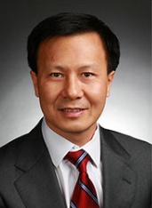 Haipeng Xiao (肖海鹏)合作论文数The First Affiliated Hospital, Sun Yat-sen University1Xiaobin Chen合作论文数1
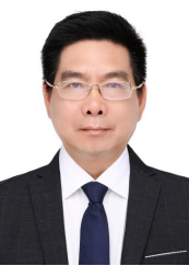 Zhangui Tang (唐瞻贵)合作论文数Hunan Xiangya Stomatological Hospital, Central South University1Ba Ruikai合作论文数1Fangqiao Niu (牛芳桥)合作论文数1Zhiwei Yan (闫志伟)合作论文数1




