Precision immunotherapy is critically hampered by the nonspecific toxicity of cGAS-STING pathway agonists. We overcome this fundamental barrier with a programmable DNA nanomaterial that operates as a logic-gated theranostic agent at the organelle level. Our nanodevice targets mitochondria and uses an integrated catalytic circuit to decipher the presence of oncogenic microRNA-21 (miR-21). Upon positive identification, it triggers the in situ architectural assembly of a physically disruptive DNA network on the mitochondrial surface. This targeted structural stress inflicts profound membrane damage, weaponizing the tumor cell's own mitochondrial DNA as a precision-guided agonist to ignite a powerful, localized STING-mediated immune assault. This strategy provides a dual function, enabling amplified diagnostic imaging of its molecular trigger while orchestrating the profound suppression of both primary and metastatic tumors in vivo with undetectable systemic toxicity. This work establishes a new design principle for intelligent therapeutics and defines a new therapeutic paradigm, the direct conversion of a fleeting molecular signal into a stable, physical, and immunomodulatory structure, forging a new frontier for dynamic materials in precision medicine.
Cancer immunotherapy holds promise for improving the efficacy of cancer treatment; however, low response rates remain a considerable challenge. Photodynamic therapy has a potential to be effective in immunotherapy, but it is limited by the inabilities to target tumor and limitation of reactive oxygen species (ROS) generation by hypoxia. Here, mitochondria-targeted zinc phthalocyanines (ZnPcs) are developed to precisely induce pyroptosis and activate immune responses. Cationic moieties incorporated in the ZnPc core allow strong localization in the mitochondrion and avoid aggregation of ZnPc, which serves as the highest site-specific production of ROS through the irradiation process. ZnPc-4 is among the synthesized derivatives that inhibit oxidative phosphorylation, relieving hypoxia and increasing type I/II ROS to cause mitochondrial dysfunction, which eventually triggers pyroptosis. Encapsulation of ZnPc-4 within DSPE-PEG2000-cRGDfk nanoparticle (ZnPc-NP) enhances its tumor-targeted capability and biocompatibility. In vivo, ZnPc-NP triggers immunogenic pyroptosis, eliciting potent anti-tumor immunity. In addition, ZnPc-NP combines with αPD-1, significantly inhibiting tumor metastasis and recurrence. This study establishes a dual-targeted photodynamic platform that overcomes microenvironmental constraints to potentiate cancer immunotherapy.
Hypochlorous acid (HOCl) serves as a vital molecule in innate immune defense, and fluctuations of lysosomal HOCl levels may be linked to a range of physiological and pathological processes, which make the quantitative detection of lysosomal HOCl a necessity. Herein, we report the design and synthesis of a novel fluorescent probe, PD-OCl, which is constructed by conjugating phenothiazine and dicyanoisophorone moieties and exhibits near-infrared (NIR) emission and large Stokes shifts. Notably, PD-OCl can function as a robust ratiometric fluorescent probe for HOCl detection, exhibiting exceptional selectivity, high sensitivity, and rapid response kinetics. Additionally, the PD-OCl has been successfully applied for monitoring both exogenous and endogenous HOCl in living cells, where it displays excellent lysosomal targeting capability, providing a visualized and ratiometric approach for tracing the variations of subcellular HOCl levels. This work establishes PD-OCl as a promising candidate for investigating lysosomal HOCl levels in LPS-induced oxidative stress state cells. A new probe, PD-OCl, with NIR emission and relatively large Stokes shifts was prepared. PD-OCl could detect HOCl with dual-channel ratiometric fluorescence analysis. The sensing was characterized with high selectivity and sensitivity, and a fast response. PD-OCl could image exogenous and endogenous lysosomal HOCl in living cells.
Tumor tertiary lymphoid structures (TLS), especially mature TLS (mTLS), have been associated with better prognosis and improved responses to immune checkpoint blockade (ICB), but the underlying mechanisms remain incompletely understood. Here, by performing single-cell RNA, antigen receptor sequencing and spatial transcriptomics on tumor tissue from head and neck squamous cell carcinoma (HNSCC) patients with different statuses of TLS, we observe that mTLS are enriched with stem-like T cells, and B cells at various maturation stages. Notably, progenitor exhausted CD4+ T cells, with features resembling follicular helper T cells, support these responses, by activating B cells to produce plasma cells in the germinal center, and interacting with DC-LAMP+ dendritic cells to support CD8+ T cell activation. Conversely, non-mTLS tumors do not promote local anti-tumor immunity which is abundant of immunosuppressive cells or a lack of stem-like B and T cells. Furthermore, patients with mTLS manifest improved overall survival and response to ICB compared to those with non-mTLS. Overall, our study provides insights into mechanisms underlying mTLS-mediated intra-tumoral immunity events against cancer.
While the mutational landscape across early T-cell precursor acute lymphoblastic leukemia (ETP-ALL) and ETP-like leukemia is known, establishing a unified framework that activates stem cell genes characteristic of these tumors remains elusive. Using complementary mouse and human models, chromatin mapping, and enhancer profiling, we show that the coactivator ZMIZ1 promotes normal and malignant ETP population growth by inducing the transcription factor MYB in feedforward circuits to convergently activate oncogenes (MEF2C, MYCN, and BCL2) through essential enhancers. A key superenhancer, the N-Myc regulating enhancer (NMRE), drives malignant ETP population growth but is dispensable for normal lymphopoiesis. This network of stem cell superenhancers identifies treatment-resistant tumors and poor survival outcomes; unifies diverse ETP-ALLs; and contributes to cardinal features of the recently genomically identified high-risk bone marrow progenitor-like (BMP-like) ETP-ALL tumor-stem cell/myeloid gene expression, inhibited NOTCH1-induced T-cell development, aggressive clinical behavior, and venetoclax sensitivity. Since ZMIZ1 is dispensable for essential homeostasis, it might be possible to safely target this network to treat high-risk diseases.
There is an urgent need to find targeted agents for T cell acute lymphoblastic leukemia (T-ALL). NOTCH1 is the most frequently mutated oncogene in T-ALL, but clinical trials showed that pan-Notch inhibitors caused dose-limiting toxicities. Thus, we shifted our focus to ETS1, which is one of the transcription factors that most frequently co-bind Notch-occupied regulatory elements in the T-ALL context. To identify the most essential enhancers, we performed a genome-wide CRISPRi screen of the strongest ETS1-dependent regulatory elements. The top-ranked element is located in an intron of AHI1 that interacts with the MYB promoter and is amplified with MYB in approximately 8.5% of patients with T-ALL. Using mouse models, we showed that this enhancer promoted self-renewal of hematopoietic stem cells and T cell leukemogenesis, maintained early T cell precursors, and restrained myeloid expansion with aging. We named this enhancer the hematopoietic stem cell MYB enhancer (H-Me). The H-Me showed limited activity and function in committed T cell progenitors but was accessed during leukemogenesis. In one T-ALL context, ETS1 bound the ETS motif in the H-Me to recruit cBAF to promote chromatin accessibility and activation. ETS1 or cBAF degraders impaired H-Me function. Thus, we identified a targetable stem cell element that was co-opted for T cell transformation.
In T-cell acute lymphoblastic leukemia (T-ALL), Notch variants are the most common oncogenic mutations, but clinical trials showed excessive toxicity of pan-Notch inhibitors. In response, we refocused to ETS1, which we and others previously showed is the top transcription factor that most frequently co-binds Notch-occupied elements in T-ALL. Here, we performed an unbiased genome-wide CRISPR-interference screen of the strongest ETS1-dependent enhancers. The #2 ranked element was the Notch-MYC enhancer (N-Me). The #1 ranked element was a +140kb Notch-bound enhancer interacting with the MYB promoter that we named the “ETS-MYB enhancer” or E-Me. MYB is an oncogene across all T-ALL subgroups. Chromatin profiling showed that the E-Me is highly active and accessible in hematopoietic stem cells (HSCs) and then silenced during differentiation. In contrast, the E-Me is highly active in Notch-type T-ALL. Therefore, we hypothesized that the E-Me is a HSC enhancer that is reactivated in T-ALL cells to drive MYB expression and promote population cell growth. To test this, we first determined the physiological role of the E-Me in hematopoietic cells using a novel E-Me conditional knockout mouse. E-Me deletion reduced Myb expression in HSCs 2-fold and increased absolute numbers 2.1-fold, whereas other hematopoietic populations were minimally affected. E-Me-deficient HSCs were defective as they reconstituted poorly in serial competitive bone marrow transplants and were depleted 6.2-fold in aged mice. Lastly, germline E-Me inactivation had no long-term effects on mouse weight or survival. These data suggest that the E-Me has limited function in committed T-cell progenitors and normal physiology but is important for HSC long-term self-renewal. We next wanted to examine E-Me function in murine and human T-ALL leukemogenesis. To do this, we first generated Notch-induced and Lmo2-induced T-ALL mouse models. E-Me deletion during initiation or maintenance reduced blast counts 38-to-105 fold and significantly prolonged survival. Next, we transduced E-Me sgRNAs into human T-ALL cell lines, which suppressed MYB expression 3.3-to-5.3-fold and inhibited cell proliferation 17-to-113-fold. These results indicate that the E-Me has major importance in murine and human leukemic cell growth and MYB induction. Following this, we wanted to understand how ETS1 induces E-Me activity. We first performed HOMER analysis of the E-Me and identified a single conserved ETS1-binding motif. Mutating this site decreased pulldown of ETS1 but no other transcription factor in reverse ChIP mass spectrometry and abrogated E-Me activity in reporter assays. In contrast, Notch inhibitors had no effect. Next, we generated a mouse model with mutation of the ETS1 site. Sequencing of ETS1 ChIP pulldowns confirmed the inability of ETS1 to bind the mutated motif. These mutant mice showed no significant alterations during steady state thymopoiesis but had impaired T-cell regeneration after sublethal irradiation. Dual reverse ChIP and co-IP mass spectrometry screens in a T-ALL cell line identified cBAF, a chromatin remodeling complex, as a top ranked ETS1 cofactor that is recruited to the E-Me. To confirm this, we genetically degraded ETS1 and found 2.5-fold reduced cBAF occupancy, 3.2-fold reduced H3K27ac signals, and 4.9-fold reduced ATAC-seq signals at the E-Me. ETS1 deprivation also reduced occupancy of Notch-associated transcriptional regulators. Lastly, AU-15330, a PROTAC degrader of cBAF, impaired E-Me H3K27ac signals 2.1-11.1-fold and reduced MYB protein levels 2.3-9.7-fold. These data suggest that ETS1 recruits cBAF after transformation to promote chromatin accessibility at the E-Me. There is an unmet need to identify the most important oncogenic enhancers and find ways to safely eject transcription factors bound to these elements as potential therapies. In addressing this, our unbiased screen revealed the top importance of a stem cell enhancer, the E-Me, which was ranked higher than the N-Me. We also suggest ways to inactivate the E-Me with cBAF and ETS1 degraders, which mouse studies predict would be safer than pan-Notch or pan-MYB inhibition. Finally, the literature provides ample examples of Notch having a central role in T-ALL. In contrast, we identify a top-ranked oncogenic enhancer that is independent of Notch but requires ETS1 to remodel chromatin to enable transcription factor complex assembly and function.
Our current understanding of the kinetics and dynamics of erythroid differentiation is based almost entirely on the ex vivo expansion of cultured hematopoietic progenitor cells. In this study, we used an erythroid-specific, inducible transgenic mouse line to investigate for the first time, the in vivo erythroid differentiation kinetics under steady-state conditions. We demonstrated that bipotent premegakaroycyte/erythroid (PreMegE) progenitor cells differentiate into erythroid-committed proerythroblast/basophilic erythroblasts (ProBasoE) after 6.6 days under steady-state conditions. During this process, each differentiation phase (from PreMegE to precolony forming unit-erythroid [PreCFU-E], PreCFU-E to CFU-E, and CFU-E to ProBasoE) took ∼2 days in vivo. Upon challenge with 5-flurouracil (5-FU), which leads to the induction of stress erythropoiesis, erythroid maturation time was reduced from 6.6 to 4.7 days. Furthermore, anemia induced in 5-FU-treated mice was shown to be due not only to depleted bone marrow erythroid progenitor stores but also to a block in reticulocyte exit from the bone marrow into the circulation, which differed from the mechanism induced by acute blood loss.
Nonsmall cell lung cancer (NSCLC) is highly malignant with limited treatment options, platinum-based chemotherapy is a standard treatment for NSCLC with resistance commonly seen. NSCLC cells exploit enhanced antioxidant defense system to counteract excessive reactive oxygen species (ROS), which contributes largely to tumor progression and resistance to chemotherapy, yet the mechanisms are not fully understood. Recent studies have suggested the involvement of histones in tumor progression and cellular antioxidant response; however, whether a major histone variant H1.2 (H1C) plays roles in the development of NSCLC remains unclear. Herein, we demonstrated that H1.2 was increasingly expressed in NSCLC tumors, and its expression was correlated with worse survival. When crossing the H1c knockout allele with a mouse NSCLC model (KrasLSL-G12D/+), H1.2 deletion suppressed NSCLC progression and enhanced oxidative stress and significantly decreased the levels of key antioxidant glutathione (GSH) and GCLC, the catalytic subunit of rate-limiting enzyme for GSH synthesis. Moreover, high H1.2 was correlated with the IC50 of multiple chemotherapeutic drugs and with worse prognosis in NSCLC patients receiving chemotherapy; H1.2-deficient NSCLC cells presented reduced survival and increased ROS levels upon cisplatin treatment, while ROS scavenger eliminated the survival inhibition. Mechanistically, H1.2 interacted with NRF2, a master regulator of antioxidative response; H1.2 enhanced the nuclear level and stability of NRF2 and, thus, promoted NRF2 binding to GCLC promoter and the consequent transcription; while NRF2 also transcriptionally up-regulated H1.2. Collectively, these results uncovered a tumor-driving role of H1.2 in NSCLC and indicate an "H1.2-NRF2" antioxidant feedforward cycle that promotes tumor progression and chemoresistance.
Activated Notch signaling is highly prevalent in T-cell acute lymphoblastic leukemia (T-ALL) but pan-Notch inhibitors were toxic in clinical trials. To find alternative ways to target Notch signals, we investigated Cell division cycle 73 (Cdc73), which is a Notch cofactor and component of transcriptional machinery, a potential target in T-ALL. While we confirmed previous work that CDC73 interacts with NOTCH1, we also found that the interaction in T-ALL was context-dependent and facilitated by the lymphoid transcription factor ETS1. Using mouse models, we showed that Cdc73 is important for Notch-induced T-cell development and T-ALL maintenance. Mechanistically, Cdc73, Ets1, and Notch intersect chromatin at promoters and enhancers to activate oncogenes and genes that are important for DNA repair and oxidative phosphorylation. Consistently, Cdc73 deletion in T-ALL cells induced DNA damage and impaired mitochondrial function. Our data suggests that Cdc73 might promote a gene expression program that was eventually intersected by Notch to mitigate the genotoxic and metabolic stresses of elevated Notch signaling. We also provide mechanistic support for testing inhibitors of DNA repair, oxidative phosphorylation, and transcriptional machinery. Inhibiting pathways like Cdc73 that intersect with Notch at chromatin might constitute a strategy to weaken Notch signals without directly targeting the Notch complex.
R-loops are regulators of many cellular processes and are threats to genome integrity. Therefore, understanding the mechanisms underlying the regulation of R-loops is important. Inspired by the findings on RNase H1-mediated R-loop degradation or accumulation, we focused our interest on the regulation of RNase H1 expression. In the present study, we report that G9a positively regulates RNase H1 expression to boost R-loop degradation. CHCHD2 acts as a repressive transcription factor that inhibits the expression of RNase H1 to promote R-loop accumulation. Sirt1 interacts with CHCHD2 and deacetylates it, which functions as a corepressor that suppresses the expression of downstream target gene RNase H1. We also found that G9a methylated the promoter of RNase H1, inhibiting the binding of CHCHD2 and Sirt1. In contrast, when G9a was knocked down, recruitment of CHCHD2 and Sirt1 to the RNase H1 promoter increased, which co-inhibited RNase H1 transcription. Furthermore, knockdown of Sirt1 led to binding of G9a to the RNase H1 promoter. In summary, we demonstrated that G9a regulates RNase H1 expression to maintain the steady-state balance of R-loops by suppressing the recruitment of CHCHD2/Sirt1 corepressors to the target gene promoter.
Non-invasive cell regulation represents a promising approach for on-site cell modulation, but is confronted with inaccuracy or poor cell selectivity. Herein, by virtue of the interfacial-activated concatenate DNA circuit, an activated recombination-to-modulation (ARM) machinery is developed to achieve accurate membrane imaging and effective cell modulation. Bioorthogonal signal transduction through the catalytic hairpin assembly circuit can continuously regenerate triggers for activating the subsequent circuit, with no activator consumption and occupation. The ARM strategy utilizes the amplified hybridization chain reaction to sensitively rearrange membrane receptors for blocking the related signaling pathways and indirectly modulating cell behaviors. Based on these two membrane receptor inputs, the controllably-activated ARM strategy enables the identification of target cells among similar interfering cells with high accuracy and sensitivity. This machinery has demonstrated good performance in the efficient inhibition of tumor metastasis via the disruption of HGF/c-Met-signaling pathway. The modular-designed ARM machinery provides a general platform for non-invasive and specific cell modulation, suggesting a broad opportunity for advanced exploration of cellular metabolism behaviors.
Herein, we developed a reliable and portable biosensor (TDR-PGM nanomachine) for the sensitive detection of microRNA by integrating an efficient toehold-mediated strand displacement reaction module (TDR) and a personal glucose meter (PGM). The system provides a versatile methodology for microRNA detection in real samples and holds broad prospects in point-of-care diagnosis.
Synthetic catalytic DNA circuits have been recognized as a promising signal amplification toolbox for sensitive intracellular imaging, yet their selectivity and efficiency are always constrained by uncontrolled off-site signal leakage and inefficient on-site circuitry activation. Thus, the endogenously controllable on-site exposure/activation of DNA circuits is highly desirable for achieving the selective imaging of live cells. Herein, an endogenously activated DNAzyme strategy was facilely integrated with a catalytic DNA circuit for guiding the selective and efficient microRNA imaging in vivo. To prevent the off-site activation, the circuitry constitute was initially caged without sensing functions, which could be selectively liberated by DNAzyme amplifier to guarantee the high-contrast microRNA imaging in target cells. This intelligent on-site modulation strategy can tremendously expand these molecularly engineered circuits in biological systems.
Surgery‐induced renal ischemia and reperfusion (I/R) injury and nephrotoxic drugs like cisplatin can cause acute kidney injury (AKI), for which there is no effective therapy. Lipid accumulation is evident following AKI in renal tubules although the mechanisms and pathological effects are unclear. Here, we report that Ehmt2‐ encoded histone methyltransferase G9a is upregulated in patients and mouse kidneys after AKI. Renal tubular specific knockout of G9a ( Ehmt2 Ksp ) or pharmacological inhibition of G9a alleviates lipid accumulation associated with AKI. Mechanistically, G9a suppresses transcription of the lipolytic enzyme Ces1 ; moreover, G9a and farnesoid X receptor (FXR) competitively bind to the same promoter regions of Ces1 . Ces1 is consistently observed to be downregulated in the kidney of AKI patients. Pharmacological inhibition of Ces1 increases lipid accumulation, exacerbates renal I/R‐injury and eliminates the beneficial effects on AKI observed in Ehmt2 Ksp mice. Furthermore, lipid‐lowering atorvastatin and an FXR agonist alleviate AKI by activating Ces1 and reducing renal lipid accumulation. Together, our results reveal a G9a/FXR‐Ces1 axis that affects the AKI outcome via regulating renal lipid accumulation.
Deoxyribozyme (DNAzyme) is single-stranded catalytic DNA that possesses various physical and chemical features, including small molecular weight, higher stability, excellent programmability, and cost-effectiveness. Considerable efforts are spent on the incorporation of RNA-cleaving DNAzyme (RCD) into biosensor and biomedical researches. However, their in vivo application is constrained by off-target activation (always turn-on) in complex biological environment. Therefore, there is a pursuit to develop spatiotemporally controlled DNAzyme in desired locations and time-points. Herein, we provide a timely and comprehensive overview of various chemical modification and structural reconfiguration methods to realize the specific stimulation/regulation of RCD activities, including metal ions, small molecules, nucleic acids, proteins, and external photo/thermal stimuli. The specific examples of using chemically and/or structurally caged RCD for stimuli-responsive biosensing and biomedical research have also been reviewed within the past five years. Lastly, the challenges and perspectives of stimuli-responsive RCD are suggested to further advance the RCD toolbox in clinical application.(c) 2022 Elsevier B.V. All rights reserved.
A non-enzymatic circuitry activation strategy can realize an orthogonally controlled catalytic DNA (CCD) circuit through multiple molecular recognition events and progressively accelerated signal amplification, as reported by Fuan Wang and co-workers in their Research Article (e202206529). The on-site activated CCD circuitry system has considerable potential for high-precision in vivo imaging of low-abundance microRNA.
The wide extracellular-intracellular distribution of microRNA requires the on-site, robust and efficient activation of catalytic DNA circuits inside live cells. Herein, we develop an efficient non-enzymatic circuitry activation strategy to realize the orthogonally controlled catalytic DNA (CCD) circuit for achieving high-fidelity in vivo microRNA imaging through multiply guaranteed molecular recognition and progressively accelerated signal amplification. For predictable on-site activation and useful catalytic efficiency, the dominating circuitry fuel strand was initially split into inactive fuel subunits that were grafted into an auxiliary catalytic circuit. There, the in-cell-specific mRNA triggered the orthogonal amplification of the active fuel strands for sensitive target detection through the chief entropy-driven catalytic DNA circuit. We believe that the on-site orthogonal circuitry activation method can contribute to clinical diagnosis and prognosis.
Mitotic catastrophe (MC) is a suppressive mechanism that mediates the elimination of mitosis-deficient cells through apoptosis, necrosis or senescence after M phase block. SIRT1 is involved in the regulation of several cellular processes, including autophagy. However, the relationship between SIRT1 and MC has been largely obscure. Our study highlights that SIRT1 might be involved in the regulation of MC. We have shown that degradation of the SIRT1 protein via proteasome and lysosomal pathway was accompanied by MC induced via BMH-21. Overexpression of SIRT1 alleviated MC by decreasing the proportion of apoptotic and multinuclear cells induced by G2/M block and triggered autophagy whereas knockdown of SIRT1 aggravated MC and repressed autophagy. Furthermore, we found that serum starvation triggered autophagy evidently generated lower MC whereas siRNA of ATG5/7 suppressed autophagy leading to higher MC. ChIP analysis revealed that SIRT1 could bind to the promoter of BubR1, a component of spindle assembly checkpoint (SAC), to upregulate its expression. Overexpression of BubR1 decreased MC whereas knockdown of BubR1 increased it. These results reveal that SIRT1 regulates MC through autophagy and BubR1 signaling, and provide evidence for SIRT1, autophagy and BubR1 being the potential cancer therapeutic targets.
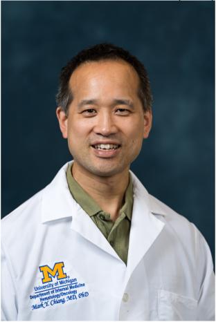 Mark Chiang合作论文数University of Michigan Cancer Center;Hematology/Oncology Division, Medical School, University of Michigan13
 Fuan Wang (王富安)合作论文数College of Chemistry and Molecular Sciences, Wuhan University13
 Ling Zheng (郑凌)合作论文数Department of Cell Biology, College of Life Sciences, Wuhan University11
 Anna C McCarter合作论文数10Kun Huang (黄昆)合作论文数华中科技大学9
 Lijia Li (李立家)合作论文数College of Life Sciences, Wuhan University9
 Russell JH. Ryan合作论文数Department of Pathology, Medical School, University of Michigan8
 Xiaoqing Liu (刘晓庆)合作论文数College of Chemistry and Molecular Sciences, Wuhan University8Yiran Liu合作论文数7
 Dong Yang (杨东)合作论文数Union Hospital, Tongji Medical College, Huazhong University of Science and Technology7
 Yan Ran合作论文数Departments of Biological Sciences, Wright State University6Ashley Melnick合作论文数5R Kuick合作论文数5Yuqiu He (何雨秋)合作论文数5Hao Zhang合作论文数5Shanshan Yu合作论文数5Yangmian Yuan合作论文数5Jinhua Shang合作论文数Wuhan Univ, Coll Chem & Mol Sci, Wuhan 430072, Peoples R China5
 Fei Gao (高飞)合作论文数School of Mathematics and Statistics, Wuhan University of Technology5Hou Haoli合作论文数5Zhou Hong合作论文数5
 Yu Zhang (张煜)合作论文数School of Basic Medical Sciences, Lanzhou University5
 Hong Chen (陈宏)合作论文数Union Hospital, Tongji Medical College of Huazhong University of Science and Technology5
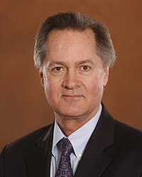 Robert Petersen合作论文数College of Medicine, Central Michigan University;Case Western Reserve University5Amparo Serna Alarcon合作论文数4Elizabeth Choe合作论文数4Nancy Pinnell合作论文数4Cher Sha合作论文数4Le Li合作论文数4
 Linda C. Samuelson合作论文数Division of Gastroenterology, Medical School, University of Michigan;Center for Cell Plasticity and Organ Design, Medical School, University of Michigan;Department of Molecular & Integrative Physiology, Medical School, University of Michigan4
 Tomasz Cierpicki合作论文数Biophysics, College of Literature, Science, and the Arts, University of Michigan-Ann Arbor4
 Ivan P. Maillard合作论文数Division of Hematology and Oncology, Department of Medicine, Perelman School of Medicine, University of Pennsylvania4
 Pu Wang (王璞)合作论文数College of Horticulture and Forestry Sciences, Huazhong Agricultural University4
 Ying Zhu (朱应)合作论文数College of Life Sciences, Wuhan University;State Key Laboratory of Virology, Wuhan University4
 Doug Engel合作论文数Medical School, University of Michigan4Chengyu Liu合作论文数3Ningjie Ma合作论文数3Fatema Akter合作论文数3Yequn Wu合作论文数3Yixue Huang合作论文数3Yunhao Xie合作论文数3Weiwei Zhao合作论文数3Nicholas Kunnath合作论文数3
 Rohan Kodgule合作论文数Sch Med, Washington Univ3Shizhen He合作论文数3Mingrui Xiong合作论文数3Mayank Gautam合作论文数3Ruomeng Li合作论文数College of Chemistry and Molecular Sciences, Wuhan University, Wuhan, 430072, P. R. China.3Erin Kim合作论文数3




